


Article Figures & Data
Figures
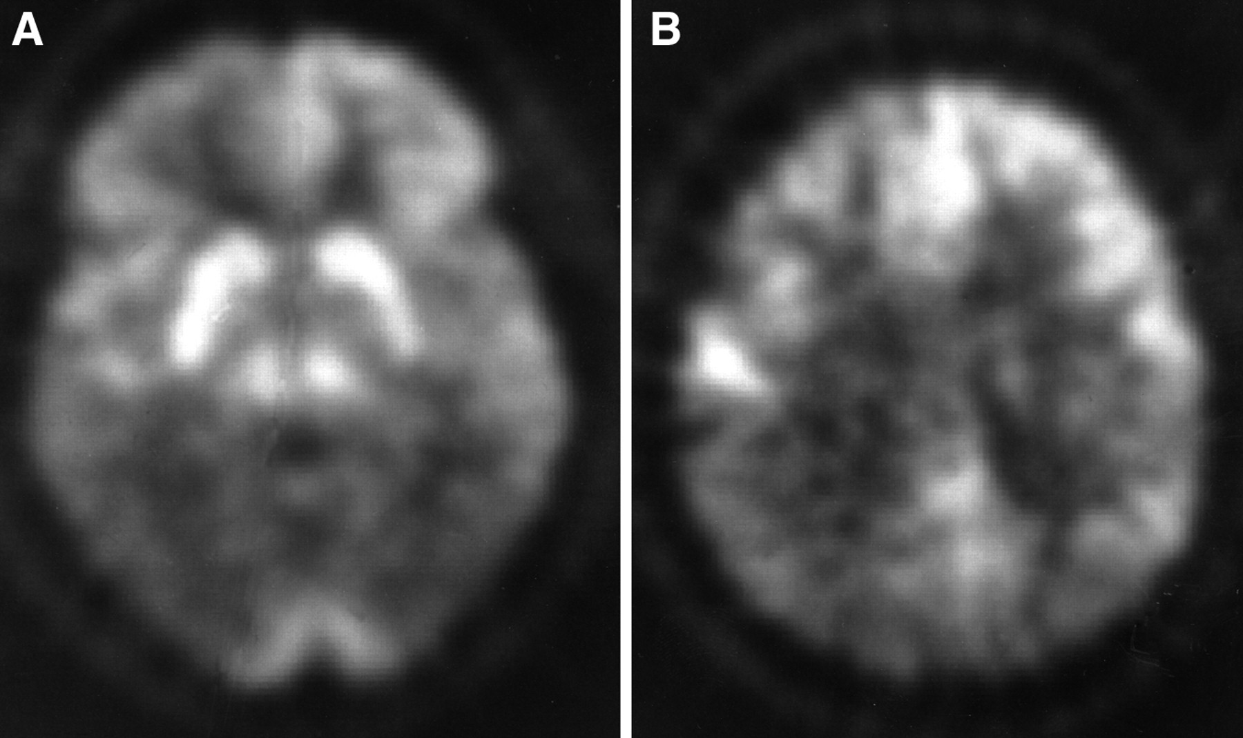
- FIGURE 1.
FDG PET scan of 54-y-old woman with progressive dementing illness (patient 7; Table 1). Note significant reduction in FDG in parietal, temporal, and frontal cortices (arrows). The metabolic pattern noted on this particular study would be considered classic for AD. The patient had AD, verified by pathological examination.
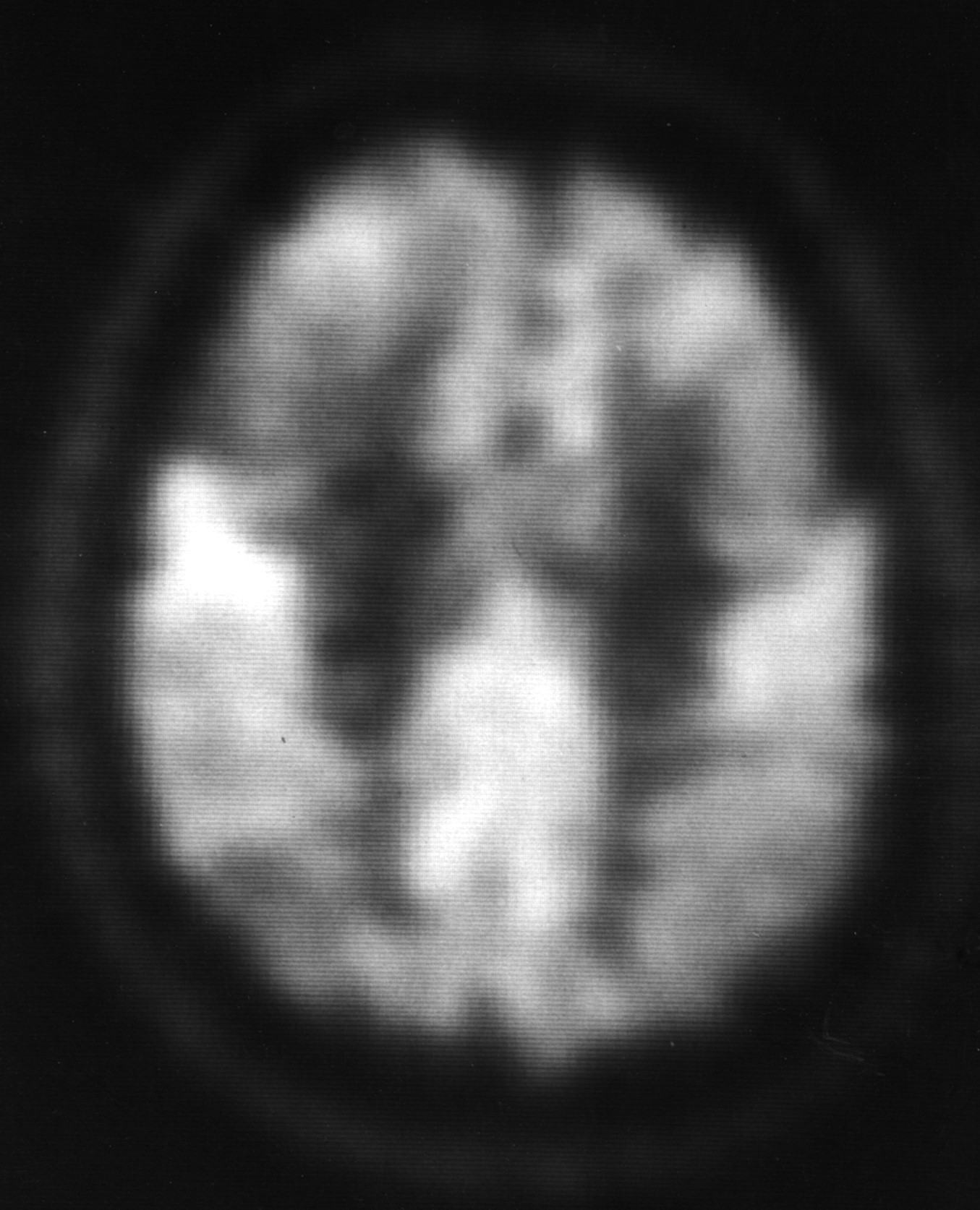
- FIGURE 2.
FDG PET scans of 60-y-old man with a rapidly progressive dementia syndrome (patient 2; Table 1). Patient had both biopsy and autopsy confirmation of diagnosis of Creutzfeldt-Jacob disease. FDG PET scans at 2 levels show marked reduction in FDG uptake in temporo-parietal (A) and parietal (B) regions bilaterally. A pattern very similar to AD has been reported in previous cases of Creutzfeldt-Jacob disease studied with FDG PET imaging (31–33).
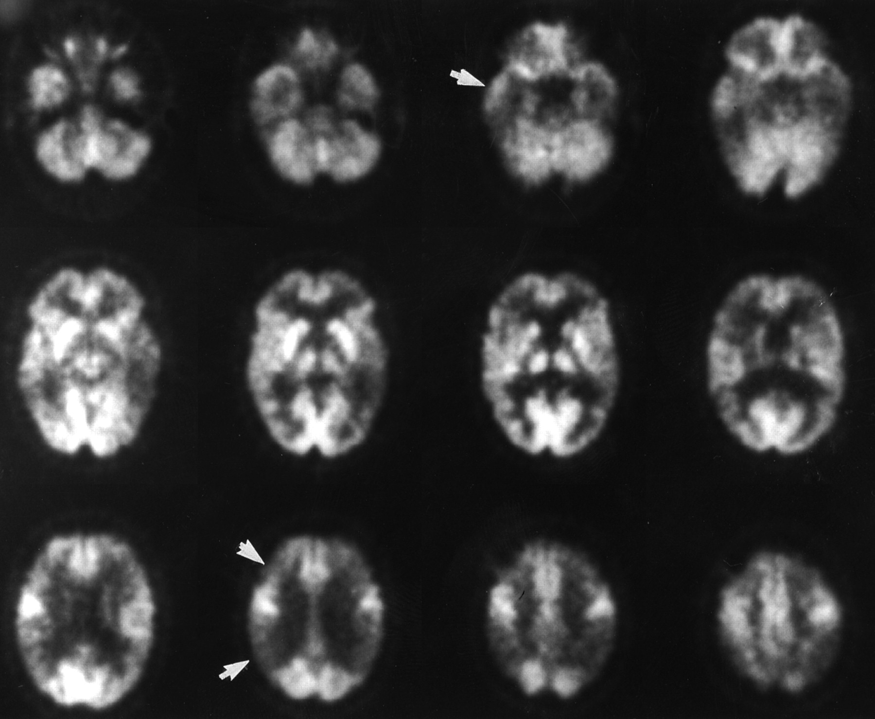
- FIGURE 3.
(A–C) FDG PET scans at 3 transaxial levels of 64-y-old man with progressive dementing illness (patient 5; Table 1). Pathologic diagnosis was nonspecific neuronal degeneration. FDG PET scan was interpreted as abnormal but not AD pattern. FDG PET images at 3 levels are shown. Note asymmetric FDG uptake, particularly in the left frontal area (arrow) on multiple imaging levels. Temporo-parietal hypometabolism was not a prominent finding in this patient with non–Alzheimer's type dementia.
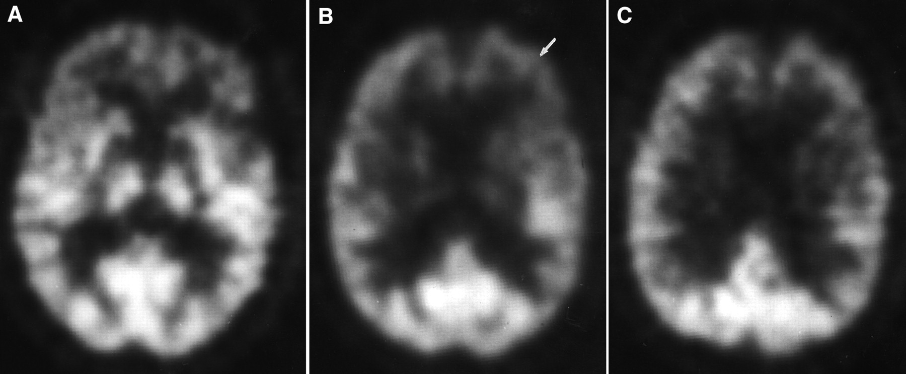
- FIGURE 4.
FDG PET scan of 66-y-old man with progressive dementia with extrapyramidal features (patient 13; Table 1). This individual had histologically proven progressive supranuclear palsy. FDG PET scan was interpreted as abnormal but not AD pattern. FDG PET image showed reduction in FDG uptake in frontal regions bilaterally. Prominent frontal hypometabolism has been previously described in patients with progressive supranuclear palsy with FDG PET (34–36).


Tables
Subject no. Sex Age at PET (y) Age at path. (y) Clinical diagnosis Path. diagnosis Biopsy or autopsy Interpreter PET grade* Interval PET to path. (mo) 1 F 56 60 Dementia AD Autopsy 3 44 2 M 60 61 CJD CJD Biopsy & autopsy 3 25 3 M 64 65 Dementia AD Autopsy 3 17 4 M 62 64 Probable AD AD Biopsy 3 −65 5 M 64 66 Dementia Neuronal deg. Autopsy 4 23 6 M 69 71 Possible AD AD Autopsy 2 28 7 F 54 54 Probable AD AD Biopsy 3 −2 8 M 73 75 Possible AD AD Autopsy 2 31 9 F 59 60 Probable AD AD Autopsy 3 16 10 F 69 71 Probable AD AD Autopsy 3 27 11 M 57 59 Probable AD AD Autopsy 3 33 12 M 64 67 Probable AD AD + Lewy Autopsy 3 44 13 M 66 69 Dementia PSP Autopsy 4 46 14 F 68 68 Dementia AD Biopsy 2 2 15 M 72 76 Probable AD AD Autopsy 3 48 16 F 74 76 PSP PSP + AD Autopsy 4 34 17 M 73 73 Probable AD AD Autopsy 3 5 18 F 80 81 PD/Lewy body Lewy body Autopsy 3 22 19 M 77 78 Probable AD AD Autopsy 4 9 20 M 72 76 Probable AD AD Autopsy 3 51 21 M 37 39 Toxic enceph. Preamyloid Autopsy 4 22 22 M 68 76 Pick's MLCD Autopsy 4 87 Path, pathologic diagnosis; CJD, Creutzfeldt-Jacob disease; Neuronal deg., neuronal degeneration; Lewy, Lewy body disease; PSP, progressive supranuclear palsy; PD, Parkinson's disease; enceph., encephalopathy; MLCD, mesio–limbo cortical dementia.
↵* See methods for description of PET score.
Results Sensitivity Specificity PV positive PV negative Accuracy Prob. AD only clinically: AD + other on path. 62.5% (10/16) 100% (6/6) 100% (10/10) 50% (6/12) 72.7% (16/22) Poss. or prob. AD clinically: AD + other on path. 75.0% (12/16) 100% (6/6) 100% (12/12) 60% (6/10) 81.8% (18/22) Prob. AD only clinically: AD only on path. 64.3% (9/14) 87.5% (7/8) 90% (9/10) 58.3% (7/12) 72.7% (16/22) Poss. or prob. AD clinically: AD only on path. 78.6% (11/14) 87.5% (7/8) 91.7% (11/12) 70% (7/10) 81.8% (18/22) PV, predictive value; Prob., probable; poss., possible; path., pathology.
Results Sensitivity Specificity PV positive PV negative Accuracy PET grade 2 or 3: path. AD only 92.9% (13/14) 62.5% (5/8) 81.3% (13/16) 83.3% (5/6) 81.8% (18/22) PET grade 2 or 3: path. AD and other 87.5% (14/16) 66.7% (4/6) 87.5% (14/16) 66.7% (4/6) 81.8% (18/22) PV, predictive value; Path., pathology.
In this issue


Jump to section
Related Articles
Cited By...
- BOLD Amplitude Correlates of Preclinical Alzheimers Disease
- Multi-transcriptomic analysis points to early organelle dysfunction in human astrocytes in Alzheimers disease
- A machine-learning approach for detection of local brain networks and marginally weak signals identifies novel AD/MCI differentiating connectomic neuroimaging biomarkers
- Clinical Neurology and Epidemiology of the Major Neurodegenerative Diseases
- Brain imaging in dementia
- Biomarkers in dementia: clinical utility and new directions
- Association of hypometabolism and amyloid levels in aging, normal subjects
- Diagnostic tests for Alzheimer disease: Judicious use can be helpful in clinical practice
- The Clinical Problem of Symptomatic Alzheimer Disease and Mild Cognitive Impairment
- Inclusion criteria provide heterogeneity in baseline profiles of patients with mild cognitive impairment: comparison of two prospective cohort studies
- Brain Imaging in Alzheimer Disease
- Effectiveness and Safety of 18F-FDG PET in the Evaluation of Dementia: A Review of the Recent Literature
- A tale of two tracers: The age of wisdom for dementia diagnosis?
- Early 11C-PIB Frames and 18F-FDG PET Measures Are Comparable: A Study Validated in a Cohort of AD and FTLD Patients
- Effects of Age on the Glucose Metabolic Changes in Mild Cognitive Impairment
- Imaging Approaches to Parkinson Disease
- What does fluorodeoxyglucose PET imaging add to a clinical diagnosis of dementia?
- Comparison of Regional Brain Volume and Glucose Metabolism Between Patients with Mild Dementia with Lewy Bodies and Those with Mild Alzheimer's Disease
- Dissociation of neuropathology from severity of dementia in late-onset Alzheimer disease
- APOE related alterations in cerebral activation even at college age
- Brain 18F-FDG PET in the Diagnosis of Neurodegenerative Dementias: Comparison with Perfusion SPECT and with Clinical Evaluations Lacking Nuclear Imaging
- Evaluating Early Dementia With and Without Assessment of Regional Cerebral Metabolism by PET: A Comparison of Predicted Costs and Benefits
- Direct Comparison of Spatially Normalized PET and SPECT Scans in Alzheimer's Disease
- SPECT Imaging in Dementias
- Practice parameter: Diagnosis of dementia (an evidence-based review): Report of the Quality Standards Subcommittee of the American Academy of Neurology
- A Tabulated Summary of the FDG PET Literature
- Evaluating Dementia Using PET: How Do We Put into Clinical Perspective What We Know to Date?