
Main menu

  • Home
  • Content
    • Current
    • Ahead of print
    • Past Issues
    • JNM Supplement
    • SNMMI Annual Meeting Abstracts
    • Continuing Education
    • JNM Podcasts
  • Subscriptions
    • Subscribers
    • Institutional and Non-member
    • Rates
    • Journal Claims
    • Corporate & Special Sales
  • Authors
    • Submit to JNM
    • Information for Authors
    • Assignment of Copyright
    • AQARA requirements
  • Info
    • Reviewers
    • Permissions
    • Advertisers
  • About
    • About Us
    • Editorial Board
    • Contact Information
  • More
    • Alerts
    • Feedback
    • Help
    • SNMMI Journals
  • SNMMI
    • JNM
    • JNMT
    • SNMMI Journals
    • SNMMI

User menu

  • Subscribe
  • My alerts
  • Log in
  • Log out
  • My Cart

Search

  • Advanced search
Journal of Nuclear Medicine
  • SNMMI
    • JNM
    • JNMT
    • SNMMI Journals
    • SNMMI
  • Subscribe
  • My alerts
  • Log in
  • Log out
  • My Cart
Journal of Nuclear Medicine

Advanced Search

  • Home
  • Content
    • Current
    • Ahead of print
    • Past Issues
    • JNM Supplement
    • SNMMI Annual Meeting Abstracts
    • Continuing Education
    • JNM Podcasts
  • Subscriptions
    • Subscribers
    • Institutional and Non-member
    • Rates
    • Journal Claims
    • Corporate & Special Sales
  • Authors
    • Submit to JNM
    • Information for Authors
    • Assignment of Copyright
    • AQARA requirements
  • Info
    • Reviewers
    • Permissions
    • Advertisers
  • About
    • About Us
    • Editorial Board
    • Contact Information
  • More
    • Alerts
    • Feedback
    • Help
    • SNMMI Journals
  • View or Listen to JNM Podcast
  • Visit JNM on Facebook
  • Join JNM on LinkedIn
  • Follow JNM on Twitter
  • Subscribe to our RSS feeds
Research ArticleCLINICAL INVESTIGATIONS

Grading of Tumors and Tumorlike Lesions of Bone: Evaluation by FDG PET

Michael Schulte, Doris Brecht-Krauss, Berno Heymer, Albrecht Guhlmann, Erich Hartwig, Michael R. Sarkar, Christoph G. Diederichs, Alexandra Von Baer, Jörg Kotzerke and Sven N. Reske
Journal of Nuclear Medicine October 2000, 41 (10) 1695-1701;
Michael Schulte
  • Find this author on Google Scholar
  • Find this author on PubMed
  • Search for this author on this site
Doris Brecht-Krauss
  • Find this author on Google Scholar
  • Find this author on PubMed
  • Search for this author on this site
Berno Heymer
  • Find this author on Google Scholar
  • Find this author on PubMed
  • Search for this author on this site
Albrecht Guhlmann
  • Find this author on Google Scholar
  • Find this author on PubMed
  • Search for this author on this site
Erich Hartwig
  • Find this author on Google Scholar
  • Find this author on PubMed
  • Search for this author on this site
Michael R. Sarkar
  • Find this author on Google Scholar
  • Find this author on PubMed
  • Search for this author on this site
Christoph G. Diederichs
  • Find this author on Google Scholar
  • Find this author on PubMed
  • Search for this author on this site
Alexandra Von Baer
  • Find this author on Google Scholar
  • Find this author on PubMed
  • Search for this author on this site
Jörg Kotzerke
  • Find this author on Google Scholar
  • Find this author on PubMed
  • Search for this author on this site
Sven N. Reske
  • Find this author on Google Scholar
  • Find this author on PubMed
  • Search for this author on this site
  • Article
  • Figures & Data
  • Info & Metrics
  • PDF
Loading

Article Figures & Data

Figures

  • Tables
  • FIGURE 1.
    • Download figure
    • Open in new tab
    • Download powerpoint
    FIGURE 1.

    A 14-y-old boy with thoracic high-grade chondroblastic osteosarcoma. (A) Radiograph shows osteolytic destruction of seventh rib. (B) MR image shows inhomogeneous uptake of gadolinium–diethylenetriaminepentaacetic acid within primary lesion and highly vascularized tumor satellite adjacent to diaphragm. (C) PET image before neoadjuvant chemotherapy shows inhomogeneous FDG uptake in primary lesion (T/B, 10.3) and homogeneous hypermetabolism of satellite (T/B, 11.7).
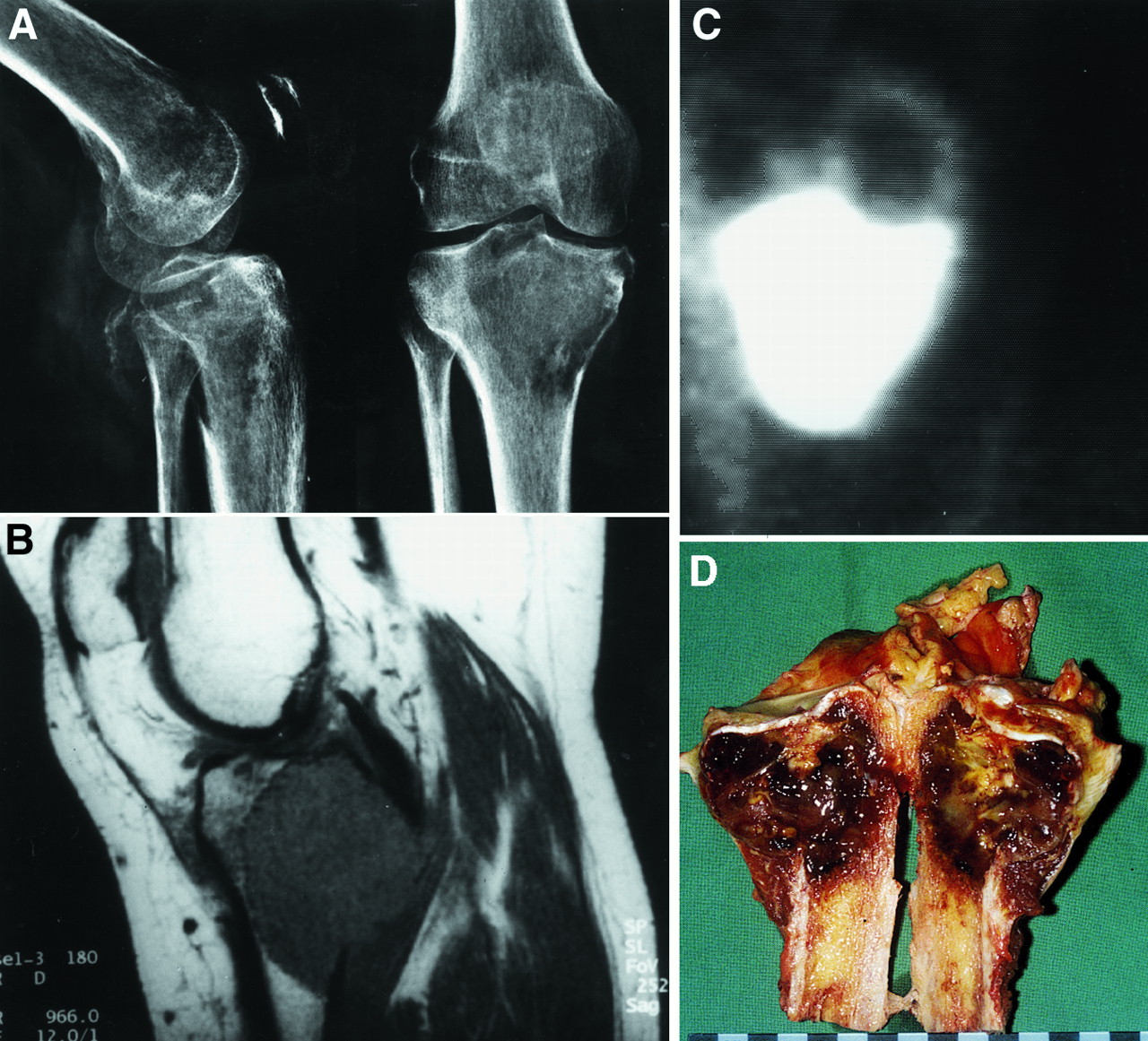
  • FIGURE 2.
    • Download figure
    • Open in new tab
    • Download powerpoint
    FIGURE 2.

    A 32-y-old woman with stage 3 giant cell tumor of tibial head. (A) Radiograph shows radiolucent, ill-defined lesion located eccentrically in epiphysis and adjacent metaphyseal region with destruction of dorsomedial cortex. (B) MR image shows homogeneous uptake of gadolinium–diethylenetriaminepentaacetic acid within lesion and tumoral infiltration of popliteus muscle. (C) PET image shows highly hypermetabolic lesion suggestive of malignant tumor (T/B, 35.0). (D) Gross specimen shows subchondral extension, cortical destruction, and typical hemorrhagic aspect of giant cell tumor.
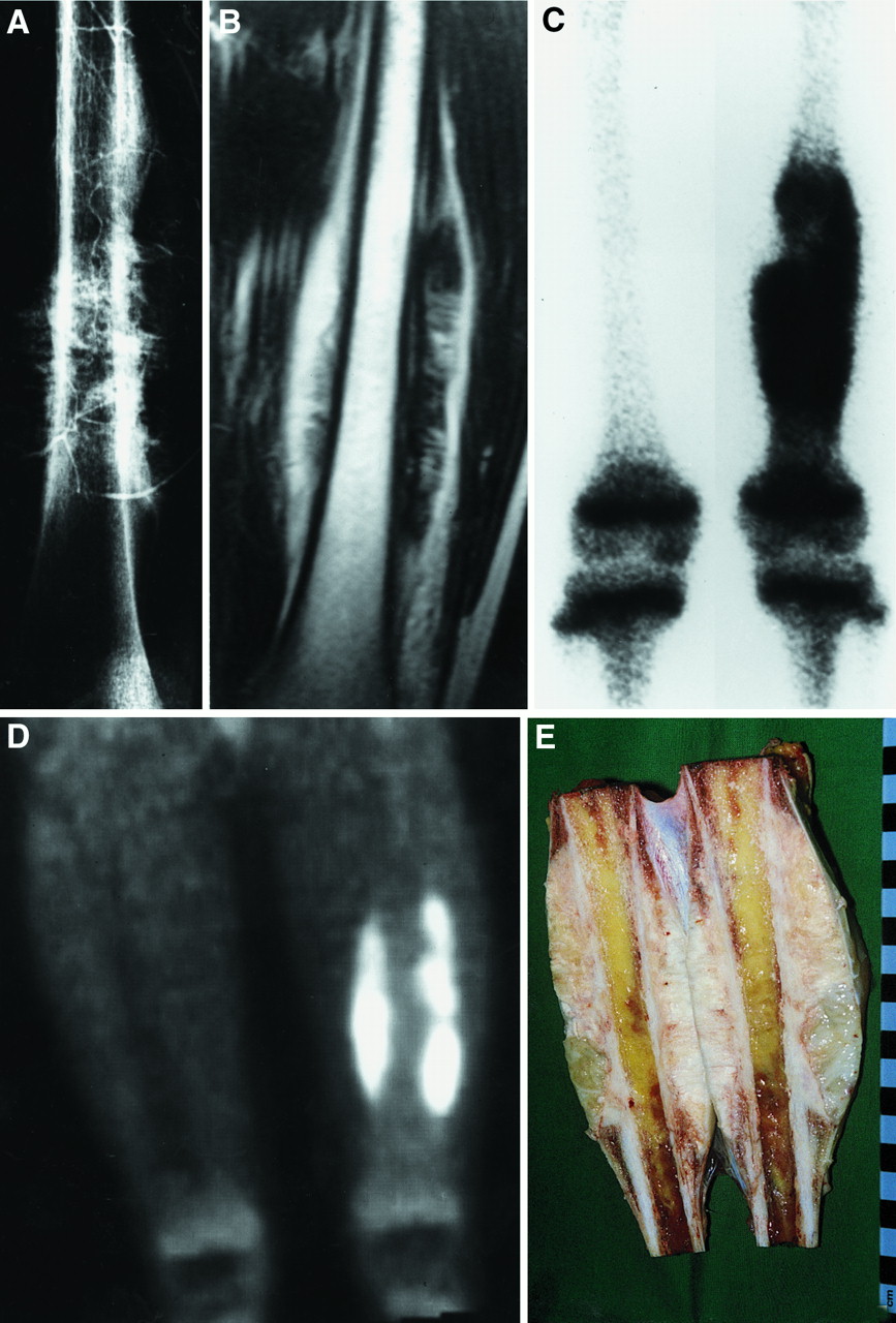
  • FIGURE 3.
    • Download figure
    • Open in new tab
    • Download powerpoint
    FIGURE 3.

    A 10-y-old girl with low-grade periosteal osteosarcoma of femur. (A) Angiogram shows moderate vascularization, saucerization of adjacent cortex, and radiating spicules. (B) MR image shows tumor located at external surface of bone without involvement of marrow cavity. (C) Bone scintigram shows hypermetabolic diaphyseal lesion without anatomic details. (D) PET image shows that tumor is surrounding femur (T/B, 6.1) and that medullary cavity is without elevated uptake, enabling definitive exclusion of classic central osteosarcoma. (E) Gross specimen shows lesion limited to external surface of bone, without medullary penetration.

Tables

  • Figures
    • View popup
    TABLE 1.

    Histologic Diagnosis and Distribution of T/B Values for 202 Skeletal Lesions

    DiagnosisnRangeMeanSDMedianP*
    Osteosarcoma443.3–33.211.17.29.1<0.001
    Ewing's sarcoma144.0–31.07.17.05.1<0.001
    Chondrosarcoma141.4–11.85.23.25.3<0.01
    Malignant fibrous histiocytoma63.3–26.013.08.614.6<0.001
    Angiosarcoma43.5–31.016.213.615.2<0.01
    Leiomyosarcoma227.2–73.0
    Other sarcomas215.9–20.5
    Chordoma53.6–12.56.93.66.6<0.01
    Giant cell tumor510.1–35.020.49.919.1<0.001
    Aneurysmatic bone cyst151.1–5.93.01.52.3NS
    Simple bone cyst91.0–3.51.70.71.5NS
    Chondroma91.4–2.82.30.52.3NS
    Osteochondroma81.0–2.92.10.62.1NS
    Fibrous dysplasia45.2–11.57.42.86.5<0.01
    Nonossifying fibroma61.2–18.66.86.94.5NS
    Desmoplastic fibroma22.2–7.0
    Chondroblastoma26.5–33.6
    Osteoid osteoma22.4–2.9
    Chondromyxoid fibroma13.0
    Osteofibrous dysplasia12.8
    Eosinophilic granuloma24.5–4.7
    Parathyroid osteopathy15.1
    Osteomyelitis111.1–24.15.67.32.4NS
    Other tumorlike lesions91.0–2.31.60.61.7NS
    Bone metastasis124.6–34.015.28.614.2<0.001
    Malignant lymphoma63.5–49.221.916.518.7<0.001
    Plasmacytoma61.3–15.18.45.69.5<0.05
    • ↵* Calculated by comparison with stage 1 and 2 benign lesions.

    • NS = not significant.

    • View popup
    TABLE 2.

    Distribution of T/B Values for Skeletal Lesions of Different Biologic Activity

    DiagnosisnRangeMeanSDMedianP*
    High-grade sarcoma703.3–73.011.910.68.1<0.001
    Low-grade sarcoma211.4–31.06.86.45.4<0.001
    Stage 3 benign lesions123.0–35.014.011.58.6<0.001
    Stage 1 and 2 benign lesions541.0–18.63.23.02.3
    • ↵* Calculated by comparison with stage 1 and 2 benign lesions.

PreviousNext
Back to top

In this issue

Journal of Nuclear Medicine
Vol. 41, Issue 10
October 1, 2000
  • Table of Contents
  • Table of Contents (PDF)
  • Index by author
Print
Download PDF
Article Alerts
Sign In to Email Alerts with your Email Address
Email Article

Thank you for your interest in spreading the word on Journal of Nuclear Medicine.

NOTE: We only request your email address so that the person you are recommending the page to knows that you wanted them to see it, and that it is not junk mail. We do not capture any email address.

Enter multiple addresses on separate lines or separate them with commas.
Grading of Tumors and Tumorlike Lesions of Bone: Evaluation by FDG PET
(Your Name) has sent you a message from Journal of Nuclear Medicine
(Your Name) thought you would like to see the Journal of Nuclear Medicine web site.
Citation Tools
Grading of Tumors and Tumorlike Lesions of Bone: Evaluation by FDG PET
Michael Schulte, Doris Brecht-Krauss, Berno Heymer, Albrecht Guhlmann, Erich Hartwig, Michael R. Sarkar, Christoph G. Diederichs, Alexandra Von Baer, Jörg Kotzerke, Sven N. Reske
Journal of Nuclear Medicine Oct 2000, 41 (10) 1695-1701;

Citation Manager Formats

  • BibTeX
  • Bookends
  • EasyBib
  • EndNote (tagged)
  • EndNote 8 (xml)
  • Medlars
  • Mendeley
  • Papers
  • RefWorks Tagged
  • Ref Manager
  • RIS
  • Zotero
Share
Grading of Tumors and Tumorlike Lesions of Bone: Evaluation by FDG PET
Michael Schulte, Doris Brecht-Krauss, Berno Heymer, Albrecht Guhlmann, Erich Hartwig, Michael R. Sarkar, Christoph G. Diederichs, Alexandra Von Baer, Jörg Kotzerke, Sven N. Reske
Journal of Nuclear Medicine Oct 2000, 41 (10) 1695-1701;
Twitter logo Facebook logo LinkedIn logo Mendeley logo
  • Tweet Widget
  • Facebook Like
  • Google Plus One
Bookmark this article

Jump to section

  • Article
    • Abstract
    • MATERIALS AND METHODS
    • RESULTS
    • DISCUSSION
    • CONCLUSION
    • Acknowledgments
    • Footnotes
    • REFERENCES
  • Figures & Data
  • Info & Metrics
  • PDF

Related Articles

  • Is There a Role for FDG PET in the Diagnosis of Musculoskeletal Neoplasms?
  • PubMed
  • Google Scholar

Cited By...

  • Metastatic Mimicker in Thyroid Cancer with Thyroglobulin Elevation and an FDG-Avid Recurrent Aneurysmal Bone Cyst
  • False-Positive Positron Emission Tomography in Patients With History of Malignancy
  • Correlation Between Glycolytic Phenotype and Tumor Grade in Soft-Tissue Sarcomas by 18F-FDG PET
  • NCCN Task Force: Clinical Utility of PET in a Variety of Tumor Types
  • Imaging Bone and Soft Tissue Tumors with the Proliferation Marker [18F]Fluorodeoxythymidine
  • PET for Sarcomas Other Than Gastrointestinal Stromal Tumors
  • Imaging of Bone Sarcomas
  • Diagnostic Value and Limitations of Fluorine-18 Fluorodeoxyglucose Positron Emission Tomography for Cartilaginous Tumors of Bone
  • PET Imaging of Osteosarcoma
  • Whole-Body 18F-FDG PET Identifies High-Risk Myeloma
  • Prognostic Significance of 18F-FDG and 99mTc-Methylene Diphosphonate Uptake in Primary Osteosarcoma
  • The Role of Quantitative 18F-FDG PET Studies for the Differentiation of Malignant and Benign Bone Lesions
  • Is There a Role for FDG PET in the Diagnosis of Musculoskeletal Neoplasms?
  • Google Scholar

More in this TOC Section

  • Feasibility of Ultra-Low-Activity 18F-FDG PET/CT Imaging Using a Long–Axial-Field-of-View PET/CT System
  • Cardiac Presynaptic Sympathetic Nervous Function Evaluated by Cardiac PET in Patients with Chronotropic Incompetence Without Heart Failure
  • Validation and Evaluation of a Vendor-Provided Head Motion Correction Algorithm on the uMI Panorama PET/CT System
Show more Clinical Investigations

Similar Articles

SNMMI

© 2025 SNMMI

Powered by HighWire