


Abstract
We demonstrated previously that human B-cell lymphomas were effectively and specifically killed in vitro by an antibody to CD74 (LL1) linked to 111In or other Auger electron emitters. This study was intended to more accurately compare the potency and specificity of 3 Auger electron emitters, 111In, 67Ga, and 125I, and to evaluate β-particle emitters, 131I and 90Y. The unique property of LL1 is its high level of intracellular uptake. Methods: Raji B-lymphoma cells were incubated with serial dilutions of the radiolabeled Abs for 2 d and then monitored for cell growth by 2 assays: a cell counting assay and a clonogenic assay. The uptake of radioactivity per cell was monitored at various time points, and the radiation dose was calculated using published S values for radioactivity located in the cytoplasm. Both specific and nonspecific toxicity were evaluated. Results: The β-particle emitters had considerably higher levels of nonspecific toxicity than the Auger electron emitters, but both 131I and 90Y, and particularly 131I, still had high levels of specificity. Both of these results were consistent with dosimetry calculations. Relative to the delivered disintegrations per cell, 131I and 67Ga were the most potent of the radionuclides tested, with 125I and 111In being significantly weaker and 90Y being intermediate. The high potency of 67Ga, together with its low nonspecific toxicity, caused this radionuclide to have the highest specificity index. Conclusion: When delivered by Ab LL1, both Auger electron and β-particle emitters can produce specific and effective toxicity. The choice of the optimal radionuclide for therapy may depend on the ease and efficiency of labeling, the specific activity obtained, the nature of the tumor being targeted, and other factors, but the high specificity indices of the Auger electron emitters may be an advantage.
We demonstrated recently that B-lymphoma cells in vitro were efficiently and specifically killed by LL1 conjugated to 3 radiolabels, namely 111In, 125I, and 99mTc (1). Cells were completely eradicated (greater than 6 logs) under conditions in which a nonreactive Ab labeled in the same way produced no significant toxicity. The toxicity is a consequence of the unusually high uptake of this Ab, with peak uptake approaching 100 counts per minute (CPM) per cell. Ab LL1 reacts with CD74, the invariant chain associated with the immature MHC class II antigen, which has normal tissue expression primarily on B-lymphocytes and macrophage-lineage cells (2). In B-cell lymphomas, this antigen is expressed at a relatively low level on the cell surface but is rapidly internalized and replaced by newly synthesized molecules, so that approximately 107 Ab molecules are taken up per cell per day (3,4). The internalized Abs are rapidly delivered to lysosomes and catabolized, but if “residualizing” radiolabels are used (which are trapped inside the cell, usually within lysosomes, after catabolism of the Ab), a large amount of radioactivity accumulates intracellularly.
The 3 radionuclides that were tested previously were selected because of their emission of Auger and conversion electrons—low-energy electrons that are expected to be most effective when emitted from an intracellular site. The radiation dose delivered, calculated from the cellular uptake and the tables in MIRD Cellular S Values (5), supported the notion that the amount of radioactivity delivered was sufficient to produce the toxicity observed. We believed that such radionuclides might have an advantage in specificity over high-energy β-particle emitters, such as 131I and 90Y, which are usually used for radioimmunotherapy; that is, the high energy of the β-particles results in delivery of a substantial radiation dose to organs that are not specifically targeted, the most important being the bone marrow, because of the presence of the radionuclide in the circulation. In contrast, Auger-emitting radionuclides are much less toxic to nontargeted cells, allowing higher doses of radioactivity to be administered (6). Thus, it seems possible that for rapidly internalized Abs such as LL1, the killing of tumor cells might be achieved at doses at which little normal tissue toxicity occurs. However, these hypotheses need to be tested, and the major purpose of this study was to determine experimentally which radionuclides are most effective and most specific in this in vitro system.
When present in sufficiently large quantities in the cytoplasm, β-particle emitters will also be toxic, as is indicated by the calculated intracellular S values for radiation emitted in the cytoplasm and targeting the nucleus (5). For a Raji cell size (diameter, 15.4 μm; nuclear diameter, 12.3 μm), these S values (in units of 10−4 Gy/Bq) are 1.54 for 131I and 0.68 for 90Y compared with 1.18 for 111In and 1.92 for 125I. The S value for 67Ga, another Auger electron emitter used in this study, is 1.33. Thus, we predicted 131I would be comparable with the Auger electron emitters whereas 90Y would be somewhat less effective. However, we expected that nonspecific toxicity from the β particles would be a significant factor at the concentrations used in our in vitro experiments, up to 1.85–3.7 MBq/mL (50–100 μCi/mL); that is, sufficiently high levels of 131I or 90Y in the medium will kill cells, independent of any antigen-specific uptake by the cells. Such nonspecific toxicity in vitro is similar in certain respects to toxicity delivered to bone marrow in vivo from circulating radioactivity and may correlate with nonspecific toxicity in vivo (although this remains to be investigated). We omitted 99mTc from this study because, although it produced a 99% kill in previous experiments (1), it was not as potent as 111In or 125I, possibly because its half-life is too short. We included 67Ga because an effective chelator was available (7).
MATERIALS AND METHODS
Tumor Cell Lines and Antibodies
Raji B-cell lymphoma cells, their culture conditions, and Ab LL1 were previously described (4). Control Abs, MN-14, and Mu-9 are murine IgG1′s, as is LL1; these were supplied by the antibody production facility at Immunomedics, Inc. (Morris Plains, NJ). Cells routinely tested negative for mycoplasma contamination using the Mycotect assay (Life Technologies, Grand Island, NY).
Radiolabeling
Conventional labeling with 125I and labeling with 111In-benzyl-DTPA (ISO-TEX, Friendswood, TX) were described previously in detail (8). We used 90Y (New England Nuclear, Boston, MA) in a similar manner to 111In to label benzyl-DTPA conjugates. Preparation of C-NOTA and its conjugation to Abs has been described previously (7). For 67Ga labeling, 18.5–37 MBq (0.5–1.0 mCi) 67Ga (Mt. Sinai Medical Center, Miami, FL) was diluted to 0.3 mL with 0.1 mol/L ammonium acetate buffer (pH, 5.5) containing 2 × 10⁻⁴ mol/L acetylacetone. Ab (0.1 mg) conjugated with C-NOTA was added and incubated for 1 hr at room temperature. Then DTPA was added to a final concentration of 1 mmol/L. If the unbound 67Ga by instant thin-layer chromatography (as described below) was >10%, which sometimes occurred, the product was purified over a PD-10 gel filtration column (Amersham Pharmacia, Piscataway, NJ) equilibrated with phosphate-buffered saline containing 1.0% human albumin. The new residualizing iodine label, IMP-R2, was described recently by Govindan et al. (9). Briefly, IMP-R2 is composed of 2 tetrapeptides containing tyrosine, made of d-amino acids to inhibit catabolism, which are linked to 2 of the carboxyl groups of DTPA. The molecule was iodinated using chloramine-T and was then conjugated to the thiol groups of mildly reduced Abs. High labeling efficiencies of up to 90% and high specific activities of up to 444 MBq/mg (12 mCi/mg) were obtained, which were comparable with the levels obtained with a chloramine-T label. The 111In Abs and 67Ga Abs also had specific activities of 370–740 MBq/mg (10–20 mCi/mg), and the 90Y Abs had specific activities of approximately 185 MBq/mg (5 mCi/mg). All labeled Abs were analyzed by either gel filtration HPLC on a Bio-Sil SEC-250 column (BIO-RAD, Hercules, CA), or by instant thin-layer chromatography on Silica gel strips (Gelman Sciences, Ann Arbor, MI), or both, and >90% of the counts were associated with Ab (usually >95%). Immunoreactivity was monitored by 2 types of cell-binding assays: (a) In our standard binding conditions, with a defined cell number and a defined Ab concentration in MBq/mL, such that neither Ab nor antigen was saturating, the level of binding provided an indication of both Ab immunoreactivity and Ab avidity (all labeled Abs showed generally similar binding in this assay); and (b) binding under conditions of antigen excess (using a large number of cells, serially diluted) demonstrated the maximum bindable CPM. Representative Ab preparations were tested in this way: the maximum bindable ranged from 53% to 65%, with no evident differences between the radiolabels used.
In Vitro Cell Toxicity
Lymphoma target cells (2.5 or 5 × 10⁵ cells per well) were plated in 24-well plates in 1.5-mL medium. Ab was added to obtain the desired concentration of radioactivity but did not exceed 5 μg/mL, a near-saturating concentration (4). Ab was kept in the medium for the duration of the experiment, but it was diluted approximately 14-fold on day 2, when the entire contents of each well were transferred to a T30 flask containing 20-mL medium. Therefore, most of the uptake was in the first 2 d. This transfer was required to maintain the cells in exponential growth. Toxicity was quantitated by viable cell counts, using Trypan blue staining to identify dead cells. Either 100 cells or all 9 large squares on the hemacytometer (for cases in which the cell count was low) were counted. The functional percentage cell kill was calculated from the growth curves. This calculation does not take into account any delay in cell division resulting from irradiation. Such division delays are known to occur in many cases (10), so the calculation may overestimate the percentage killed; therefore, the calculated value is designated the “functional” cell kill. More specifically, the time required for 16-fold cell multiplication was determined in control and treated wells. The value from control wells, in each experiment, was used to calculate the “doubling time” (this value ranged from 20–28 hr). The time required for treated cells to multiply 16-fold was expressed in doubling times and is designated “time required” (TR). The fraction surviving (FS) equaled ½(TR−4). In certain cases, in which partial toxicity occurred, the medium turned yellow, and cell growth slowed before 16-fold multiplication was attained. In such cases, additional medium was added to maintain the cells in exponential growth.
A limiting dilution clonogenic assay was used for more precise quantitative analysis of toxicity. After the same 2-d exposure to the labeled Ab, cells from 1 well of a 24-well plate were serially diluted, using 8 serial 4-fold dilutions. Each dilution was plated in 48 wells of a 96-well plate. To achieve high cloning efficiency (>50%), it was necessary to use feeder cells, namely mitomycin C-treated Raji cells. Cells at a concentration of 107 cells/mL were treated with mitomycin C at 50 μg/mL for 45 min at 37oC, washed, and plated at 104 feeder cells per well. There was no growth of the feeder cells alone, which was tested in every experiment. The fraction of wells with growing clones was determined at day 14 after plating. (Control clones were large and countable after 12 d, but the irradiated cells grew more slowly, so 2 additional days were allotted.) Dilutions at which 10%–90% of the wells were negative were used for calculations. If FN equals the fraction of wells that are negative, then the average number of cells per well is –ln(FN) (11).
Ab Uptake Experiments
Cell-bound CPM was determined under identical conditions to those used for the toxicity experiments, using replicate wells. At various time points, in duplicate, cells were pelleted, washed 3 times, and the CPM determined. Cell counts were obtained before harvesting. In the clonogenic assay, time points after day 2 were prepared differently, because the cells were cloned at this time. Aliquots of cells were diluted with 10-mL culture medium into T25 flasks, to maintain the cells under optimal growth conditions and to prevent significant additional uptake of Ab. At various times, from day 3 to day 6, cells from each flask were counted, washed, and assayed for radioactivity as described above.
Dosimetry
From the curve of bound CPM versus time and the γ-counter efficiency, the disintegrations per cell over various time intervals were calculated. The γ-counter efficiencies were determined by comparison with the Nuclear Associates Deluxe Isotope Calibrator II (Victoreen, Cleveland, OH) and were 76.5% for 125I, 70.9% for 111In, 65.5% for 67Ga, and 51.7% for 131I. The γ-counter efficiency for 90Y (which is a complex issue (12)) was determined by comparison with a Capintec CRC-15R dose calibrator (Ramsey, NJ), with a calibration factor of 48 × 10. The sample in the dose calibrator was in a 2-mL NENSURE vial (New England Nuclear, Boston, MA), as recommended by the supplier, and all γ-counter samples consisted of 50 μL in a particular plastic tube. The γ-counter efficiency for 90Y was 21.5%, which was similar to that reported by others (12). Multiplying the disintegrations by the S value yielded the dose in rads (cGy). The S values were obtained from Goddu et al. (5). The variables that must be entered; the radii of the cell, RC; and the nucleus, RN, were determined previously (1). Raji cells increase markedly in size after lethal irradiation (1), but this was not taken into consideration for purposes of dosimetry calculations.
The radiation dose from radioactivity in the medium, which corresponds to nonspecific irradiation, was estimated for 131I and 90Y. We assumed 100% absorption of the emitted electrons, and used S values (Δ) from Weber et al. (13) for the electron energy emitted per decay. Because the cells were at the bottom of the well, the dose at this location was considered to be half the dose within the medium. For the Auger electron emitters used, the dose delivered to the nucleus from the medium was not significant.
RESULTS
Specific and Nonspecific Toxicities of 5 Radionuclides Conjugated to LL1 or a Control, Nonreactive Ab
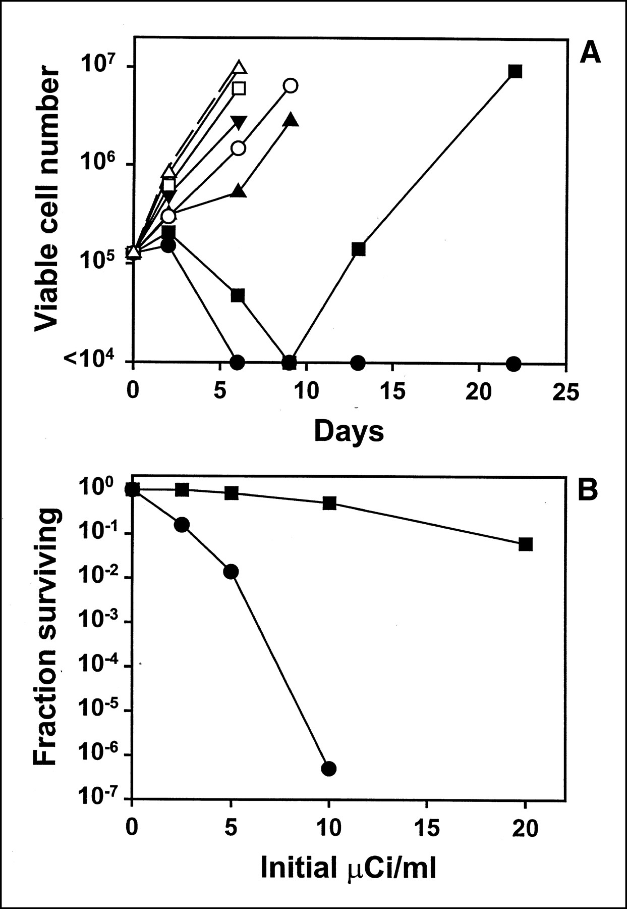
These experiments were intended to determine whether the LL1 conjugates could kill cells and to what extent the killing was antigen specific. Nonspecific toxicity was determined with a nonreactive control Ab, labeled in the same way as LL1. Figure 1A is an example of the experiments performed, in this case with 90Y-labeled Abs. LL1 produced a 100% kill rate at the highest concentration tested, 0.74 MBq/mL (20 μCi/mL). A concentration of 0.37 MBq/mL (10 μCi/mL) also produced strong killing results, with a surviving fraction of 1.6 × 10⁻⁶; 0.185 MBq/mL (5 μCi/mL) produced approximately 98.6% killing; and 0.093 MBq/mL (2.5 μCi/mL) produced 84% killing. The nonspecific toxicity from the labeled control Ab was much less but still substantial. Figure 1B shows a plot of FS versus initial μCi/mL Although the nonspecific Ab did not kill strongly, even at the highest concentration tested here, we estimate that LL1 was 4- to 8-fold more toxic than the nonreactive Ab.

(A) Toxicity of 90Y-LL1 for Raji cells (filled symbols) compared with nonreactive control Ab MN-14 labeled in same way (open symbols). Cells were incubated for 2 d with radiolabeled Ab at starting concentration of 0.74 MBq/mL (20 μCi/mL) (circles), 0.37 MBq/mL (10 μCi/mL) (squares), 0.185 MBq/mL (5 μCi/mL) (triangles), or 0.095 MBq/mL (2.5 μCi/mL) (inverted triangles). Growth rate of control, untreated cells is also shown (dotted line without symbols). Data shown are cell counts obtained at various times and are representative of 2 experiments, each done in duplicate. Cells treated with highest concentration of LL1 were 100% killed, because no viable cells were detected after day 6, and growth of a single viable cell would be readily detected in 22 d. (B) Fraction surviving was calculated from growth curves, as described in Materials and Methods section, and plotted versus the initial μCi/mL for LL1 (filled circle) or the nonreactive control Ab (filled square). One hundred percent killing cannot be shown on exponential y-axis, but next-higher concentration of 90Y-LL1, 20 μCi/mL, produced 100% killing. Results shown are representative of 2 experiments, each done in duplicate.
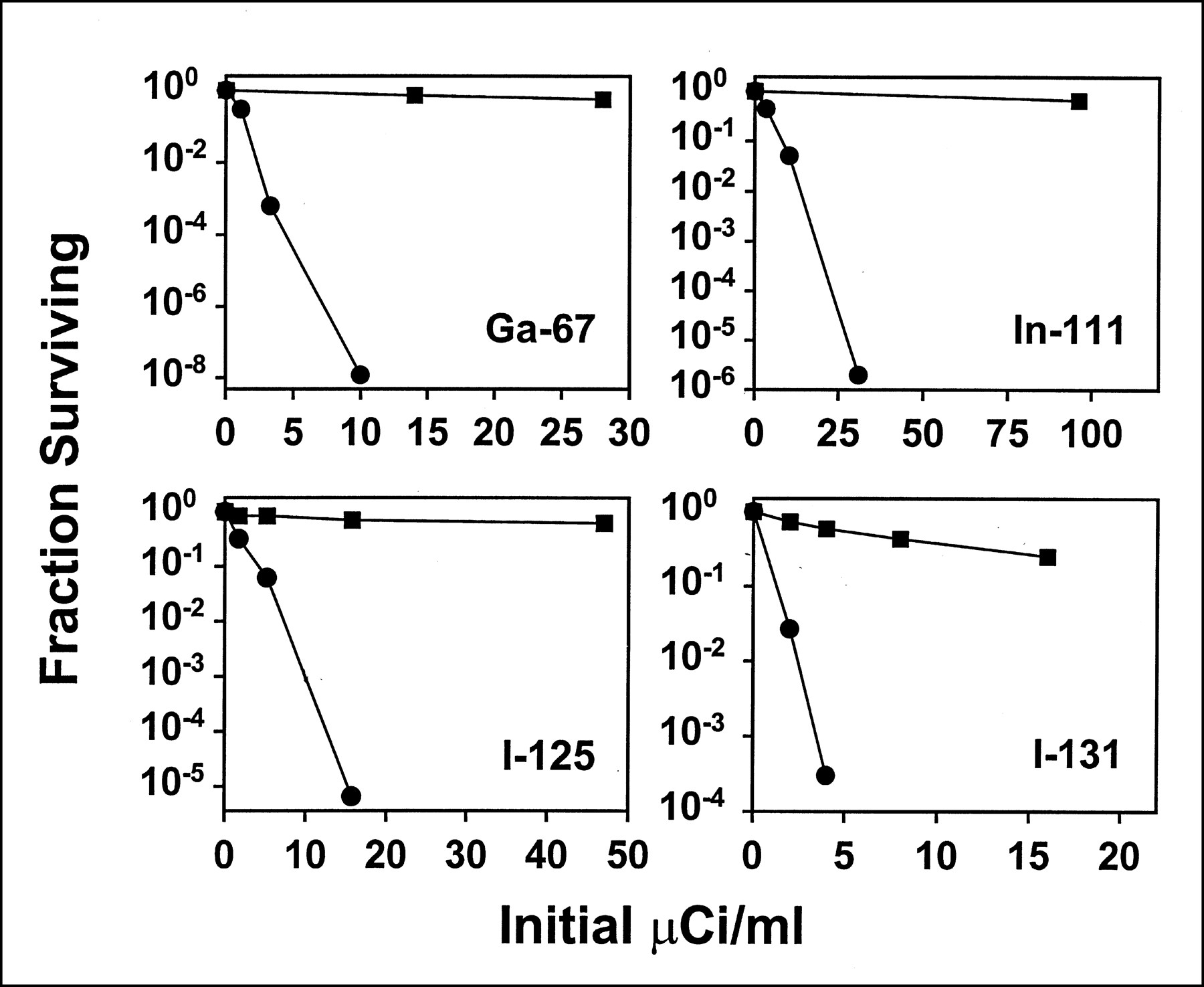
Figure 2 summarizes the data obtained with the 4 other radionuclides tested. As shown, all of the radionuclides tested produced strong, antigen-specific toxicity. However, considerable nonspecific toxicity was observed only with the β-particle emitters. Thus, 90Y on a nonreactive Ab, produced 94% cytotoxicity at the highest concentration tested, 0.74 MBq/mL (20 μCi/mL) (Fig. 1), and 131I on a nonreactive Ab produced 75% toxicity at 0.59 MBq/mL (16 μCi/mL) (Fig. 2). Such levels of killing were substantial, yet relatively minor in this context. For example, 94% killing would result in a growth delay of only approximately 4 d. In contrast, the Auger electron emitters produced no significant toxicity at the concentrations used in these experiments, which were selected because LL1 conjugates at the same concentration produced 100% kill.

Toxicity of Ab LL1 conjugated to 4 radionuclides for Raji B-lymphoma cells (filled circles). Results are also shown for a nonreactive control Ab (filled squares). Cells were incubated for 2 d with indicated starting concentration of Ab, then diluted and counted at various times. Fraction surviving was calculated from growth curves as described in Materials and Methods section. One hundred percent killing cannot be shown on the exponential y-axis, but all the LL1 conjugates used produced 100% killing at next-higher concentration, which was 2 or 3 times higher than highest concentration shown. Results shown are representative of at least 2 experiments performed with each radiolabel and Ab, each done in duplicate. Note different scales on x-axis. Similar experiments with 90Y label are shown in Figure 1.
To better determine the level of nonspecific killing, similar experiments were performed with higher concentrations of radioactivity on the control Ab. Figure 3 shows the level of nonspecific killing observed with the radionuclides used in this study and demonstrates that nonspecific killing was much greater with the β-particle emitters than with the Auger electron emitters. The data are plotted in 2 ways. Panel A shows the starting μCi/mL on the x-axis. Because of the large variation in half-life of the isotopes tested (range, 64–1443 h), the total disintegrations per well, over the 2-d period of incubation, was considerably higher for the isotopes with longer half-lives. Therefore, Panel B shows the total disintegrations per well on the x-axis. The only substantial difference between these 2 plots is a small rightward shift of the 125I curves. Note that 125I appears to have displayed significantly less nonspecific toxicity than the other Auger electron emitters, 111In or 67Ga, probably because of the lack of higher energy electrons (>30 keV) from 125I. Table 1 lists the estimated initial radionuclide concentration required to achieve 99% killing of Raji cells nonspecifically. This was calculated from a semilog plot of FS versus initial concentration, by interpolation or extrapolation; in those cases in which extrapolation was required, the lowest percentage killing was 94%, so the extrapolation was not far. Although 90Y was slightly more toxic than 131I, and 111In and 67Ga were somewhat more toxic than 125I, the major difference was between the β-particle emitters as a group and the Auger electron emitters as a group. The mean radionuclide concentration required for 99% kill was 1.81 ± 1.00 MBq/mL (48.9 ± 26.9 μCi/mL) for the β-particle emitters (131I and 90Y) and 15.95 ± 9.21 MBq/mL (431.0 ± 248.8 μCi/mL) for the Auger electron emitters (111In, 67Ga, and 125I), and this 8.8-fold difference was statistically significant (P < 0.01). At very high concentrations, efficient killing was obtained with the Auger electron emitters: nearly complete killing was observed with these radionuclides at 18.5–37.0 MBq/mL (500–1000 μCi/mL).
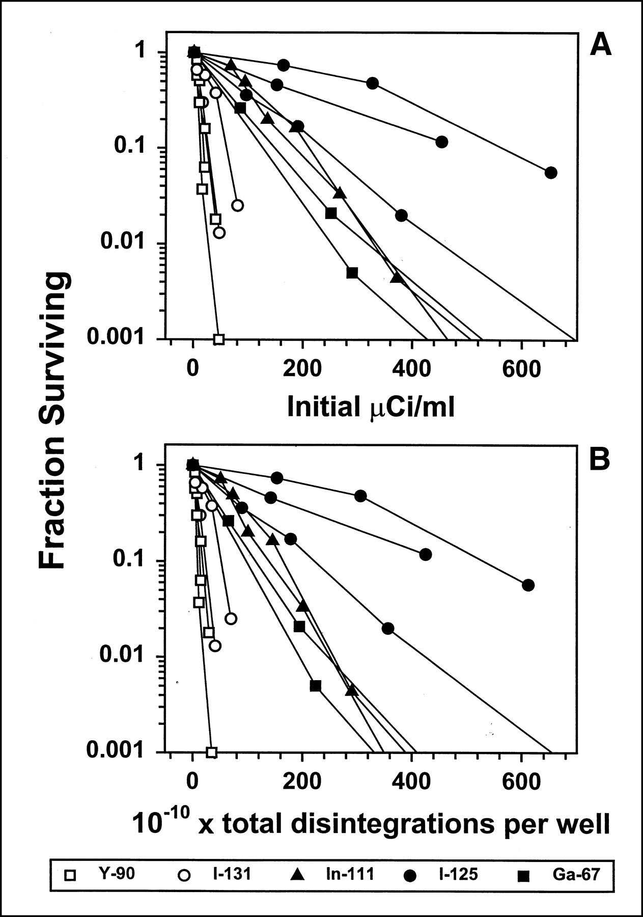
Nonspecific toxicity of 5 radionuclides conjugated to control nonreactive Ab. Experimental protocol was as described in Figure 1, except that higher concentrations of radioactivity were used. Results are shown for 90Y (open squares), 131I (open circles), 125I (filled circles), 111In (filled triangles), and 67Ga (filled squares). Note that β-particle emitters have open symbols, whereas Auger electron emitters have filled symbols. Separate curves indicate different experiments with same radiolabel. Iodine labels used a chloramine-T label, rather than IMP-R2 label used with LL1, because there was virtually no nonspecific uptake of radionuclide by cells (1), and therefore there was no need to use residualizing label. (A) x-axis shows initial μCi/mL; (B) x-axis is total disintegrations per well (total volume, 1.5 mL) in 2 d. Only substantial difference between parts A and B is small rightward shift of 125I curves.
Specific and Nonspecific Cytotoxicity of 5 Radionuclides for Raji Cells
One of the goals of this study was to determine a specificity index for the 5 LL1 conjugates, meaning the level of antigen-specific toxicity relative to nonspecific toxicity, and defined as the ratio: (concentration of nonspecific Ab required for a particular percentage kill)/(concentration of LL1 required for the same percentage kill). Specificity indices determined at the level of 99% kill are summarized in Table 1. This ratio is a constant, independent of the percentage kill chosen, if the semilog toxicity curve is linear, as appears to be the case. Table 1 also shows the concentration of LL1 conjugate required for 99% kill, for each radiolabel; there are significant differences between the radionuclides tested. Thus, a group of 3 radionuclides, 131I, 90Y, and 67Ga, required only 0.11–0.16 MBq/mL (2.9–4.4 μCi/mL), whereas 125I required 0.34 MBq/mL (9.1 μCi/mL) and 111In required 0.50 MBq/mL (13.6 μCi/mL). The potency of the various radionuclides is compared more definitively below, where we describe the results of directly assaying disintegrations per cell. The calculated specificity index was 7.7 for 90Y, which was considerably lower than for the other radionuclides tested. 131I and 111In had similar specificity indices of 25–30. The value for 125I was higher, approximately 75, which can be attributed primarily to its very low nonspecific toxicity, because the specific toxicity was somewhat lower, in terms of MBq/mL required, than for 131I or 90Y. The value for 67Ga was also high at approximately 95; this high value was from both potent specific killing and relatively low nonspecific killing. In conclusion, when conjugated to Ab LL1, the β-particle emitters 131I and 90Y are more potent at killing cells than the Auger electron emitters 125I and 111In, but the disadvantage of the β-emitters is their higher nonspecific toxicity. Because 131I-LL1 is almost 6 times more potent than 111In-LL1, its specificity index is similar to that of 111In. The apparent exceptional radionuclide is 67Ga, which kills as potently as the β-particle emitters, yet has the low nonspecific toxicity of the Auger electron emitters, resulting in a considerably higher specificity index than any of the other radionuclides tested.
Was Cross-Fire a Significant Factor in Toxicity with β-Particle Emitters?
It seemed possible that there might be significant cross-fire between cells, meaning that a significant fraction of irradiation hitting each cell might derive from a radionuclide bound to adjacent cells. This possibility arose because the cells settled at the bottom of the 24-well plates, and by day 2, at the time of transfer to T30 flasks, the cells were confluent and contained a large fraction of the total CPM in the well, as much as one third of the total (1). However, this effect was limited by the geometry: Because the cells formed a monolayer, only a very small fraction of the β-particles emitted from 1 cell would hit the nucleus of adjacent cells. To evaluate the magnitude of this factor, experiments were set up at 2 different cell concentrations. In addition to the normal concentration, other wells contained 50-fold fewer cells but were otherwise identical. If cross-fire was significant, there would be much less killing in the cells plated at a low concentration. However, the toxicity was very similar at both cell concentrations for both 131I-IMP-R2 and 90Y (data not shown), indicating that cross-fire was not significant and that the radiation killing the cells came primarily from the same individual cell. This experiment was performed previously with 111In-LL1, with similar results (1).
Comparison of the Potency of 5 Radionuclides Conjugated to Ab LL1
Although the specificity index is in many respects the most important criterion for therapeutic use, the potency of the various radionuclides is also important. This is true because, in vivo, some cells may not be fully saturated with Ab, and it is important to effectively kill those cells with less uptake of radioactivity. Thus, experiments were designed to determine which radionuclides were most potent per decay. The data presented in Table 1 provide some indication of relative potency, but that data include only the initial concentration of radioactivity in the wells. Although this concentration is, in general, closely related to the actual uptake, the relationship can vary depending on the specific activity of the conjugates, the immunoreactivity of the conjugates, the half-life of the radionuclides, and other factors. Therefore, in experiments with all 5 radionuclides, we determined actual CPM per cell at various time points, calculated the cumulative disintegrations, and compared this with the percentage cell kill determined in the same experiments. In these experiments, cell kill was monitored by a clonogenic assay, rather than by determination of the growth rate, because this was considered to provide a more accurate estimate of the percentage kill and because preliminary experiments indicated that differences between some of the radionuclides were relatively small. The cloning efficiency of control Raji cells was 60.3% ± 22.9% (mean ± SD) in the experiments performed. These experiments were designed to have levels of cell kill of approximately 99%–99.9%, to provide more accurate comparisons between the radionuclides than would have been achieved with higher levels of cell kill. The highest concentrations used, determined from the earlier experiments described above, were as follows: 111In, 0.59 MBq/mL (16 μCi/mL); 125I, 0.44 MBq/mL (12 μCi/mL); 67Ga, 0.30 MBq/mL (8 μCi/mL); and 131I and 90Y, 0.22 MBq/mL (6 μCi/mL). Two 2-fold dilutions of each of these concentrations were also tested.
There are 2 possible ways of presenting these data: by disintegrations per actual cell number or by disintegrations per initial cell number. Although superficially it might seem that use of the actual cell number is most meaningful, in fact there are problems with this approach, and the use of the initial cell number is preferable. This issue was discussed previously, and data were presented showing the differences between the methods (1). Therefore, even though we determined the actual cell number in every experiment at every time point, Figure 4 shows the use of disintegrations per initial cell number. In any case, it should be noted that although control cells, of course, multiplied considerably over the first 2 d, the irradiated cells showed very little cell division, as a consequence of irradiation at these fairly high levels (10,14). Therefore, over the first 2 d, the actual cell number was never substantially higher than the initial cell number.

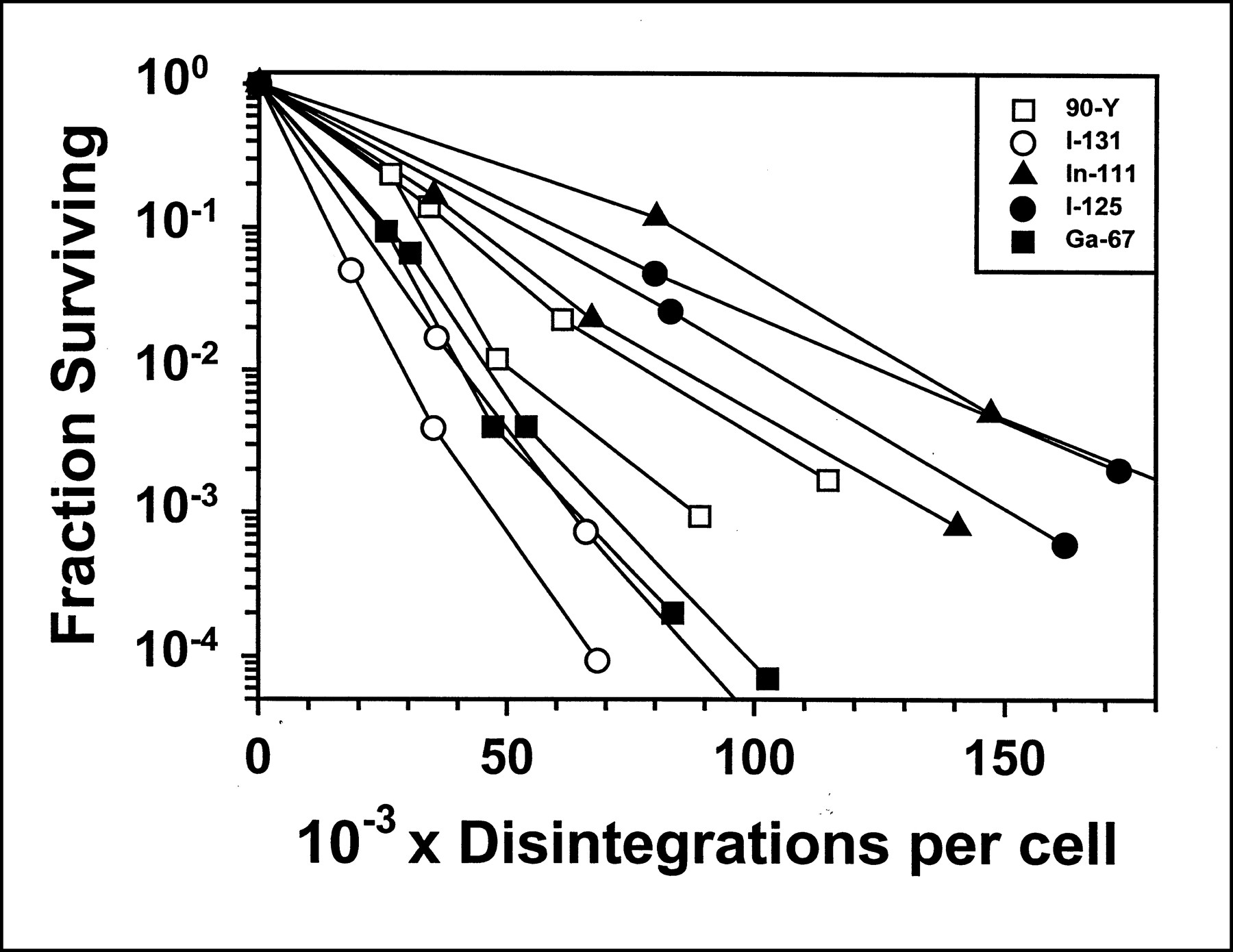
Relationship between fraction surviving and total disintegrations per cell. Raji cells were incubated with radiolabeled LL1 for 2 d, and fraction surviving was determined by clonogenic assay. Cell-bound CPM was determined at days 1, 2, 3, and 6, or days 1, 2, and 5, and cumulative disintegrations per initial cell number were calculated. Results are shown for 90Y (open squares), 131I (open circles), 125I (filled circles), 111In (filled triangles), and 67Ga (filled squares). Note that β-particle emitters have open symbols, whereas Auger electron emitters have filled symbols. Separate curves indicate different experiments with same radiolabel.
As shown in Figure 4, there were significant differences between the radionuclides in the dose–response curve. 125I and 111In were less potent than the other 3 radionuclides tested. 67Ga was more potent than the other Auger electron emitters and comparable with 131I, which was the most potent radionuclide tested. This finding, then, is consistent with the data shown in Table 1, which were calculated from entirely different experiments, using different assays to measure FS. D0 was calculated from these curves, from a straight line calculated by linear regression: This is the dose required to kill 63% of the cells. The mean values, in disintegrations per cell, were 15,110 for 90Y; 9,359 for 131I; 23,598 for 111In; 24,924 for 125I; and 10,165 for 67Ga.
Dosimetry
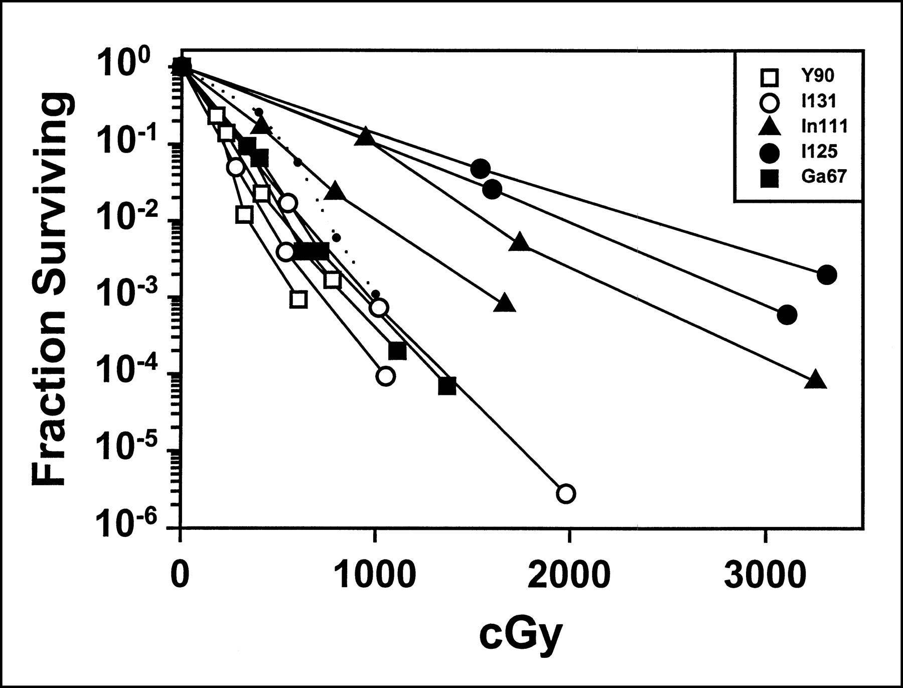
There are several complications in the calculation of the radiation dose delivered, in cGy, most of which have been discussed above or previously (1). A basic problem is uncertainty regarding when the cells are reproductively dead, considering that the radionuclides remain inside the cells for many days. That is, cells may be reproductively dead at day 2, but still intact and counted as viable; any radiation delivered after this time would be irrelevant. Still, it is useful to calculate the estimated cGy dose delivered, to determine, at least, if the values are reasonable. Moreover, if the S values provided by MIRD Cellular S Values (5) are applicable to our experimental system, a plot of FS versus calculated absorbed cGy dose should show similar curves for all the radionuclides tested. As shown in Figure 5, this is the case for LL1 conjugated to 67Ga, 131I, and 90Y, and the toxicity curves with these 3 radionuclides are very similar to the curve obtained previously with irradiation by 137Cs. Considering that the 137Cs dose was administered over a few minutes, whereas the Ab dose was administered over 5–6 d, the similarity in these curves is remarkable. In contrast, the toxicity mediated by 125I and 111In appears to be significantly less than that predicted from the calculated cGy dose.

Relationship between fraction surviving and calculated cGy dose. Using data presented in Figure 4, cGy dose per cell was calculated from disintegrations per cell. Separate curves indicate different experiments with same radiolabel. Dotted line shows toxicity caused by irradiation from 137Cs, which was determined previously (1).
DISCUSSION
We previously described the cytotoxicity mediated by LL1 conjugated to 125I and 111In (1). As a continuation of this study, we have herein similarly demonstrated toxicity with 67Ga, 131I, and 90Y. 67Ga is another Auger electron emitter, whereas 131I and 90Y are β-particle emitters. All of these radionuclides were able to effectively and specifically kill B-lymphoma, with 100% kill (>6 logs). These results are generally consistent with radiation dose calculations that are based on the uptake of CPM/cell and the intracellular S values of the radionuclides (5). In this study, we attempted to determine which of the radionuclides has the highest level of potency and specificity. Although significant differences between the radionuclides were evident, it should be noted that these differences were not large, and the selection of the optimal radionuclide for therapy depends also on other important factors (discussed below).
A major difference between β-particle emitters and Auger electron emitters, in this experimental system, is the higher level of nonspecific killing observed with the former. It is possible that this type of nonspecific toxicity in vitro may correlate with bone marrow toxicity in vivo, and in fact considerable evidence demonstrates that Auger electron emitters are much less toxic that β-particle emitters in mice (6). This, of course, may represent an advantage of Auger electron emitters for cancer therapy. However, there are several reasons why β-particle emitters, particularly 131I, should not be eliminated as candidates for clinical use in LL1 conjugates. First, 131I was as potent in specific killing as any of the other radionuclides tested, as shown in Table 1 and Figure 4, and significantly more potent than most of the Auger electron emitters. Therefore, the specificity index for 131I was comparable with that for 111In, although it was lower than that for 125I or 67Ga. Second, because of their higher energy, β-particles are able to kill cells that are close to antigen-positive cells but are not directly reached by Ab. These may be cells that are antigen negative or antigen low or cells that are not efficiently reached by Abs (because of an inadequate blood supply). Thus, it could be argued that β-particle emitters are more versatile in their ability to kill both single cells and large tumor masses.
Although 90Y produced slightly greater nonspecific toxicity than 131I, the difference was not as large as expected, on the basis of dosimetry calculations. With an initial concentration of 1.85 MBq/mL (50 μCi/mL), which is close to the maximum values used in Figure 3 with the β-particle emitters, the dose over 2 d was calculated to be 447 cGy for 131I and 1862 cGy for 90Y. Given a D0 for Raji cells of approximately 90 cGy, and an extrapolation number of 1.31, which were previously determined by irradiation with 137Cs (1), the predicted FS is 0.009 and 1.4 × 10⁻⁹, respectively. This value is reasonably close to the observed value for 131I, but the killing by 90Y is much less than predicted. This can be attributed to 2 factors, primarily. First, the assumption of 100% absorption is not true for the high-energy electrons of 90Y; the total volume of medium, 1.5 mL, would absorb approximately 73% of the electron energy, if it were spherical (15). Second, cells at the edges of the well receive a lower dose than cells in the center, by at factor of at least 2, and cells tend to be most dense at the edges. Although we have not attempted to develop a model that incorporates these factors, because the geometry is complex, these simple corrections would predict a dose from 90Y of 680 cGy at the edges of the well and a survival fraction of 6.9 × 10⁻⁴, which is relatively close to the actual value.
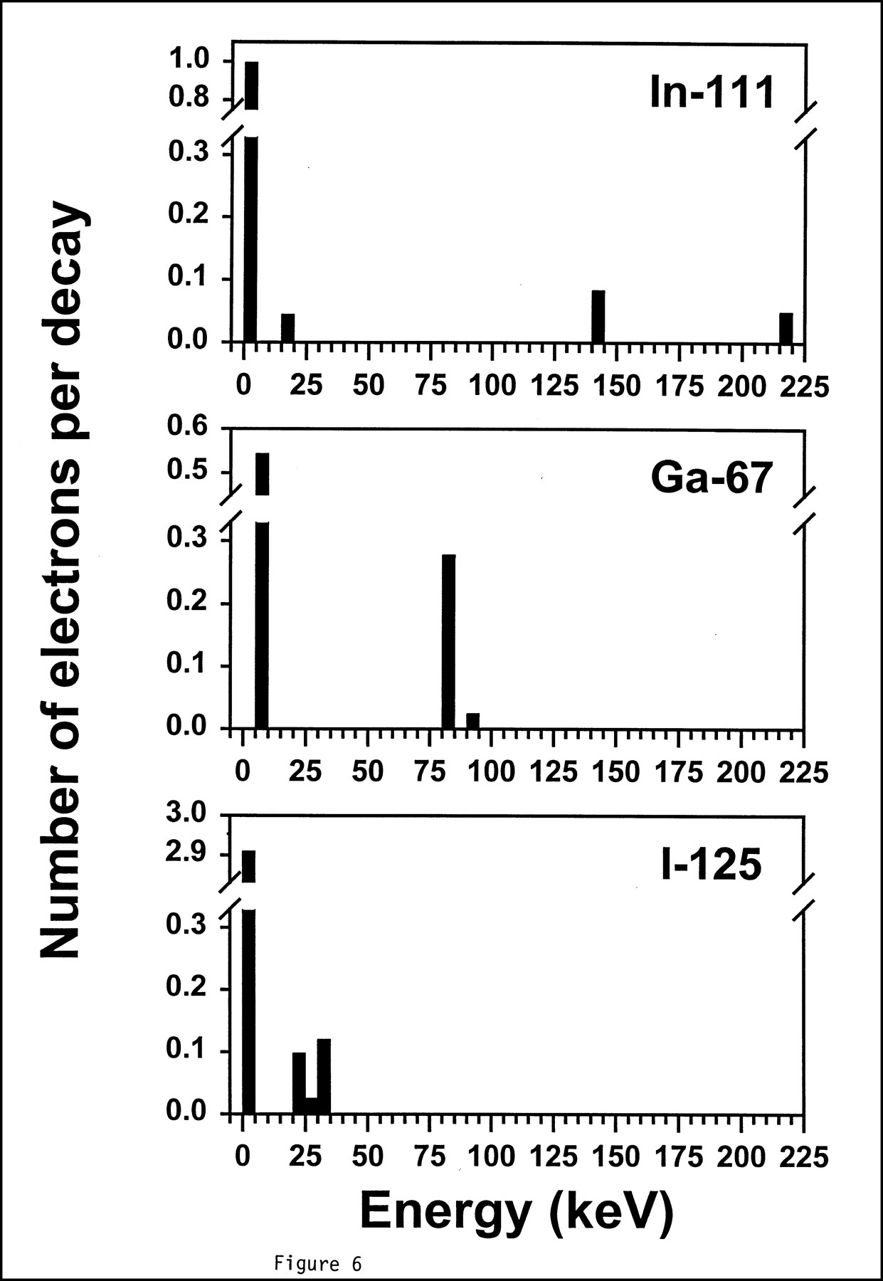
It should be noted that there are many assumptions involved in the dose calculations that make them only approximations. More specifically, the calculations assume that the nucleus and the cell are concentric spheres and that the radioactivity is homogeneously distributed in the cytoplasm. Although it is well established that the great majority of the radioactivity delivered by 111In- or 125I-IMP-R2-LL1 is retained in lysosomes (4,16), the exact location of the lysosomes within the cytoplasm is not known, and it seems likely that lysosomes are not randomly distributed. Because many of the Auger electrons emitted have a range in tissue of <1.5 μm (the average distance from the nuclear membrane to the cell membrane in Raji cells), the frequency of lysosomes close to the nuclear membrane significantly affects the radiation dose delivered to DNA. Thus, certain radionuclides may be more or less potent than would be indicated by their calculated S values. We speculate that this factor may explain the relatively low cytotoxic activity of 125I and 111In, relative to the calculated cGy dose (Fig. 5). These 2 radionuclides only, of the 5 tested, emitted large amounts of very-low-energy electrons, <5 keV (Fig. 6). These electrons may have had less of an impact than expected, possibly because of the presence of few lysosomes close enough to the nuclear membrane.

Energy spectrum of electrons emitted by 125I, 111In, and 67Ga. Electrons are grouped by energy in increments of 5 keV. Note break in the scale of y-axis and different scales used.
The emergence of 67Ga as the optimal radionuclide was unexpected. 67Ga had little nonspecific toxicity, like the other Auger electron emitters, but its potency in specific killing was considerably higher than that of the other Auger electron emitters, being virtually identical to that of 131I, which had the highest potency of the radionuclides tested. This may be explained by a comparison of the electrons emitted by the various radionuclides, which is shown in Figure 6. 67Ga has 2 properties that distinguish it from 125I and 111In. First, it emits a relatively abundant electron of 84 keV. Second, the low-energy electrons are in the range of 5–10 keV, rather than 0–5 keV. It is likely that the potency of 67Ga can be attributed to 1 or both of these properties. Chemical, heavy-metal toxicity of released free gallium is unlikely to play a role, because the stability of the chelator is very high, with approximately 1% release of free gallium per day at 37°C (7), which was confirmed under our conditions of tissue culture (unpublished data).
Our method of 67Ga labeling produced a specific activity of approximately 370 MBq/mg (10 mCi/mg), which is high enough for most purposes, but it should be noted that 111In conjugates can be prepared at much higher specific activities, up to 2960 MBq/mg (100 mCi/mg), which can be considered an advantage of 111In. On a practical level, this means that less Ab conjugate is required for particular experiments. The reason for the greater labeling capacity with 111In is not known, but several possibilities should be considered: (a) 111In as supplied may have fewer interfering metal contaminants; the 111In used is a high-purity grade from its supplier; and (b) the macrocyclic Ga chelator NOTA may be less readily entered than the open In chelator benzyl-DTPA. Indeed, the macrocyclic In chelator DOTA does not allow 111In labeling to as high a specific activity as does benzyl-DTPA, in our experience (unpublished data). Other open-chain Ga chelators have also been used to label Abs. The chelator HBED-CC appears not to be a residualizing label (17), so is not useful for this purpose. Deferoxamine has been used with a carrier molecule of dialdehyde starch to obtain a chelator:Ab ratio of 6:1 and a specific activity of 11,766 MBq/mL (318 mCi/mg) (18).
Inasmuch as this study used 2 different chelators, as well as a novel ligand for iodination, it is necessary to consider whether differences in these labeling methods may have contributed to the results. All of the chelators used are known to be very stable, with <1% release of the radioligand per day (7,8,19), and this stability was confirmed in our standard Ab processing experiments, which were performed with all of the conjugates tested except for 90Y-DTPA (8,9, and unpublished data for 67Ga-NOTA). The immunoreactivity of all radioconjugates was similar, as stated under the Materials and Methods section, and the level of dissociation of intact Ab, in Ab processing experiments, was similar for all radiolabels, which indicates no substantial damage to 1 of the 2 binding sites on the Ab (20). Regarding retention of catabolites by the cells, this depends on the inability of the molecules to cross cell membranes and, therefore, is similar for a wide variety of hydrophilic moieties. Similar retention of 111In-DTPA, 67Ga-NOTA, and iodinated IMP-R2 was demonstrated in direct comparisons in vitro (8,9, and unpublished data for 67Ga-NOTA). Although 90Y-DTPA was not tested in vitro, this label was tested extensively in vivo, in tumor localization experiments, and behaved similarly to other residualizing radiolabels (21). Moreover, we expected that 90Y-DTPA would be retained as well as 111In-DTPA, given that the chelation is stable, which was demonstrated in other experiments (19).
We continue to consider 111In to be a useful radionuclide for future studies, even though it appeared to be significantly less potent than most of the other radionuclides tested, with a lower specificity index than 67Ga and 125I. This is because the labeling method for 111In is simple (once the DTPA conjugate is prepared), and high specific activities of up to 3600 MBq/mg (100 mCi/mg) are readily achieved. Under the conditions we have used, we were unable to obtain such high specific activities with any of the other radiolabels. Furthermore, the iodination procedure used here was considerably more complex than radiometal labeling. The radiation half-life is another important factor in regard to radionuclide selection. The 60-d half-life of 125I must be considered a disadvantage, because most of the isotope injected will not decay over a reasonable therapeutic interval, even though 125I performed very well in this study. The other radionuclides tested have 3–8-d half-lives, which seem more appropriate.
Although it is clear that the cytotoxicity with radiolabeled LL1 was caused by the very high cellular uptake of this Ab, it seems possible that a similar approach may be effective with other Abs that are not internalized at such a high level. By using Abs labeled to a higher specific activity, it is possible to deliver more radioactivity with less Ab. The experiments reported here used 111In-LL1 with a specific activity of 370–740 MBq/mg (10–20 mCi/mg), but if each Ab molecule was conjugated with a single 111In atom, on average, the specific activity would be 8547 MBq/mg (231 mCi/mg). Although we are not aware of Abs being labeled to this specific activity, it seems feasible; we have reached 3700 MBq/mg (100 mCi/mg) while maintaining a high radiolabeling efficiency. Moreover, it has been demonstrated that up to 5 chelating groups can be conjugated to an Ab without affecting immunoreactivity (22). Therefore, Abs reacting with only 2 × 10⁵ sites per cell can potentially deliver a toxic dose of radiation. In addition, there are many Auger electron emitting radionuclides that are expected to be much more potent than those used herein, as judged by their cytoplasmic S values (5). It is unclear at this time whether internalization of bound Ab is a critical factor. Judging from the calculated S values (5), radionuclides on the cell surface are almost as potent as internalized radionuclides, with the difference being less than 2-fold. These factors suggest that this general approach may be applicable to many other Abs.
CONCLUSION
Given sufficiently high Ab uptake, as occurs with LL1, it is possible to efficiently kill tumor cells with radiolabeled Ab. In this study, we obtained complete cell killing (approximately 6 logs). All of the radionuclides tested, including 3 Auger electron emitters and 2 β-particle emitters, were effective, but there were significant differences between the radionuclides. The β-particle emitters showed more nonspecific toxicity than the Auger electron emitters. Such conjugates seem promising for the therapy of B-cell lymphomas and other tumors expressing the antigen recognized.
Acknowledgments
The authors are grateful to Dr. Habibe Karacay, Thomas Jackson, Philip Andrews, and Anthony Zarcone for assistance with radiolabeling. Sammy Elsamra was supported by the Undergraduate Projects in Technology and Medicine program of the Stevens Institute of Technology. This work was supported in part by National Institutes of Health grants CA39841 and CA72324.
Footnotes
Received Oct. 29,1999; revision accepted Jun. 8, 2000.
For correspondence or reprints contact: M. Jules Mattes, PhD, Center for Molecular Medicine and Immunology, 520 Belleville Ave., Belleville, NJ 07109.
REFERENCES
In this issue


Jump to section
Related Articles
Cited By...
- PARP-1-Targeted Auger Emitters Display High-LET Cytotoxic Properties In Vitro but Show Limited Therapeutic Utility in Solid Tumor Models of Human Neuroblastoma
- Auger Radiopharmaceutical Therapy Targeting Prostate-Specific Membrane Antigen
- Monte Carlo Evaluation of Auger Electron-Emitting Theranostic Radionuclides
- A Universally Applicable 68Ga-Labeling Technique for Proteins
- Antitumor Effects and Normal-Tissue Toxicity of 111In-Nuclear Localization Sequence-Trastuzumab in Athymic Mice Bearing HER-Positive Human Breast Cancer Xenografts
- CD74: A New Candidate Target for the Immunotherapy of B-Cell Neoplasms
- Nuclear Localizing Sequences Promote Nuclear Translocation and Enhance the Radiotoxicity of the Anti-CD33 Monoclonal Antibody HuM195 Labeled with 111In in Human Myeloid Leukemia Cells
- Effective therapy of human lymphoma xenografts with a novel recombinant ribonuclease/anti-CD74 humanized IgG4 antibody immunotoxin
- Trifunctional Somatostatin-Based Derivatives Designed for Targeted Radiotherapy Using Auger Electron Emitters
- Anti-CD74 Antibody-Doxorubicin Conjugate, IMMU-110, in a Human Multiple Myeloma Xenograft and in Monkeys
- In vitro cytotoxicity of carcinoma cells with 111In-labeled antibodies to HER-2
- Therapy of Small Subcutaneous B-Lymphoma Xenografts with Antibodies Conjugated to Radionuclides Emitting Low-Energy Electrons
- Antiproliferative activity of a humanized anti-CD74 monoclonal antibody, hLL1, on B-cell malignancies
- In vitro Toxicity of A-431 Carcinoma Cells with Antibodies to Epidermal Growth Factor Receptor and Epithelial Glycoprotein-1 Conjugated to Radionuclides Emitting Low-Energy Electrons
- Cure of SCID Mice Bearing Human B-Lymphoma Xenografts by an Anti-CD74 Antibody-Anthracycline Drug Conjugate
- Antitumor Effects and Normal Tissue Toxicity of 111In-Labeled Epidermal Growth Factor Administered to Athymic Mice Bearing Epidermal Growth Factor Receptor-Positive Human Breast Cancer Xenografts
- Targeting Primary Human Ph+ B-Cell Precursor Leukemia-Engrafted SCID Mice Using Radiolabeled Anti-CD19 Monoclonal Antibodies
- A Comparison of 4 Radionuclides Conjugated to Antibodies for Single-Cell Kill
- Improved Iodine Radiolabels for Monoclonal Antibody Therapy
- Cooperative Effect of Radioimmunotherapy and Antiangiogenic Therapy with Thalidomide in Human Cancer Xenografts
- Intracellular Accumulation of the Anti-CD20 Antibody 1F5 in B-Lymphoma Cells
- Therapy of Disseminated B-Cell Lymphoma Xenografts in Severe Combined Immunodeficient Mice with an Anti-CD74 Antibody Conjugated with 111Indium, 67Gallium, or 90Yttrium
- Single-cell Cytotoxicity with Radiolabeled Antibodies