


Abstract
Our objective was to evaluate the toxicity of the anti-CD33 monoclonal antibody HuM195 modified with peptides (CGYGPKKKRKVGG) harboring the nuclear localizing sequence (NLS; underlined) of simian virus 40 large T antigen and labeled with 111In against acute myeloid leukemia (AML) cells. Methods: HuM195 was derivatized with sulfosuccinimidyl-4-(N-maleimidomethyl)-cyclohexane-1-carboxylate (sulfo-SMCC) to introduce maleimide groups for reaction with NLS-peptides and then conjugated with diethylenetriaminepentaacetic acid for labeling with 111In. The immunoreactivity of NLS-HuM195 was evaluated by its ability to displace the binding of 111In-HuM195 to HL-60 leukemia cells. Nuclear localization was measured in HL-60 cells by subcellular fractionation. The antiproliferative effects of 111In-NLS-HuM195 and 111In-HuM195 on HL-60, U937, or K562 cells with high, intermediate, or minimal CD33 expression, respectively, were studied. The survival of HL-60 cells or patient AML specimens treated with 111In-NLS-HuM195 or 111In-HuM195 was studied. Normal tissue toxicity was evaluated in BALB/c mice injected intravenously with of 3.7 MBq (22 μg) of 111In-NLS-HuM195 or 111In-HuM195. Results: NLS-HuM195 exhibited relatively preserved CD33 binding affinity (dissociation constant [Kd] = 4.3 ± 1.7 × 10⁻⁹ mol/L to 6.9 ± 1.3 × 10⁻⁹ mol/L). Nuclear uptake increased from 10.5% ± 0.5% for 111In-HuM195 to 28.5% ± 4.1% or 65.9% ± 1.5% for 111In-HuM195 substituted with 4 or 8 NLS-peptides, respectively. The inhibitory concentrations of 50% (IC50) and 90% (IC90) for HL-60 cells treated with 111In-NLS-HuM195 were 37 kBq per 103 cells and 77–81 kBq per 103 cells, respectively. The IC50 and IC90 values for 111In-HuM195 were 92 kBq per 103 cells and 203 kBq per 103 cells. Growth inhibition was correlated with the level of CD33 expression. The survival of HL-60 cells was reduced from 232 ± 22 colonies (control) to 7 ± 1 colonies with 1.48 mBq per cell of 111In-NLS-HuM195; no colonies were found at 3.33 mBq per cell. The surviving fraction decreased >2-fold in 7 of 9 AML specimens treated with an excess of 111In-NLS-HuM195 and >10-fold in 2 of these specimens. There were no decreases in body weight or hematologic parameters or increases in alanine aminotransferase or creatinine in mice administered 3.7 MBq (22 μg) of 111In-NLS-HuM195 or 111In-HuM195. There was no morphologic damage to the liver or kidneys. Conclusion: We conclude that NLS-peptides routed 111In-HuM195 to the nucleus of AML cells, where the emitted Auger electrons were lethal. 111In-NLS-HuM195 is a promising targeted radiotherapeutic agent for AML.
- acute myelogenous leukemia
- 111In
- nuclear localization sequences
- simian virus 40 large T antigen
- Auger electrons
Acute myelogenous leukemia (AML) accounts for about 40% of all adult leukemias but the incidence of the disease has been rising, especially in the elderly (1). Induction chemotherapy achieves complete remission (CR) in 60%–80% of patients <60 y old and in 40%–65% of older patients (2). However, relapse rates are high, particularly in older patients, and new therapies are urgently needed. Furthermore, the remission rate in relapsed AML treated with chemotherapy is only 10%–15% (2). This is increased to 10%–41% by hematopoietic stem cell transplant (HSCT) (2), but elderly patients often are not eligible because of the associated high mortality and morbidity risks.
Strategies to develop targeted and potentially less toxic treatments for patients with AML have focused on the CD33 epitope displayed in 80%–90% of cases and on normal myeloid cells, but not by granulocytes or early myeloid progenitors (3). One such approach is the use of anti-CD33-calicheamicin immunoconjugates (gemtuzumab ozogamicin; Mylotarg, Wyeth-Ayerst). Early clinical trials of gemtuzumab demonstrated eradication of leukemia cells from the blood and bone marrow producing CR in 20%–30% of patients with refractory/relapsed AML (4). This agent was approved by the U.S. Food and Drug Administration in 2000 as a treatment for elderly patients with relapsed AML. However, most patients treated with gemtuzumab relapse; this drug was subsequently found to be a substrate for the multidrug resistance transporter pgp-170 and was ineffective for killing CD33-positive leukemia cells that overexpress the MDR1 gene (5). Furthermore, a lack of CR in patients treated with gemtuzumab has been correlated with pgp-170 expression in leukemia blasts (6).
An alternative strategy for treatment of refractory/relapsed AML is radioimmunotherapy (RIT). RIT of AML has focused mainly on the murine anti-CD33 monoclonal antibody (mAb) M195 or its humanized IgG1 form, HuM195 (7). RIT with nonmyeloablative doses of 131I-M195 produced potent antileukemia effects in patients, killing >99% of blasts in some cases (8), whereas higher doses combined with HSCT produced CR in 28 of 30 (93%) patients (7). The dose-limiting toxicity was myelosuppression, which was, in part, due to cross-fire from the long-range (2 mm; 200 cell diameters) β-particles emitted by 131I from targeted leukemia cells in the bone marrow on hematopoietic stem cells. In an attempt to increase its potency and minimize nonspecific hematopoietic toxicity, HuM195 was conjugated with the short-range (50–100 μm; 5–10 cell diameters) α-emitters 213Bi (9) or 225Ac (10). In particular, the use of 225Ac-HuM195 appeared highly promising, as 225Ac generates daughter decay products that are themselves α-emitters (221Fr, 217At, 213Bi, or 213Po) or β-emitters (213Bi, 209Tl, or 209Pb), thus amplifying DNA damage and killing of malignant cells (“atomic nanogenerator”) (10). However, it was subsequently reported that the decay products of 225Ac-HuM195 redistributed, causing severe renal tubular damage and anemia in nonhuman primates, thus seriously limiting its use for treatment of AML in humans (11).
In this study, we describe an analogous targeted radiotherapeutic strategy for AML—but one which may be less damaging to normal tissues—that uses HuM195 labeled with the nanometer-to-micrometer range Auger electron-emitter 111In. The decay product of 111In is cadmium, a stable element, thus obviating the radiotoxicity of daughter decay products observed with 225Ac. In addition, the linear energy transfer (LET) of Auger electron-emitters such as 111In approaches that of α-emitters (∼100 keV/μm), rendering them very damaging to DNA and highly toxic if they decay in the cytoplasm and, especially, if they decay in the cell nucleus (12). Indeed, our group showed that human epidermal growth factor labeled with 111In (111In-hEGF) was translocated to the nucleus of MDA-MB-468 breast cancer cells overexpressing EGF receptors, where the emitted Auger electrons killed >95% of cells at only 74–111 mBq per cell (2−3 pCi per cell) (13). Furthermore, 111In-hEGF strongly inhibited the growth of MDA-MB-468 breast cancer xenografts in athymic mice administered subcutaneously in 5 weekly doses of 18.5 MBq (500 μCi) (14). In this study, we report that the CD33-mediated internalization of HuM195 by leukemia cells rendered the antibody lethal when labeled with 111In. We further demonstrate that 13-mer peptides harboring the nuclear localizing sequence (NLS) of simian virus 40 (SV-40) large T antigen (15) conjugated to 111In-HuM195 efficiently routed the antibodies to the nucleus of leukemia cells, where the cytotoxicity was enhanced.
MATERIALS AND METHODS
Human Leukemia and Lymphoma Cell Lines and Specimens
HL-60 human AML cells, K562 human chronic myeloid leukemia (CML) cells, and U937 human histiocytic lymphoma cells were obtained from the American Type Culture Collection. HL-60 cells were CD33 positive by flow cytometry (corresponding to 4 × 10⁴ receptors/cell for 111In-HuM195 (16)), whereas K562 cells displayed minimal CD33 expression. U937 cells have intermediate CD33 expression, displaying about one-third CD33 epitopes compared with HL-60 cells at 37°C (17). All cells were cultured in RPMI 1640 medium (R-8758; Sigma) supplemented with 10% fetal calf serum (FCS) at 37°C, 5% CO2. AML specimens from patient bone marrow or peripheral blood samples were collected under a protocol (no. 02-0763-C) approved by the Human Subjects Research Ethics Board at the Princess Margaret Hospital, University Health Network. All specimens demonstrated CD33 positivity in at least 95% of blast cells by flow cytometry.
Peptides Containing NLS
A 13-mer peptide, CGYGPKKKRKVGG containing the NLS of SV-40 large T antigen (underlined), was synthesized (Advanced Protein Technology Center, Hospital for Sick Children, Toronto, ON, Canada) and stored at −20°C. The purity of the peptides was >95% determined by reversed-phase high-performance liquid chromatography, amino acid analysis, and mass spectrometry (observed molecular weight [Mr], 1,418.81; calculated Mr, 1,419.63). The N-terminal cysteine of the peptides was acetylated to prevent dimerization. The presence of the N-terminal cysteine and absence of peptide dimers formed through disulfide bond cross-linking was confirmed by a thiol assay using Ellman's reagent (Pierce Chemical Co.). The peptides were synthesized with a tyrosine residue (boldface) for labeling with 123I. Radioiodination was performed by dispensing 100 μg of peptides into a glass tube precoated with 50 μg of 1,3,4,6-tetrachloro-3α,6α-diphenylglycouril (Sigma-Aldrich Co.) and adding 7.4 MBq (5 μL) of 123I sodium iodide (MDS-Nordion Inc.). The mixture was incubated at room temperature for 1 min; then the 123I-labeled peptides were purified on a Sephadex G-10 minicolumn (exclusion limit, 700 Da; Pharmacia). The radiochemical purity of 123I-NLS-peptides was >95% determined by paper chromatography developed in 85% methanol (Rf 123I-NLS-peptides, 0.0; Rf 123I−, 1.0).
Conjugation of HuM195 with NLS-Containing Peptides
The anti-CD33 humanized mAb HuM195 (IgG1; Protein Design Laboratories, Inc.) was conjugated with NLS-peptides by reaction of maleimide-derivatized HuM195 with the N-terminal cysteine of the peptides. Maleimide groups were introduced into HuM195 by reaction of 0.5−2 mg of antibodies (8 mg/mL in phosphate-buffered saline [PBS], pH 7.6) with a 10- to 1,200-fold molar excess of sulfosuccinimidyl-4-(N-maleimidomethyl)-cyclohexane-1-carboxylate (sulfo-SMCC; Pierce Chemical Co.) at room temperature for 1 h. Maleimide-derivatized HuM195 was purified on a PD-10 column (exclusion limit, 5 kDa; Pharmacia) eluted with PBS, pH 7.0. The fractions containing maleimide-HuM195 were pooled and transferred to a Centricon YM-100 ultrafiltration device (Mr cutoff, 100 kDa; Amicon). The device was centrifuged at 1,000g to reconcentrate maleimide-HuM195 to 10 mg/mL, which was then reacted with a 100-fold molar excess of NLS-peptides (10 mg/mL in PBS, pH 7.0) containing a tracer amount (1−2 MBq; 1 μg) of 123I-peptides for 18 h at 4°C. NLS-HuM195 was purified from excess NLS-peptides and reconcentrated to 5 mg/mL in PBS, pH 7.4, by ultrafiltration through a Centricon YM-100 device.
NLS-HuM195 immunoconjugates were analyzed by sodium dodecyl sulfate− polyacrylamide gel electrophoresis (SDS–PAGE) under nonreducing conditions on a 5% Tris-HCl ready-minigel (Bio-Rad) stained with Coomassie R-250 brilliant blue. The migration distance in the gel relative to the bromphenol blue dye front (Rf) was measured and the number of NLS-peptides introduced into HuM195 was estimated by reference to a plot of the logarithm of Mr versus 1/Rf for broad-range Mr standards (Precision Plus Standards, Mr 10–250 kDa; Bio-Rad) electrophoresed under identical conditions. In some cases, the number of NLS-peptides introduced were quantified by adding tracer quantities of 123I-peptides, measuring the proportion of radioactivity incorporated and multiplying by the peptides-to-HuM195 molar ratio in the reaction. The correlation between the 2 methods of estimating peptide substitution was examined.
Radiolabeling of NLS-HuM195 and HuM195 with 111In
HuM195 and NLS-HuM195 (5 mg/mL in 150 mmol/L NaCl buffer, pH 7.6) were reacted with a 10-fold molar excess of diethylenetriaminepentaacetic acid (DTPA) dianhydride (Sigma-Aldrich) for labeling with 111In. DTPA-derivatized HuM195 or NLS-HuM195 was purified on a Sephadex G-50 (exclusion limit, 30 kDa; Pharmacia) minicolumn eluted with PBS, pH 7.0. DTPA-conjugated HuM195 or NLS-HuM195 was labeled by incubation of 50 μg of the immunoconjugates with 37 MBq of 111In-acetate for 30 min at room temperature. 111In-Acetate was prepared by mixing equal volumes of 111In- chloride (MDS-Nordion, Inc.) with 1 mol/L sodium acetate buffer, pH 6.0. 111In-labeled HuM195 or NLS-HuM195 was purified on a Sephadex G-50 minicolumn. The final radiochemical purity was >95% determined by instant thin-layer silica gel chromatography (ITLC-SG; Pall Corp.) developed in 100 mmol/L sodium citrate buffer, pH 5.0 (Rf 111In-NLS-HuM195 or 111In-HuM195, 0.0; Rf free 111In, 1.0).
CD33 Binding Affinity of HuM195 and NLS-HuM195
The CD33 binding affinity of DTPA-derivatized NLS-HuM195 was compared with that of unmodified HuM195 using a competition binding assay using HL-60 cells and 111In-HuM195. Using a direct binding assay, we previously determined that 111In-HuM195 exhibited specific binding to HL-60 cells (association constant [Ka] = 2 × 10⁹ L/mol; maximum number of binding sites [Bmax] = 4 × 10⁴ binding sites per cell) (16). For the competition assay, 2 × 10⁶ HL-60 cells were incubated with 12 ng (10 kBq) of 111In-HuM195 in microtubes in the presence of increasing concentrations of HuM195 or NLS-HuM195 (containing 4−12 NLS-peptides) in 200 μL of PBS, pH 7.4, at 4°C for 1 h. The cells were recovered and centrifuged at 500g for 5 min, and the supernatant was removed and resuspended in PBS, pH 7.4. This process was repeated 3 times. Finally, the cell pellets were transferred to γ-counting tubes and cell-bound radioactivity was measured in a γ-counter (Cobra II Auto-γ-model 5003; Packard Instrument Co.). The proportion of 111In-HuM195 displaced from HL-60 cells by increasing concentrations of HuM195 or NLS-HuM195 was plotted using Prism Version 3.02 software (Graphpad Software) and the resulting curve was fitted to a 1-site competition binding model. Dissociation constant (Kd) values were estimated.
Nuclear Localization of 111In-HuM195 and 111In-NLS-HuM195
The nuclear localization in HL-60 cells of 111In-NLS-HuM195 was measured by subcellular fractionation and compared with that of 111In-HuM195. Briefly, 111In-NLS-HuM195 or 111In-HuM195 (20 nmol/L) was incubated with agitation at 37°C for 1 h with 1 × 10⁷ HL-60 cells in 500 μL of PBS, pH 7.4, in microtubes. The tubes were centrifuged at 500g for 5 min, the supernatant was removed, and the cell pellet was resuspended in ice-cold PBS, pH 7.4. This procedure was repeated twice. Finally, the cell pellets were resuspended twice in 500 μL of Nuclei EZ Lysis buffer (Sigma-Aldrich) on ice for 10 min and then centrifuged at 1,000g for 5 min to separate nuclear from cytoplasmic and cell membrane fractions (supernatant). The radioactivity in the nuclear fraction was measured in a γ-counter and the percentage of nuclear uptake relative to total cellular radioactivity was calculated. We previously determined by Western blot for the abundent cytoplasmic protein calpain that this method results in a very pure nuclear fraction (18). Nuclear localization in HL-60 cells was also examined qualitatively by fluorescence microscopy using fluorescein-labeled HuM195 and NLS-HuM195.
Antiproliferative Effects of 111In-HuM195 and 111In-NLS-HuM195
The antiproliferative effects of 111In-HuM195 and 111In-NLS-HuM195 toward HL-60, U937, or K562 cells with high, intermediate, or minimal CD33 expression, respectively, were evaluated using the WST-1 cell viability assay (Boehringer-Mannheim). The WST-1 assay relies on the mitochondrial conversion of WST-1 reagent in viable cells to a colored complex, which can be measured at 450 nm. Approximately 1 × 10³ HL-60, K562, or U937 cells were dispensed in triplicate into wells in a 96-well culture plate (Nunclon; Canadian Life Technologies). The cells were cultured for 24 h; then the growth medium was replaced by fresh medium containing increasing concentrations (3–100 ng/mL) of 111In-NLS-HuM195 (1.25 MBq/μg; 188.2 GBq/μmol) or 111In-HuM195 (2.03 MBq/μg; 304.5 GBq/μmol). Control wells contained cells incubated in growth medium alone. The cells were cultured for an additional 7 d; then WST-1 reagent (10 μL) was added to each well and the dishes were incubated at 37°C for 1 h. The absorbance of the wells was measured in a microplate reader (model Elx800; Bio-Tek). The concentration (kBq per well) of 111In-HuM195 or 111In-NLS-HuM195 required to inhibit cell growth by 50% (IC50) or 90% (IC90) was estimated from the growth curves.
Survival of Leukemia Cells Exposed to 111In-HuM195 or 111In-NLS-HuM195
The clonogenic survival of HL-60 cells was measured by incubating increasing amounts of 111In-NLS-HuM915 in PBS, pH 7.4, with 1 × 10⁶ HL-60 cells in microtubes at 37°C for 45 min. The cell suspensions were centrifuged at 500g for 5 min. The supernatant (containing unbound 111In-NLS-HuM195) was removed and the cells were resuspended in PBS, pH 7.4. This procedure was repeated 2 times. The cells were assayed in a radioisotope calibrator (model CRC-12; Capintec) and the amount of 111In bound per cell (mBq per cell) was estimated. The cells were finally resuspended in RPMI 1640 medium containing 10% FCS and 1 × 10⁶ cells in 300 μL were mixed with 2.7 mL of MethoCult 4230 (Stem Cell Technology). About 1 × 10³ to 1 × 10⁴ HL-60 cells in 1.0 mL of RPMI 1640 medium/MethoCult 4230 mixture were plated in triplicate into 6-well culture plates (Sarstedt). The cells were cultured at 37°C, 5% CO2, for 10 d, and the number of colonies (>100 cells) was counted using an inverted microscope (Televal model 31; Carl Zeiss) and compared with cells treated with PBS, pH 7.4. Identical assays were conducted for AML patient bone marrow or peripheral blood specimens. AML cells were exposed to an excess (5–20 MBq; 3.7–15 μg) of 111In-NLS-HuM195 containing 7 or 8 NLS-peptides or 111In-HuM195 and then centrifuged to separate the cells from the supernatant (containing excess unbound radioligand). The cells were rinsed 3 times with RPMI 1640 medium containing 10% FCS as described for HL-60 cells and, finally, 5 × 10⁵ cells were plated in triplicate and cultured for 14 d. Control experiments included AML cells treated with 111In-trastuzumab (Herceptin; Roche Pharmaceuticals, Inc.), which recognizes the HER2/neu epitope not displayed by AML cells (19) or cells treated with PBS, pH 7.4. The surviving fraction was calculated by dividing the number of colonies formed for treated specimens by that for untreated AML specimens.
Normal Tissue Toxicity of 111In-NLS-HuM195 and 111In-HuM195
Normal tissue toxicity was evaluated in BALB/c mice after intravenous (tail vein) injection of 3.7 MBq (22 μg) of 111In-NLS-HuM195, 111In-HuM195 (without NLS), or an equivalent amount of unlabeled NLS-HuM195 or HuM195. The amounts of radioactivity administered to mice corresponded to human doses of 12,950 MBq (77 mg) scaled on a MBq/kg (mg/kg) basis, assuming a body weight of 70 kg and 20 g for a human and mouse, respectively. Control mice received injections of normal saline. Body weight was monitored every 2–4 d for 15 d. At the end of the observation period, the mice were killed by cervical dislocation and samples of blood were collected into ethylenediaminetetraacetic acid–coated microtubes for biochemistry (plasma alanine aminotransferase [ALT] and creatinine [Cr]) and hematology analyses (leukocyte, platelet, and erythrocyte counts; hematocrit; and hemoglobin) conducted by the Core Laboratory at the Hospital for Sick Children, Toronto. In addition, samples of liver and kidney were removed, fixed, sectioned, and stained with hematoxylin and eosin. These sections were examined for evidence of morphologic damage by light microscopy by a clinical pathologist. All animal studies were conducted under a protocol (no. 04-025) approved by the Animal Care Committee at the University Health Network and following Canadian Council on Animal Care regulations.
Statistical Analysis
Values are expressed as mean ± SEM and statistical significance was determined using the Student t test (P < 0.05).
RESULTS
Characterization of HuM195 Conjugated with NLS-Peptides
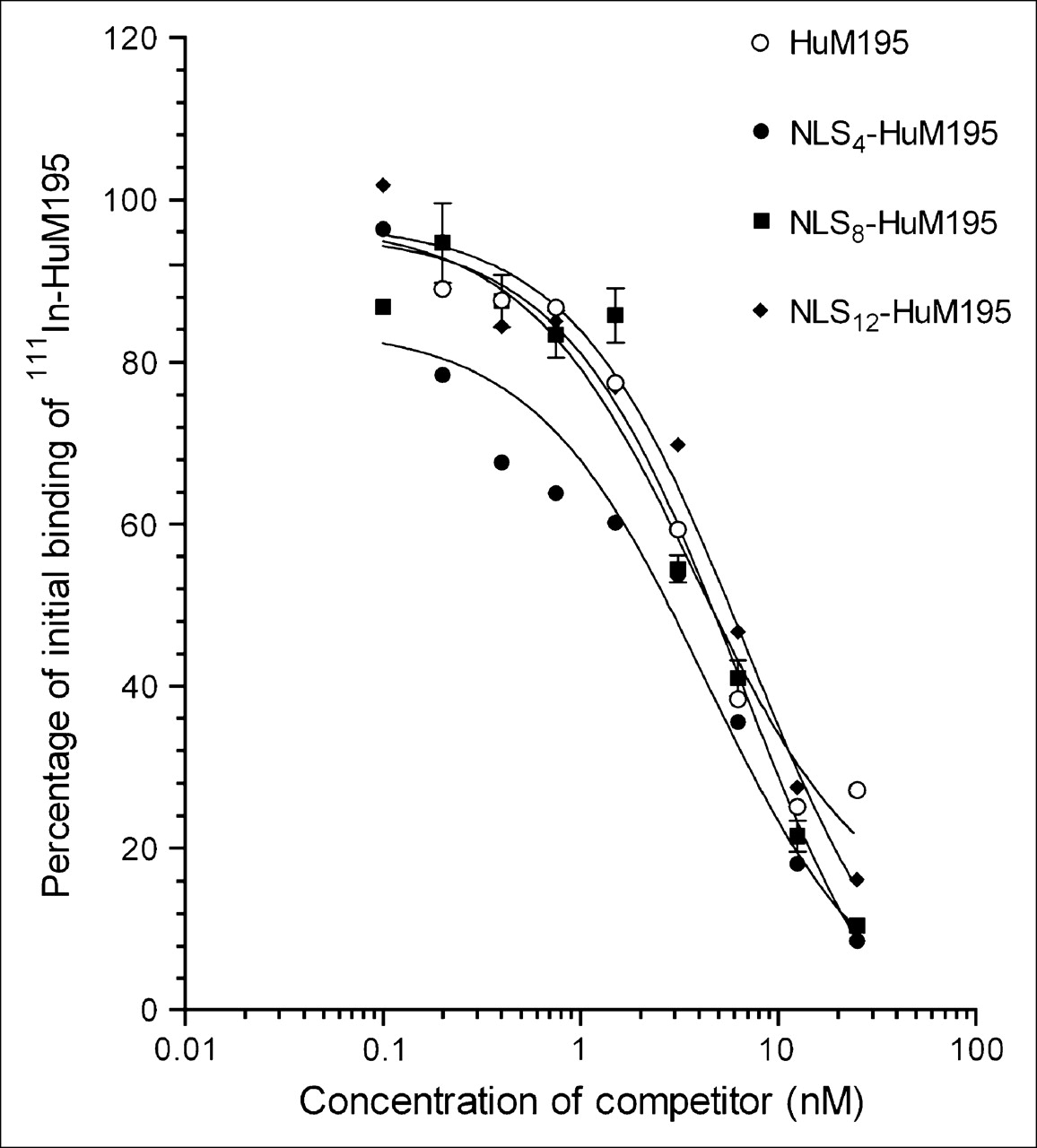
The number of NLS-peptides incorporated into each molecule of HuM195 ranged from 4 to 5 peptides at an SMCC-to-HuM195 ratio of 10:1 to 20:1 to as many as 83 peptides per molecule at a ratio of 1,200:1, estimated by the apparent increase in Mr from 158 kDa for HuM195 to 164−276 kDa for NLS-HuM195 by SDS−PAGE analysis (Fig. 1A). The Mr of NLS-peptides was 1.42 kDa. There was an excellent correlation (r2 = 0.981) between the number of NLS-peptides introduced into HuM195 by SDS−PAGE analysis or by measuring the proportion of tracer 123I-NLS-peptides incorporated into the antibodies (Fig. 1B). HuM195 derivatized with 4, 8, or 12 NLS-peptides competed with 111In-HuM195 for binding to CD33 on HL-60 cells (Fig. 2). There was a slight, but insignificant, increase in the Kd as the substitution level increased from 4 peptides (Kd = 4.3 ± 1.7 × 10⁻⁹ mol/L) to 8 or 12 peptides per molecule (Kd = 6.3 ± 1.3 × 10⁻⁹ mol/L and 6.9 ± 1.3 × 10⁻⁹ mol/L, respectively). The Kd for unconjugated HuM195 was 3.9 ± 1.4 × 10⁻⁹ mol/L.

(A) SDS–PAGE analysis of HuM195 and HuM195 derivatized with SMCC at increasing SMCC-to-HuM195 ratios to introduce maleimide groups for reaction with NLS-containing peptides. Gradual increase in apparent Mr was evident for HuM195 from 158 to 276 kDa as SMCC-to-HuM195 ratio was increased, indicating peptide substitution. (B) Good correlation was found between NLS-peptide substitution ratio determined by SDS–PAGE and that estimated by incorporating tracer amounts of 123I-NLS-peptides into conjugation reaction.

Displacement of binding of 111In-HuM195 to HL-60 myeloid leukemia cells by increasing concentrations of HuM195 or NLS-conjugated HuM195. Kd value for HuM195 was 3.9 ± 1.4 × 10⁻⁹ mol/L, and Kd value for NLS-HuM195 substituted with 4, 8, or 12 NLS-peptides was 4.3 ± 1.7 × 10⁻⁹ mol/L, 6.3 ± 1.3 × 10⁻⁹ mol/L, or 6.9 ± 1.3 × 10⁻⁹ mol/L, respectively.
Nuclear Localization of 111In-NLS-HuM195 and 111In-HuM195
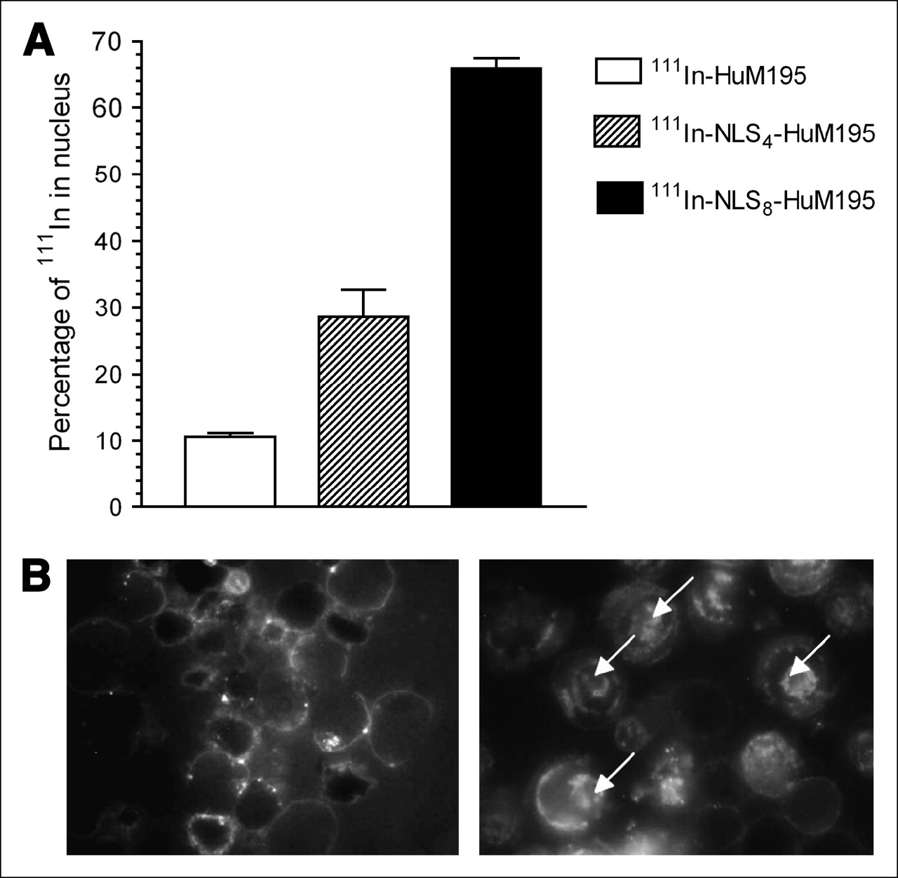
There was a strong and direct correlation between the number of NLS-peptides introduced into 111In-HuM195 and the proportion of radioactivity imported into the nucleus of HL-60 cells (Fig. 3A). Nuclear uptake was 28.5% ± 4.1% for 111In-HuM195 conjugated to 4 NLS-peptides and 65.9% ± 1.5% conjugated to 8 NLS-peptides compared with 10.5% ± 0.5% for unmodified 111In-HuM195. Fluorescence microscopy also revealed nuclear localization in HL-60 cells incubated with fluorescein-labeled NLS-HuM195 but only membrane staining for fluorescein-HuM195 not containing NLS-peptides (Fig. 3B).

(A) Nuclear accumulation of radioactivity in HL-60 cells incubated for 1 h at 37°C with 111In-NLS-HuM195 substituted with 4 or 8 NLS-peptides or with unmodified 111In-HuM195 determined by subcellular fractionation. (B) Fluorescence microscopy of HL-60 cells incubated with fluorescein-labeled HuM195 (left) or NLS-HuM195 (right). Note nuclear localization (arrows) observed for NLS-HuM195 compared with cell membrane localization for unmodified HuM195.
Antiproliferative Effects of 111In-NLS-HuM195 and 111In-HuM195 on HL-60 Cells
The growth of HL-60 cells cultured for 7 d in the presence of increasing amounts of 111In-NLS-HuM195 substituted with 12 or 15 NLS-peptides or 111In-HuM195 is shown in Figure 4A. The IC50 for 111In-NLS-HuM195 was 37 kBq per well (seeded with 1 × 10³ cells) and the IC90 was 77−81 kBq per well. The IC50 and IC90 for 111In-HuM195 were 92 and 203 kBq per well, respectively. The percentage growth of HL-60 cells incubated with 44.4 kBq per well of 111In-NLS-HuM195 containing 12 or 15 NLS-peptides compared with that of untreated cells was 21.8% ± 1.5% and 24.6% ± 3.2%, respectively. These values were significantly lower than that for HL-60 cells incubated with 55.5 kBq per well of 111In-HuM195 (75.7% ± 2.5%). The effect of 111In-NLS-HuM195 substituted with 8 NLS-peptides on the growth of 1 × 10³ HL-60, U937, or K562 cells with high, intermediate, or minimal CD33 expression, respectively, is shown in Figure 4B. At the highest amount of 111In-NLS-HuM195 studied (24.5 kBq per well) growth over 7 d compared with untreated cells was 83.1% ± 8.5%, 52.8% ± 3.4%, or 13.1% ± 3.9% for K562, U937, or HL-60 cells, respectively.
Clonogenic Survival of Leukemia Cells Treated with 111In-NLS-HuM195
Because hematologic malignancies are functionally heterogeneous, any novel therapeutic agent must target the clonogenic cells, as these are the progenitors that sustain the proliferating pool. Clonogenic progenitors for leukemia cell lines or primary AML samples can be assayed on the basis of colony formation in methylcellulose cultures. There was a strong dose-dependent decrease in colony formation of HL-60 cells treated with increasing amounts of 111In-NLS-HuM195 substituted with 8 NLS-peptides (Fig. 5A), with 232 ± 22 colonies formed with no treatment compared with only 7 ± 1 colonies formed at 1.48 mBq per cell and no colonies formed at 3.33 mBq per cell of 111In-NLS-HuM195. The surviving fraction of leukemia cells in 9 AML patient specimens treated with PBS, pH 7.4, 111In-trastuzumab (anti-HER2/neu), 111In-HuM195, or 111In-NLS-HuM195 is shown in Figure 5B. In 7 of 9 specimens, 111In-NLS-HuM195 decreased the surviving fraction >2-fold and in 2 of these, survival was decreased >10-fold. 111In-HuM195 without NLS reduced the surviving fraction >2-fold in 5 of 9 specimens and >10-fold in 1 of these specimens. In 7 of 9 specimens, there was good discrimination between the toxicity of 111In-NLS-HuM195 or 111In-HuM195 and 111In-trastuzumab (anti-HER2/neu). However, in 2 of 9 specimens, treatment with 111In-NLS-HuM195 or 111In-HuM195 was less effective at killing the cells, and the differentiation with 111In-trastuzumab was not good. In 1 specimen, 111In-NLS-HuM195 and 111In-HuM195 were both effective, but 111In-trastuzumab was also cytotoxic.

(A) Growth of 1 × 10³ HL-60 cells cultured in presence of increasing amounts of 111In-HuM195 or 111In-NLS-HuM195 substituted with 12 or 15 NLS-peptides for 7 d compared with untreated cells. (B) Growth of 1 × 10³ HL-60, U937, or K562 cells with high, intermediate, or minimal CD33 expression in presence of increasing amounts of 111In-NLS-HuM195 substituted with 8 NLS-peptides for 7 d compared with untreated cells.

(A) Clonogenic survival of HL-60 cells treated with increasing amounts of 111In-NLS-HuM195 substituted with 8 NLS-peptides. (B) Clonogenic survival of AML specimens treated with 111In-HuM195 or 111In-NLS-HuM195 substituted with 7 or 8 NLS-peptides. Control experiments consisted of specimens treated with 111In-trastuzumab (anti-HER-2/neu) or PBS, pH 7.4.
Normal Tissue Toxicity of 111In-NLS-HuM195 and 111In-HuM195
There were no increases in serum ALT or Cr at 15 d after intravenous administration of 3.7 MBq (22 μg) of 111In-HuM195 or 111In-NLS-HuM195 (Table 1). There were also no major decreases in leukocyte, erythrocyte, or platelet counts or in hemoglobin (Hb) or hematocrit (Hct). There were no decreases in body weight of BALB/c mice at 8 or 15 d after injection of PBS, HuM195, 111In-HuM195, NLS-HuM195, or 111In-NLS-HuM195 (Table 2); all mice gained weight over the 15-d period. Examination of the liver and kidneys by light microscopy confirmed no evidence of morphologic damage (results not shown).
Clinical Biochemistry and Hematology Parameters in BALB/c Mice Intravenously Administered PBS (pH 7.4), 111In-HuM195, or 111In-NLS-HuM195
Body Weights of BALB/c Mice Intravenously Administered PBS (pH 7.4), HuM195, 111In-HuM195, NLS-HuM195, or 111In-NLS-HuM195
DISCUSSION
In this study, we demonstrated that the humanized anti-CD33 mAb HuM195, labeled with the Auger electron-emitter 111In, was toxic to HL-60 human myeloid leukemia cells and to leukemia cells in specimens from AML patients. Furthermore, the nuclear translocation and cytotoxicity of 111In-HuM195 was enhanced by its conjugation to 13-mer peptides (CGYGPKKKRKVGG) harboring the NLS (underlined) of SV-40 large T antigen (21). Auger electron-emitters such as 111In are most damaging to DNA if their decay occurs in close proximity to the cell nucleus (22) because of their nanometer-to-micrometer range (23). It has been estimated that the nuclear radiation-absorbed dose from the decay of 111In is amplified 2-fold when the radionuclide decays in the cytoplasm compared with decay on the cell surface, and the dose is magnified >34-fold when 111In decays in the nucleus itself (23).
HuM195 and its murine form, M195, are rapidly internalized by leukemia cells after binding to CD33 epitopes on the cell surface (24). Humanization of M195 was expected to enhance antibody-dependent cytotoxicity (ADCC) and complement-mediated cytotoxicity (CMC) toward leukemia cells through the humanized Fc domain (25), but only modest levels of ADCC were noted against HL-60 cells and no CDC was found (25). In addition, HuM195 produced minimal antileukemia effects in a phase II trial of 50 patients with relapsed or refractory AML (26) and did not improve remission rates or survival combined with chemotherapy in a phase III study in relapsed/refractory AML (27). The suboptimal killing of leukemia cells was believed to be due to low levels of CD33 and rapid internalization of HuM195. However, CD33-mediated internalization renders 111In-HuM195 an attractive agent for targeted Auger electron radiotherapy of leukemia, particularly if the antibody can be routed to the nucleus, where the electrons are most damaging to DNA.
The nuclear pore complex (NPC) regulates the nuclear import of proteins from the cytoplasm to the nucleus. The NPC is composed of a complex of nucleoporins with an inner channel with a diameter of approximately 9 nm that allows passive diffusion into the nucleus of molecules with Mr < 40−60 kDa but requires active transport of larger proteins mediated by NLS (21). NLS are recognized by importin α-β heterodimers, which shuttle the proteins across the NPC into the nucleus (28). In this study, peptides harboring the NLS from SV-40 large T antigen (15) efficiently routed 111In-HuM195 to the nucleus of HL-60 cells. Nuclear uptake was increased 3-fold when 111In-HuM195 was conjugated with 4 NLS-peptides and >6-fold when conjugated with 8 NLS-peptides (Fig. 3). Substitution of HuM195 with multiple NLS-peptides did not have a major deleterious effect on its ability to bind CD33 on HL-60 cells. The Kd values for HuM195 substituted with 4 NLS-peptides (4.3 ± 1.7 × 10⁻⁹ mol/L), 8 NLS-peptides (6.3 ± 1.3 × 10⁻⁹ mol/L), or 12 NLS-peptides (6.9 ± 1.3 × 10⁻⁹ mol/L) (Fig. 2) were not significantly different than that of 111In-HuM195 without NLS-peptides (Kd, 5.0 × 10⁻⁹ mol/L) (16). The good preservation of immunoreactivity of NLS-HuM195 may be due to the fact that only 1 of 5 lysines in the VL domain and 2 of 6 lysines in the VH domain are required for CD33 recognition (29). Nevertheless, the Kd values for 111In-NLS-HuM195 and 111In-HuM195 were about 25- to 40-fold higher than those reported for unmodified HuM195 (Kd, 1.7 × 10⁻¹⁰ mol/L) (29).
mAbs have been previously conjugated with Auger electron-emitters (e.g., 111In, 67Ga, 125I, or 99mTc) for single cell killing of malignant B-cell lymphomas (30–32) and solid tumors (33,34) but, to our knowledge, the application of NLS-containing peptides to insert radiolabeled antibodies into the nucleus of cancer cells for Auger electron radiotherapy has not been reported previously. Our work confirms a recently published study that demonstrated that the [99mTc(OH2)3(CO)3]+ complex linked to a peptide containing the NLS of SV-40 large T antigen routed the complex to the nucleus of melanoma cells, where the emitted Auger electrons were lethal to the cells (35). In this study, the 99mTc-peptide conjugates were not specific for melanoma cells, however, and a comparison of the cytotoxicity was made only against free 99mTcO4−, which does not readily enter cells. In our study, NLS-mediated nuclear localization enhanced the specific toxicity of 111In-NLS-HuM195 compared with 111In-HuM195 after its CD33-mediated internalization in HL-60 cells as well as in AML patient specimens (Figs. 4 and 5). Moreover, on a molar concentration basis, the IC50 values against HL-60 cells of 111In-NLS-HuM195 (1.2 × 10⁻⁸ mol/L) and 111In-HuM195 (2.8 × 10⁻⁸ mol/L) were 100- to 200-fold lower than we measured for nonradiolabeled HuM195 (IC50, 1.7 × 10⁻⁶ mol/L) (16).
The toxicity of 111In-HuM195 and 111In-NLS-HuM195 toward AML specimens was quite heterogeneous (Fig. 5B). In 7 of 9 specimens, 111In-NLS-HuM195 decreased the survival of AML cells >2-fold and, in 2 of these specimens, survival was decreased >10-fold. 111In-HuM195 was less effective, decreasing the survival >2-fold in 5 of 9 specimens and >10-fold in 1 of 9 specimens. The specificity of 111In-HuM195 and 111In-NLS-HuM195 for CD33-positive leukemia cells was illustrated by their differential toxicity toward HL60, U937, or K562 cells with high, intermediate, or minimal CD33 expression (Fig. 4B), respectively, and by the lower radiotoxicity of 111In-trastuzumab (anti-HER2/neu) in 7 of 9 AML specimens (Fig. 5B). The decreased survival of leukemia cells in 1 specimen (patient 9) treated with 111In-trastuzumab was unexpected. Buhring et al. (19) found that none of 30 blood or bone marrow specimens from AML patients expressed messenger RNA for HER2/neu but 3 of 4 patients with CML in B-lymphoid blast crisis were HER2/neu positive. Given this very low HER2/neu expression in AML, a possible explanation for the observed cytotoxicity is that the long-range γ-emissions from 111In (Eγ, 171 and 245 keV) conjugated to trastuzumab may have nonspecifically killed AML cells in this specimen. It was previously reported that 111In-labeled LL1 mAb specific for CD74 was 24-fold more radiotoxic to CD74-positive Raji B-cell lymphoma cells than an 111In-labeled nonspecific antibody, but the nonspecific antibody killed lymphoma cells at higher concentrations, thought to be due to the low LET γ-emissions from 111In (30). The resistance to 111In-HuM195 or 111In-NLS-HuM195 in 2 of 9 specimens (Fig. 5B) may be attributed to a lower density of CD33 on the blast cells, but all AML specimens tested contained >95% CD33-positive cells. Thus, it is probable that other resistance mechanisms could also account for the lack of response to 111In-HuM195 or 111In-NLS-HuM195, such as enhanced DNA repair or resistance to apoptosis via caspase inhibition (36). Elucidation of the mechanisms would require more detailed analysis of these cells. Interestingly, resistance to gemtuzumab not correlated with CD33 expression or multidrug resistance transporters was reported in 1 of 4 human myeloid leukemia cell lines and 3 of 10 AML specimens in another study (37).
An important finding was that there was no evidence of normal tissue toxicity in mice administered 3.7 MBq (22 μg) of 111In-HuM195 or 111In-NLS-HuM195. These single doses of 111In-NLS-HuM195 or 111In-HuM195 corresponded to 185 MBq/kg and were almost 500-fold higher than the cumulative dose found to cause severe anemia and renal tubular damage in monkeys administered 225Ac-HuM195 (377 kBq/kg) (38). Only single doses of <28 kBq/kg of 225Ac-HuM195 were found to be safe. The toxic effects of 225Ac-HuM195 on normal tissues were thought to be due to redistribution of 225Ac decay products, particularly 213Bi. It was recently shown that pretreatment with the metal chelator 2,3-dimercapto-1-propanesulfonic acid or meso-2,3-dimercaptosuccinic acid reduced the renal uptake of 213Bi in mice and monkeys given 225Ac-HuM195 (39). Administration of furosemide or chlorothiazide or competitive inhibition with bismuth subnitrate was similarly effective (39). These pharmacologic interventions may ultimately permit the use of 225Ac-HuM195 in humans, but our results suggest that targeted Auger electron radiotherapy of AML using 111In-HuM195 or 111In-NLS-HuM195 may be an alternative treatment that is potentially less harmful to normal tissues. The in vitro efficacy of eradicating clonogenic progenitors combined with the lack of in vivo toxicity set the stage to perform definitive experiments to determine if this novel therapy will eradicate the AML stem cell, which originates and sustains the AML clone. The NOD/SCID (nonobese diabetic/severe combined immunodeficiency) mouse leukemia initiating cell assay (40) provides an important preclinical tool to determine whether antileukemia therapeutics such as 111In-NLS-HuM195 eradicate the leukemia stem cell.
CONCLUSION
We conclude that 13-mer synthetic peptides harboring the NLS of SV-40 large T antigen conjugated to 111In-labeled anti-CD33 mAb HuM195 efficiently routed the antibodies to the nucleus of AML cells, where the nanometer-to-micrometer range Auger electrons were lethal. 111In-NLS-HuM195 was not toxic to hematopoietic or other normal tissues in BALB/c mice. These results suggest that 111In-NLS-HuM195 may be a promising novel targeted radiotherapeutic agent for CD33-positive AML in humans.
Acknowledgments
This research was supported by a grant from the Canadian Institutes of Health Research (grant 53156) and by funds donated by the Bank of Montreal. The authors acknowledge the supply of HuM195 mAbs provided by Protein Design Laboratories, Inc., Fremont, CA.
References
- Received for publication August 8, 2005.
- Accepted for publication September 27, 2005.


Jump to section
Related Articles
Cited By...
- HOXA/PBX3 knockdown impairs growth and sensitizes cytogenetically normal acute myeloid leukemia cells to chemotherapy
- Experimental Radionuclide Therapy of HER2-Expressing Xenografts Using Two-Step Targeting Nuclisome Particles
- Auger Electron Radioimmunotherapeutic Agent Specific for the CD123+/CD131- Phenotype of the Leukemia Stem Cell Population
- In Vivo SPECT Quantification of Transplanted Cell Survival After Engraftment Using 111In-Tropolone in Infarcted Canine Myocardium
- Trastuzumab-Resistant Breast Cancer Cells Remain Sensitive to the Auger Electron-Emitting Radiotherapeutic Agent 111In-NLS-Trastuzumab and Are Radiosensitized by Methotrexate
- Drug-Resistant AML Cells and Primary AML Specimens Are Killed by 111In-Anti-CD33 Monoclonal Antibodies Modified with Nuclear Localizing Peptide Sequences
- Epidermal Growth Factor Receptor Inhibition Modulates the Nuclear Localization and Cytotoxicity of the Auger Electron Emitting Radiopharmaceutical 111In-DTPA Human Epidermal Growth Factor
- 111In-Labeled Trastuzumab (Herceptin) Modified with Nuclear Localization Sequences (NLS): An Auger Electron-Emitting Radiotherapeutic Agent for HER2/neu-Amplified Breast Cancer
- Carbon Nanotubes: Potential Benefits and Risks of Nanotechnology in Nuclear Medicine
- [Lys40(Ahx-DTPA-111In)NH2]Exendin-4, a Very Promising Ligand for Glucagon-like Peptide-1 (GLP-1) Receptor Targeting
- Nuclear Localizing Sequences: An Innovative Way to Improve Targeted Radiotherapy