
Main menu

  • Home
  • Content
    • Current
    • Ahead of print
    • Past Issues
    • JNM Supplement
    • SNMMI Annual Meeting Abstracts
    • Continuing Education
    • JNM Podcasts
  • Subscriptions
    • Subscribers
    • Institutional and Non-member
    • Rates
    • Journal Claims
    • Corporate & Special Sales
  • Authors
    • Submit to JNM
    • Information for Authors
    • Assignment of Copyright
    • AQARA requirements
  • Info
    • Reviewers
    • Permissions
    • Advertisers
  • About
    • About Us
    • Editorial Board
    • Contact Information
  • More
    • Alerts
    • Feedback
    • Help
    • SNMMI Journals
  • SNMMI
    • JNM
    • JNMT
    • SNMMI Journals
    • SNMMI

User menu

  • Subscribe
  • My alerts
  • Log in
  • My Cart

Search

  • Advanced search
Journal of Nuclear Medicine
  • SNMMI
    • JNM
    • JNMT
    • SNMMI Journals
    • SNMMI
  • Subscribe
  • My alerts
  • Log in
  • My Cart
Journal of Nuclear Medicine

Advanced Search

  • Home
  • Content
    • Current
    • Ahead of print
    • Past Issues
    • JNM Supplement
    • SNMMI Annual Meeting Abstracts
    • Continuing Education
    • JNM Podcasts
  • Subscriptions
    • Subscribers
    • Institutional and Non-member
    • Rates
    • Journal Claims
    • Corporate & Special Sales
  • Authors
    • Submit to JNM
    • Information for Authors
    • Assignment of Copyright
    • AQARA requirements
  • Info
    • Reviewers
    • Permissions
    • Advertisers
  • About
    • About Us
    • Editorial Board
    • Contact Information
  • More
    • Alerts
    • Feedback
    • Help
    • SNMMI Journals
  • View or Listen to JNM Podcast
  • Visit JNM on Facebook
  • Join JNM on LinkedIn
  • Follow JNM on Twitter
  • Subscribe to our RSS feeds
Meeting ReportPhysics, Instrumentation & Data Sciences

Evaluation of molecular imaging PSMA score (PROMISE) in the context of lesion size: do we need a correction factor?

Claudia Ortega, Josh Schaefferkoetter, Reut Anconina, Asmaa Hawsawy, Douglas Hussey, Patrick Veit-Haibach and Ur Metser
Journal of Nuclear Medicine May 2019, 60 (supplement 1) 262;
Claudia Ortega
2Join Department Medical Imaging University Health Network Toronto ON Canada
  • Find this author on Google Scholar
  • Find this author on PubMed
  • Search for this author on this site
Josh Schaefferkoetter
1Join Department Medical Imaging Siemens Healthcare Toronto ON Canada
  • Find this author on Google Scholar
  • Find this author on PubMed
  • Search for this author on this site
Reut Anconina
3Joint Department Medical imaging University Health Network Toronto ON Canada
  • Find this author on Google Scholar
  • Find this author on PubMed
  • Search for this author on this site
Asmaa Hawsawy
2Join Department Medical Imaging University Health Network Toronto ON Canada
  • Find this author on Google Scholar
  • Find this author on PubMed
  • Search for this author on this site
Douglas Hussey
2Join Department Medical Imaging University Health Network Toronto ON Canada
  • Find this author on Google Scholar
  • Find this author on PubMed
  • Search for this author on this site
Patrick Veit-Haibach
3Joint Department Medical imaging University Health Network Toronto ON Canada
  • Find this author on Google Scholar
  • Find this author on PubMed
  • Search for this author on this site
Ur Metser
3Joint Department Medical imaging University Health Network Toronto ON Canada
  • Find this author on Google Scholar
  • Find this author on PubMed
  • Search for this author on this site
  • Article
  • Figures & Data
  • Info & Metrics
Loading

Abstract

262

Objectives: In order to standardize interpretation of PSMA PET, a molecular imaging PSMA scoring system (miPSMA) was recently introduced, considering degree of radiotracer uptake, in relation to reference structures [1]. To allow accurate quantification of radiotracer uptake on PET, several corrections are needed including partial volume effect (PVE) [2]. PVE originates from the finite spatial resolution of the PET and causes underestimation of the measured activity concentration especially for lesions at or below the resolution of the scanner [3]. Clinically, this may become relevant when assessing radiotracer uptake in small lesions. In the setting of staging/restaging prostate cancer (PCa), the identification of all disease sites may have significant implications to tailoring the most appropriate therapeutic approach. The aim of the current study was to investigate the impact of PVE on the miPSMA score in PCa metastases detected in patients with biochemical recurrence with negative or equivocal CT and bone scintigraphy. This was performed as part of an ongoing PSMA PET registry.

Methods: A total of 27 patients who completed 18F-DCFPyL (PSMA) PET/CT scans for assessment of recurrent PCa (mCT40, Siemens Healthcare). Of these patients, 7 where excluded (no metastatic disease), and 1 who has lesions which could not be accurately contoured on the unenhanced CT, resulting in a final cohort of 20 patients with 71 suspected metastases. Of the 71 lesions included, the majority were small lymph nodes (n=68), and the remaining lesions were bone metastases (n=2) and a lung deposit (n=1). All lesions were manually contoured on CT and PET separately, using Mirada Vision workstation (Mirada Medical) and calculated volume (cm3), SUV max and SUV mean recorded. For the purpose of reference, the SUV max/mean of parotid gland, liver and blood pool were also obtained. PET images were corrected by segmented anatomical CT, smoothed by the simulated PET scanner point-spread function. Dividing the smoothed volume by the original mask, while considering the appropriate lesion contrast, provides a correction map, which was transformed into the PET image voxel space and applied to the PET images. The result was a new PET image with the same total activity as the original, but with local, deconvolution-like PVE correction [Fig.1]. The measured vs corrected SUVmax and SUVmean were compared. Furthermore, the prospectively assigned miPSMA score was compared to the score obtained from the corrected dataset. All quantitative and qualitative assessment was correlated for lesion volume.

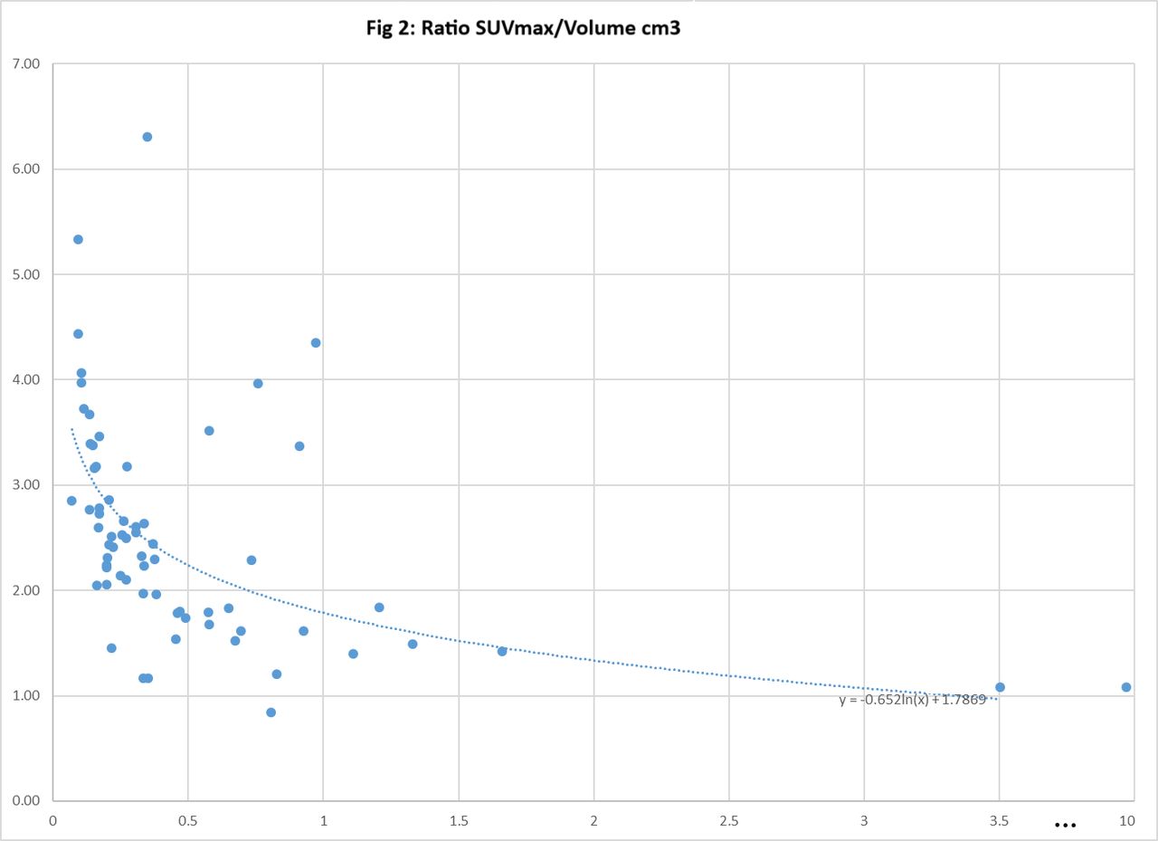
Results: Of the 71 available lesions, 4 lymph nodes were excluded from final analysis due to PVE correction errors (usually due to proximity to ureter). There were 67 corrected lesions with a measured contoured volume of 0.31 cm3 (median; range: 0.06-10.35 cm3). Before and after PVE correction, SUVmax was 13.0 (median; range 1.8-89.5) vs 29.3 (median; range 6-182.2), respectively; SUVmean was 6.5 (median; range 1.4-31.3) vs 11.7 (median; range 2.9-55.3), respectively; average miPSMA score was 1.98 and 2.53, respectively. The estimated SUVmax correction factor for this cohort was 2.52 [Fig.2]. After PVE correction, the corrected miPSMA score were upgraded in 33 lesions (50%) from score 1 to 2 in 10 lesions (30%); from score 1 to 3 in 4 lesions (12%) and from score 2 to 3 in 19 lesions (58%). When assessing lesions with a volume < 0.3 cm3 (volume of a sphere with a diameter of 8mm), 2/3 of lesions (21/32; 66%) had a higher miPSMA score after correction, compared to 1/3 (12/34; 35%) of lesions with volume ≥ 0.3 cm3. [Fig.3-4] Conclusions: Partial volume effects have an impact on quantitative assessment of metastatic lesions on PSMA PET. For small lesions, and especially those with a volume < 0.3 cm3, this also impacts qualitative assessment, resulting in a higher miPSMA score in nearly 2/3 of lesions. This should be considered in future revised versions of the miPSMA score.

Figure
  • Download figure
  • Open in new tab
  • Download powerpoint
Figure
  • Download figure
  • Open in new tab
  • Download powerpoint
Figure
  • Download figure
  • Open in new tab
  • Download powerpoint
Figure
  • Download figure
  • Open in new tab
  • Download powerpoint
Previous
Back to top

In this issue

Journal of Nuclear Medicine
Vol. 60, Issue supplement 1
May 1, 2019
  • Table of Contents
  • Index by author
Article Alerts
Sign In to Email Alerts with your Email Address
Email Article

Thank you for your interest in spreading the word on Journal of Nuclear Medicine.

NOTE: We only request your email address so that the person you are recommending the page to knows that you wanted them to see it, and that it is not junk mail. We do not capture any email address.

Enter multiple addresses on separate lines or separate them with commas.
Evaluation of molecular imaging PSMA score (PROMISE) in the context of lesion size: do we need a correction factor?
(Your Name) has sent you a message from Journal of Nuclear Medicine
(Your Name) thought you would like to see the Journal of Nuclear Medicine web site.
Citation Tools
Evaluation of molecular imaging PSMA score (PROMISE) in the context of lesion size: do we need a correction factor?
Claudia Ortega, Josh Schaefferkoetter, Reut Anconina, Asmaa Hawsawy, Douglas Hussey, Patrick Veit-Haibach, Ur Metser
Journal of Nuclear Medicine May 2019, 60 (supplement 1) 262;

Citation Manager Formats

  • BibTeX
  • Bookends
  • EasyBib
  • EndNote (tagged)
  • EndNote 8 (xml)
  • Medlars
  • Mendeley
  • Papers
  • RefWorks Tagged
  • Ref Manager
  • RIS
  • Zotero
Share
Evaluation of molecular imaging PSMA score (PROMISE) in the context of lesion size: do we need a correction factor?
Claudia Ortega, Josh Schaefferkoetter, Reut Anconina, Asmaa Hawsawy, Douglas Hussey, Patrick Veit-Haibach, Ur Metser
Journal of Nuclear Medicine May 2019, 60 (supplement 1) 262;
Twitter logo Facebook logo LinkedIn logo Mendeley logo
  • Tweet Widget
  • Facebook Like
  • Google Plus One
Bookmark this article

Jump to section

  • Article
  • Figures & Data
  • Info & Metrics

Related Articles

  • No related articles found.
  • Google Scholar

Cited By...

  • No citing articles found.
  • Google Scholar

More in this TOC Section

Physics, Instrumentation & Data Sciences

  • Keel-Edge Height Selection for Improved Multi-Pinhole 123I Brain SPECT Imaging
  • Ultra-Fast Reconstruction of Short List-Mode PET Data Frames for Real-Time Visualization and Processing
  • Deep Learning Based 3D Dose Estimation from Prompt Gamma Distribution for Proton Therapy Monitoring
Show more Physics, Instrumentation & Data Sciences

Cardiac and Cancer Imaging (Data Management, PIDS)

  • A 4D Statistical Atlas of the Human Heart from Gated PET Myocardial Perfusion Imaging
  • SPECT/CT studies for dosimetry after PRRT: impact of reduced number of studies on organ dose calculation and patient management
  • On the Accuracy of Voxel-Based Kidney Dosimetry
Show more Cardiac and Cancer Imaging (Data Management, PIDS)

Similar Articles

SNMMI

© 2025 SNMMI

Powered by HighWire