


Abstract
A simplified technique using 123I-N-isopropyl-p-iodoamphetamine (123I-IMP) autoradiography (ARG) with SPECT has been proposed recently for quantifying regional cerebral blood flow (rCBF). To validate the accuracy of 123I-IMP-ARG for quantifying regional cerebrovascular reactivity (rCVR) to acetazolamide, we compared rCVR determined using 123I-IMP-ARG with that determined using H215O PET. Methods: Thirty-nine patients with chronic stenoocclusive disease in a unilateral major cerebral artery underwent SPECT and PET studies before and after intravenous administration of acetazolamide. The rCBF images in the 4 conditions in each patient were calculated according to the ARG method. The same standard input function and the same distribution volume of 35 mL/mL were used in the calculation of rCBF images using the 123I-IMP-ARG method at resting state and with acetazolamide challenge. One large cortical region of interest (ROI) for a unilateral middle cerebral artery territory was bilaterally determined on each standardized summed rCBF image. On the basis of the rCBF values in each ROI, rCVR to acetazolamide was calculated as follows: rCVR (%) = ([acetazolamide challenge rCBF − resting rCBF]/resting rCBF) × 100. Results: Significant correlation was observed between rCVR values obtained using 123I-IMP-ARG and H215O PET methods in the 78 ROIs examined in the 39 patients (r = 0.820; P < 0.0001). When a rCVR lower than the mean − 2 SD of values obtained in healthy volunteers (18.4% for 123I-IMP-ARG and 18.2% for H215O PET) was defined as reduced, and when the H215O PET method was assumed to represent the true determinant of rCVR, 123I-IMP-ARG was 90% sensitive and 92% specific and displayed an 87% positive predictive value for detecting patients with reduced rCVR. Conclusion: These findings demonstrate that 123I-IMP-ARG methods accurately quantify rCVR and can adequately define subgroups of patients with reduced rCVR.
- cerebral blood flow quantitation
- cerebrovascular reactivity
- brain SPECT
- 123I-N-isopropyl-p-iodoamphetamine
Evaluation of the regional cerebrovascular reactivity (rCVR) to a cerebral vasodilatory stimulus is important in the investigation of patients with ischemic cerebrovascular disease. Qualitative or quantitative measurement of regional cerebral blood flow (rCBF) using SPECT with 99mTc- hexamethylpropyleneamine oxime, 99mTc-ethylcysteinate dimer, 123I-N-isopropyl-p-iodoamphetamine (123I-IMP), or 133Xe has been widely used for assessing rCVR (1–6). Recent prospective studies have demonstrated that rCVR to acetazolamide determined quantitatively by 133Xe SPECT can predict the outcome of major cerebral arterial occlusive disease (7,8), whereas Yokota et al. (9) reported a prospective study using qualitative measurement of rCVR to acetazolamide with 123I-IMP SPECT that failed to find an association between hemodynamic failure and stroke risk. As Yonas et al. (10,11) indicated, qualitative assessment of acetazolamide reactivity is known to have low sensitivity and specificity for detecting patients with a compromised reserve. These authors reported that the positive predictive value of the qualitative methods was 50%. Therefore, the possibility arises that the acetazolamide reactivity of some patients has been incorrectly classified by qualitative analysis.
Recently, a simplified technique known as 123I-IMP–autoradiography (ARG), which requires only 1-point arterial blood sampling and the acquisition of a single static scan, has been developed to quantify rCBF using 123I-IMP and SPECT (12,13). The 123I-IMP-ARG method is based on the 2-compartment model for tracer kinetics. The method uses a standard arterial input calibrated by the radioactivity of a single arterial whole-blood sample, a standard lipophilic fraction of 123I-IMP in whole blood, and a fixed distribution volume (Vd) of 123I-IMP. Previous studies have demonstrated a good correlation between rCBF at the resting state measured by PET with H215O and that measured by the 123I-IMP-ARG method (12,14).
The aim of this study was to validate the accuracy of the 123I-IMP-ARG method for quantifying rCVR to acetazolamide. We compared rCVR with acetazolamide determined quantitatively by the 123I-IMP-ARG method using SPECT with that determined quantitatively by the H215O autoradiographic method using PET in patients who had chronic stenoocclusive disease in a unilateral major cerebral artery.
MATERIALS AND METHODS
Subjects
Healthy Volunteers.
We studied 14 male and 6 female healthy volunteers between 30 and 68 y old (mean ± SD, 53 ± 11 y old) to measure the normal values of rCBF by SPECT or PET. Screening of the health status included a medical review of past history, a physical examination, and neurologic and mental tests. Subjects having a past history of hypertension, diabetes mellitus, atrial fibrillation, or pulmonary disease were excluded. Before the SPECT study or the PET study, brain CT was performed to rule out organic lesions of the brain. Subjects with leukoaraiosis or asymptomatic lacunar infarction were excluded from this study. These healthy volunteers were divided into 2 groups; the first group (7 men, 3 women; mean age, 52 ± 12 y; range, 30–68 y) participated in the SPECT study and the second group (7 men, 3 women; mean age, 53 ± 11 y; range, 35–66 y) participated in the PET study.
Stroke Patients.
From March 1997 to September 1998, 157 patients with unilateral chronic major cerebral artery stenoocclusive disease and a history of minor past strokes or transient ischemic attacks (TIAs) were admitted to our institute for evaluation of rCBF. Of the 157 patients, 39 (27 men, 12 women; mean age, 62 ± 12 y; range, 35–76 y) who underwent both SPECT and PET studies were included in the study. All patients had their last cerebral ischemic event >2 mo before entry into the study. No patient had pulmonary disease. Brain CT or MRI and cerebral angiography were performed before PET and SPECT studies in all patients. No infarction or border zone infarction or lacunar infarction in the basal ganglia or deep white matter was observed in any of the patients. Twenty-two of the 39 patients examined had minor past strokes with definite cerebral infarctions on CT or MRI, and 10 had only TIAs with definite cerebral infarctions. The remaining 7 patients had TIAs without definite cerebral infarctions. Unilateral atherosclerotic vascular lesions were noted on the trunk of the middle cerebral artery (MCA) in 16 patients (10 occlusions, 6 stenoses) and the internal carotid artery (ICA) in 23 patients (13 occlusions, 10 stenoses).
Informed consent was obtained from all participants and the study was approved by our Ethics Committee.
123I-IMP SPECT Study
SPECT studies were performed using a ring-type SPECT scanner, a Headtome-SET080 (Shimadzu Corp., Kyoto, Japan), which provides 31 tomographic images simultaneously. The spatial resolution of the scanner with a low-energy, all-purpose collimator was 13-mm full width at half maximum at the center of the field of view, and the slice thickness was 25-mm full width at half maximum at the center of the field of view. Image slices were taken at 5-mm center-to-center spacing parallel to the orbitomeatal line. The images were reconstructed using the weighted-filtered backprojection technique, in which attenuation correction was made by detecting the edge of the object. An attenuation coefficient of 0.065 cm−1, a Butterworth filter (cutoff, 0.45 cycle/cm; order, 3), and a ramp filter were used for image reconstruction.
The 123I-IMP SPECT study was performed as described (12,13). After a 1-min intravenous infusion of 222 MBq of 123I-IMP (5-mL volume) at a constant rate of 5 mL/min and a 1-min infusion of physiologic saline at the same rate, data acquisition was performed at a midscan time of 30 min after the 123I-IMP administration for a scan duration of 20 min.
At 10 min after the beginning of the 123I-IMP infusion, arterial blood (2 mL) was taken from the brachial artery. The whole-blood radioactivity of 1 mL of each blood sample obtained was measured using a well counter that was cross-calibrated to the SPECT scanner. The arterial partial pressures of O2 (Pao2) and CO2 (Paco2) and the blood pH were also measured in the remaining blood samples using a blood gas tension analyzer.
Two days after the measurement of the rCBF at the resting state, subjects underwent SPECT with acetazolamide challenge. Acetazolamide (1,000 mg; range, 13–19 mg/kg body weight) was given intravenously 10 min before 123I-IMP administration, and the SPECT study was performed by the same procedure as for the resting state.
All reconstructed SPECT images were corrected for the radioactive decay of 123I back to the 123I-IMP injection start time, normalized by the data collection time and cross-calibrated to the well counter system. The rCBF images were calculated according to the 123I-IMP-ARG method (12). The whole-blood radioactivity counts of the single blood sample were refereed to the standard input function. The same standard input function at the resting state was used in the calculation of rCBF with acetazolamide challenge (15). The Vd value was assumed to be 35 mL/mL in the calculation of rCBF images both at resting state and with acetazolamide challenge (16,17).
H215O PET Study
All patients underwent PET 2 d before SPECT at the resting state. We used a 4-ring, 7-slice PET scanner (Headtome-IV; Shimadzu Corp.) with in-plane and axial resolutions of 8 and 10 mm, respectively (18). The PET scanner provides 14 tomographic images with 6.5-mm intervals by the continuous axial motion of the gantry. The image slices were parallel to the orbitomeatal line (same as for the SPECT studies).
Before emission scanning, a transmission scan using a 68Ga-68Ge line source was obtained to correct tissue attenuation. The rCBF was calculated using the ARG method with 90-s scanning after an intravenous bolus injection of 1,110 MBq H215O (19). Continuous arterial blood sampling and β-ray monitoring with a scintillator were conducted throughout PET scanning using a catheter implanted in the radial artery to obtain the arterial input function. Pao2 and Paco2 and the blood pH were also measured in the same blood samples.
The PET studies at the resting state and with acetazolamide challenge were performed on the same day. Ten minutes after the resting rCBF measurement, acetazolamide (1,000 mg; range, 13–19 mg/kg body weight) was given intravenously. Fifteen minutes later, the rCBF was measured by the same procedure as for the resting state.
Blood pressure was measured by auscultation twice for each CBF measurement. The average values of mean blood pressure in each CBF measurement were used to assess the change in mean blood pressure.
Data Analysis
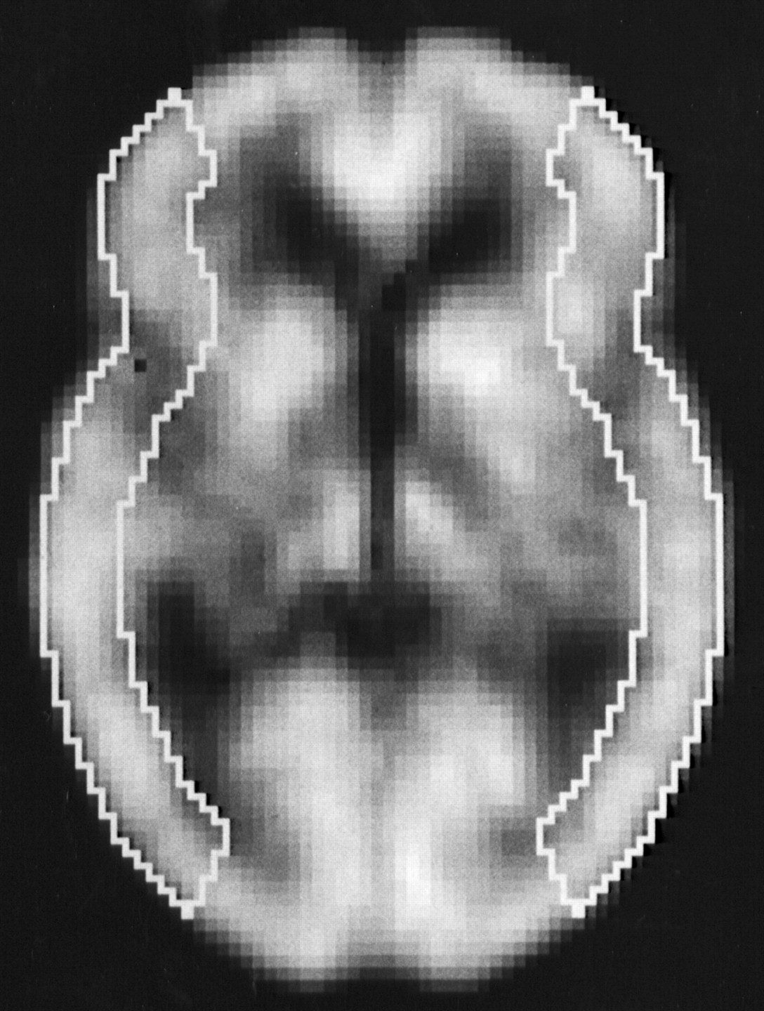
All SPECT and PET images obtained were analyzed as follows. Coregistration and anatomic standardization were performed on a 128 × 128 × 60 matrix (2.25 × 2.25 × 2.25 mm) for each rCBF image, using part of a program set within NEUROSTAT (20–22). Slices between the anterior commissure–posterior commissure level (AC–PC level) − 11.25 mm and the AC-PC level + 9 mm of the standardized images, which had nearly identical cortical shapes, were summed for region-of-interest (ROI) analysis. The outer cortical border was automatically drawn on the section, and the inner cortical border was drawn 15.75 mm (7 pixels × 2.25 mm) further inward. The cortical ribbon was then divided into twelve 30° sectors in a clockwise fashion, and we defined 30° to 150° sectors as the left MCA territory and 210° to 330° sectors as the right MCA territory (Fig. 1). Thus, 1 large cortical ROI for each unilateral hemisphere was determined on each standardized summed rCBF image. Furthermore, the ROI was set bilaterally in all subjects.

Regions of interest in image slice standardized and summed using 3-dimensional stereotactic surface projections.
On the basis of the rCBF values in each ROI, rCVR to acetazolamide was calculated as follows: rCVR (%) = ([acetazolamide challenge rCBF − resting rCBF]/resting rCBF) × 100.
For statistical analysis, the data were expressed as the mean ± SD, and differences among the 4 groups were examined by repeated-measures ANOVA. Correlation between various parameters was determined by linear regression analysis. Statistical significance was set at the P < 0.05 level.
RESULTS
The values of rCBF at the resting state and with acetazolamide challenge and rCVR obtained by the 123I-IMP-ARG method in 20 ROIs of the first group (10 healthy volunteers) were 35.9 ± 4.4 mL/100 g/min, 48.7 ± 5.0 mL/100 g/min, and 36.8% ± 9.2%, respectively. The same values obtained by the H215O PET method in 20 ROIs of the second group (10 healthy volunteers) were 43.5 ± 3.1 mL/100 g/min, 58.7 ± 5.8 mL/100 g/min, and 34.8% ± 8.3%, respectively. For the 123I-IMP-ARG method and the H215O PET method, rCVR in the healthy volunteers was not age dependent.
Table 1 shows the average values of the physiologic variables measured in the 39 patients during SPECT and PET scanning at the resting state and after acetazolamide administration. No significant difference in PaO2, PaCO2, blood pH, or mean blood pressure was observed among the 4 conditions (repeated-measures ANOVA).
Physiologic Data of 39 Patients Measured During SPECT and PET at Rest and with Acetazolamide Challenge
Figure 2 shows comparisons of rCBF values obtained by the 123I-IMP-ARG method and those by the H215O PET method (resting state and acetazolamide challenge, respectively) in 78 ROIs of 39 patients. In both conditions, the fits to the regression lines were significant (P < 0.0001), with the correlation coefficients being 0.808 and 0.872, respectively. In particular, rCBF values obtained by the 123I-IMP-ARG method were highly consistent with those obtained by the H215O PET method in the hypoperfusion areas. However, the rCBF values obtained by the 123I-IMP-ARG method were underestimated with increase in rCBF. Representative images are shown in Figure 3.

Correlations of rCBF values calculated by 123I-IMP-ARG method with those calculated by H215O PET method. (A) Resting state. (B) Acetazolamide challenge. Significant correlation was observed in both conditions. Dashed straight line denotes line of identity.

Functional rCBF images calculated by 123I-IMP-ARG method and those calculated by H215O PET method. Data were obtained from patient with left ICA occlusion. Same color scale is used to display these 2 quantitative rCBF images. rCBF images obtained by 123I-IMP-ARG method are reduced in high rCBF areas compared with PET images.
Figure 4 shows comparisons of rCVR values obtained by the 123I-IMP-ARG method and those by the H215O PET method in 78 ROIs of 39 patients. Significant correlation was observed between the 2 methods (r = 0.820; P < 0.0001). When a rCVR lower than the mean − 2 SD of the values obtained in healthy volunteers (18.4% for the 123I-IMP-ARG method and 18.2% for the H215O PET method, respectively) was defined as reduced, and when the H215O PET method was assumed to be the true determinant (or gold standard) of rCVR, 4 of 31 ROIs (13%) in which the 123I-IMP-ARG method identified reduced rCVR were considered false-positives. The values of rCVR obtained by the 123I-IMP-ARG method in these 4 ROIs with false-positives were 13.1%–17.8%. No ROIs with rCVR lower than the mean − 3 SD of the values obtained in healthy volunteers (9.2%) exhibited false-positives. Conversely, the H215O PET method detected reduced rCVR in 3 of the 47 ROIs (6%) identified as having no reduced rCVR using the 123I-IMP-ARG method (false-negatives). The values of rCVR obtained by the 123I-IMP-ARG method in these 3 ROIs with false-negatives were 20.9%–25.5%. No ROIs with rCVR higher than the mean − 1 SD of the values obtained in healthy volunteers (27.6%) exhibited false-negative findings. Both analytic approaches obtained identical results in 91% of the ROIs; 35% (27/78 ROIs) were true-positives and 56% (44/78 ROIs) were true-negatives. Sensitivity, specificity, and predictive values were calculated for the 123I-IMP-ARG method of assessing rCVR. The 123I-IMP-ARG and H215O PET methods will identify the same patients as positive 90% of the time (sensitivity). The 2 methods will identify the same patients as negative 92% of the time (specificity). A patient that the 123I-IMP-ARG method identifies as positive will be positive according to the H215O PET method 87% of the time (positive predictive value). A patient that the 123I-IMP-ARG method identifies as negative will be negative according to the H215O PET method 94% of the time (negative predictive value).

Correlation of rCVR values calculated by 123I-IMP-ARG method with those calculated by H215O PET method. Significant correlation was observed between these 2 values. Plot of relationship between these 2 values revealed 4 groups of results: (a) those with reduced rCVR (true-positive; ○); (b) those that only H215O PET method identified as reduced rCVR (false-negative; •); (c) those without reduced rCVR (true-negative; ○); and (d) those considered reduced rCVR only by 123I-IMP-ARG method (false-positive;  ). Dashed horizontal and vertical lines denote mean − 2 SD of rCVR values obtained in healthy volunteers by 123I-IMP-ARG method and by H215O PET method, respectively.
DISCUSSION
Our findings indicated that the 123I-IMP-ARG method accurately quantifies rCVR to acetazolamide, with a high correlation (r = 0.820) between rCVR values obtained by the 123I-IMP-ARG method and those obtained by the H215O PET method. On the other hand, recent prospective studies have demonstrated that rCVR lower than the mean − 2 SD or the 95% confidence limit of the values quantitatively obtained in healthy volunteers using 133Xe SPECT is significantly associated with an increased risk of stroke recurrence in patients with symptomatic MCA or ICA occlusion (7,8). Therefore, we defined rCVR lower than the mean − 2 SD of the values obtained in healthy volunteers as reduced—that is, being at high risk of stroke recurrence. We also assumed the H215O PET method to be the true determinant (or gold standard) of rCVR. As a result, our study showed that the 123I-IMP-ARG method is 90% sensitive and 92% specific for detecting patients with reduced rCVR. The positive predictive value using the 123I-IMP-ARG method is 87%, and the negative predictive value is 94%. These findings suggest that the subgroup of patients at increased risk for stroke recurrence can be adequately defined by rCVR quantitatively measured using the 123I-IMP-ARG method.
This study also showed the high correlation of rCBF values obtained by the 123I-IMP-ARG method with those obtained by the H215O PET method. In particular, the 2 values coincided strongly in the hypoperfusion areas. These findings are noteworthy because correct quantification of rCBF values in the ischemic territories is the greatest clinical problem. On the other hand, the rCBF values obtained by the 123I-IMP-ARG method were underestimated as rCBF increased, which resulted in the slopes of the linear regression lines being significantly smaller than that of the identity line. This may be due to errors in the SPECT reconstruction, such as imperfect attenuation correction, scatter, and limited spatial resolution of the scanner (partial-volume effect). Another possible explanation for the systematic underestimation at high flow may be due to the limited first-pass extraction fraction of 123I-IMP in the brain (23). The use of a fixed Vd may also be one of the reasons why the rCBF values obtained by the 123I-IMP-ARG method were underestimated as rCBF increased. Because we performed coregistration and anatomic standardization for each rCBF image, and there was no significant difference in Pao2, Paco2, blood pH, or mean blood pressure among each study, factors such as misalignment of ROIs and physiologic change in rCBF are unlikely to have contributed to the observed difference in rCBF values between the 123I-IMP-ARG method and the H215O PET method.
This study possesses several limitations regarding the 123I-IMP-ARG method. First, the method uses the same standard arterial input function both at the resting state and after acetazolamide administration instead of the measurement of arterial radioactivity. Hauge et al. (24) examined the pharmacologic effects on the cerebral and systemic conditions of acetazolamide and concluded that cardiac output and total peripheral resistance were unaltered during acetazolamide loading. Takeuchi et al. (25) also monitored cardiac output and systemic blood pressure during acetazolamide administration in healthy volunteers using an ultrasound Doppler system, with no significant changes observed. Furthermore, Ogura et al. (15) compared the percentage difference in the area under the curve of the estimated from the real arterial input functions between the resting and acetazolamide analyses of 19 cases. No significant difference in the area under the curve was observed between the 2 study groups. The integration period was 40 min, and the standardized input function was calibrated using a single sample at 10 min. Thus, arterial input after acetazolamide administration could be regarded as being the same as that at the resting state. Second, the Vd was assumed to be the same value (35 mL/mL) at the resting state and after acetazolamide administration. The mean Vd value of 123I-IMP for the normal cortex is 37.5 ± 3.6 mL/g and is independent of age and sex (16). In addition, Toyoda et al. (17) demonstrated that the optimal values for Vd at the resting state and after acetazolamide administration were both in the range of 30–35 mL/mL and the difference between the values at the resting state and after acetazolamide administration was very small. Therefore, the Vd could be set as the constant value of 35 mL/mL both at the resting state and after acetazolamide administration.
CONCLUSION
The 123I-IMP-ARG method can be performed using conventional SPECT scanners (12,14) and does not require frequent arterial blood sampling. This allows the technique to be used much more readily in assessing patients with major cerebral artery occlusive disease in routine clinical practice, although 123I-IMP is now unavailable in the West because of the cost. Our study suggests that the ability of the 123I-IMP-ARG method to detect reduced rCVR is almost identical to that of the H215O PET method and that the subgroup of patients at increased risk for stroke recurrence can be adequately defined by rCVR quantitatively measured using the 123I-IMP-ARG method. Whether extracranial–intracranial arterial bypass surgery, which improves impaired cerebral hemodynamics (26,27), reduces the risk of stroke in this subgroup remains unclear. Preoperative low rCVR quantitatively obtained using SPECT has been shown to be significantly associated with an increased risk of hyperperfusion syndrome after carotid endarterectomy (28,29). Standardization of hemodynamic compromise measurement and further investigation to assess the usefulness of extracranial–intracranial arterial bypass surgery or the risk of hyperperfusion syndrome after carotid endarterectomy would be of great benefit.
Acknowledgments
The authors thank Prof. Satoshi Minoshima, University of Washington Medical School, for the use of 3-dimensional stereotactic surface projections (NEUROSTAT) and his helpful suggestions. This work was supported in part by Grants-in-Aid for Advanced Medical Scientific Research by the Ministry of Science, Education, Sports and Culture, Japan.
Footnotes
Received Jul. 25, 2002; revision accepted Oct. 31, 2002.
For correspondence or reprints contact: Kuniaki Ogasawara, MD, Department of Neurosurgery, Iwate Medical University, 19-1 Uchimaru, Morioka, 020-8505 Japan.
E-mail: kuogasa{at}iwate-med.ac.jp
REFERENCES
In this issue


Jump to section
Related Articles
Cited By...
- Robust estimation of dynamic cerebrovascular reactivity using breath-holding fMRI: application in diabetes and hypertension
- Overview of Imaging Modalities in Stroke
- Acetazolamide-Loaded Dynamic 7T MR Quantitative Susceptibility Mapping in Major Cerebral Artery Steno-Occlusive Disease: Comparison with PET
- Preoperative Cerebral Oxygen Extraction Fraction Imaging Generated from 7T MR Quantitative Susceptibility Mapping Predicts Development of Cerebral Hyperperfusion following Carotid Endarterectomy
- Preoperative Central Benzodiazepine Receptor Binding Potential and Cerebral Blood Flow Images on SPECT Predict Development of New Cerebral Ischemic Events and Cerebral Hyperperfusion After Carotid Endarterectomy
- Central Benzodiazepine Receptor Binding Potential and CBF Images on SPECT Correlate with Oxygen Extraction Fraction Images on PET in the Cerebral Cortex with Unilateral Major Cerebral Artery Occlusive Disease
- Multicenter Evaluation of a Standardized Protocol for Rest and Acetazolamide Cerebral Blood Flow Assessment Using a Quantitative SPECT Reconstruction Program and Split-Dose 123I-Iodoamphetamine
- The Acetazolamide Challenge: Techniques and Applications in the Evaluation of Chronic Cerebral Ischemia
- Precision of Cerebrovascular Reactivity Assessment with Use of Different Quantification Methods for Hypercapnia Functional MR Imaging
- Simple Assessment of Cerebral Hemodynamics Using Single-Slab 3D Time-of-Flight MR Angiography in Patients with Cervical Internal Carotid Artery Steno-Occlusive Diseases: Comparison with Quantitative Perfusion Single-Photon Emission CT
- Postoperative Cortical Neural Loss Associated With Cerebral Hyperperfusion and Cognitive Impairment After Carotid Endarterectomy: 123I-iomazenil SPECT Study
- Tracer Delay-Insensitive Algorithm Can Improve Reliability of CT Perfusion Imaging for Cerebrovascular Steno-Occlusive Disease: Comparison with Quantitative Single-Photon Emission CT
- Preoperative Cerebral Hemodynamic Impairment and Reactive Oxygen Species Produced During Carotid Endarterectomy Correlate With Development of Postoperative Cerebral Hyperperfusion
- Quantitative Assessment of Cerebral Hemodynamics Using Perfusion-Weighted MRI in Patients With Major Cerebral Artery Occlusive Disease: Comparison With Positron Emission Tomography
- Reduced Blood Flow and Preserved Vasoreactivity Characterize Oxygen Hypometabolism Due to Incomplete Infarction in Occlusive Carotid Artery Diseases
- Oxygen extraction fraction and acetazolamide reactivity in symptomatic carotid artery disease
- Differences in Vasodilatory Capacity and Changes in Cerebral Blood Flow Induced by Acetazolamide in Patients with Cerebrovascular Disease
- Qualitative versus Quantitative Assessment of Cerebrovascular Reactivity to Acetazolamide Using iodine-123-N-Isopropyl-p-Iodoamphetamine SPECT in Patients with Unilateral Major Cerebral Artery Occlusive Disease