


Article Figures & Data
Figures
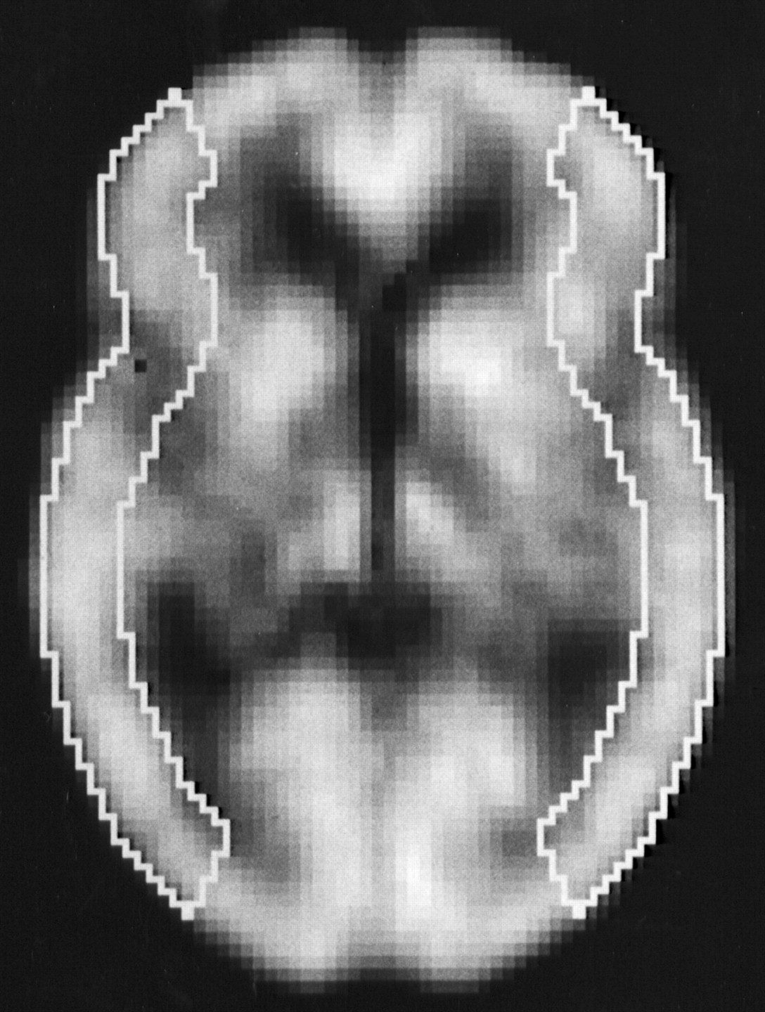
- FIGURE 1.
Regions of interest in image slice standardized and summed using 3-dimensional stereotactic surface projections.
- FIGURE 2.
Correlations of rCBF values calculated by 123I-IMP-ARG method with those calculated by H215O PET method. (A) Resting state. (B) Acetazolamide challenge. Significant correlation was observed in both conditions. Dashed straight line denotes line of identity.
- FIGURE 3.
Functional rCBF images calculated by 123I-IMP-ARG method and those calculated by H215O PET method. Data were obtained from patient with left ICA occlusion. Same color scale is used to display these 2 quantitative rCBF images. rCBF images obtained by 123I-IMP-ARG method are reduced in high rCBF areas compared with PET images.
- FIGURE 4.
Correlation of rCVR values calculated by 123I-IMP-ARG method with those calculated by H215O PET method. Significant correlation was observed between these 2 values. Plot of relationship between these 2 values revealed 4 groups of results: (a) those with reduced rCVR (true-positive; ○); (b) those that only H215O PET method identified as reduced rCVR (false-negative; •); (c) those without reduced rCVR (true-negative; ○); and (d) those considered reduced rCVR only by 123I-IMP-ARG method (false-positive;
 ). Dashed horizontal and vertical lines denote mean − 2 SD of rCVR values obtained in healthy volunteers by 123I-IMP-ARG method and by H215O PET method, respectively.


Tables
- TABLE 1
Physiologic Data of 39 Patients Measured During SPECT and PET at Rest and with Acetazolamide Challenge
Parameter SPECT PET Rest ACZ Rest ACZ pH 7.39 ± 0.03 7.40 ± 0.03 7.40 ± 0.04 7.41 ± 0.03 Paco2 (mm Hg) 41.8 ± 3.0 40.1 ± 2.8 40.6 ± 2.9 39.9 ± 3.1 Pao2 (mm Hg) 94.1 ± 2.9 95.9 ± 4.3 94.8 ± 3.1 96.8 ± 4.1 Mean blood pressure (mm Hg) 95.1 ± 8.1 96.6 ± 8.2 95.9 ± 7.6 97.1 ± 9.8 ACZ = acetazolamide; Paco2 = arterial carbon dioxide tension; Pao2 = arterial oxygen tension.
In this issue


Jump to section
Related Articles
Cited By...
- Robust estimation of dynamic cerebrovascular reactivity using breath-holding fMRI: application in diabetes and hypertension
- Overview of Imaging Modalities in Stroke
- Acetazolamide-Loaded Dynamic 7T MR Quantitative Susceptibility Mapping in Major Cerebral Artery Steno-Occlusive Disease: Comparison with PET
- Preoperative Cerebral Oxygen Extraction Fraction Imaging Generated from 7T MR Quantitative Susceptibility Mapping Predicts Development of Cerebral Hyperperfusion following Carotid Endarterectomy
- Preoperative Central Benzodiazepine Receptor Binding Potential and Cerebral Blood Flow Images on SPECT Predict Development of New Cerebral Ischemic Events and Cerebral Hyperperfusion After Carotid Endarterectomy
- Central Benzodiazepine Receptor Binding Potential and CBF Images on SPECT Correlate with Oxygen Extraction Fraction Images on PET in the Cerebral Cortex with Unilateral Major Cerebral Artery Occlusive Disease
- Multicenter Evaluation of a Standardized Protocol for Rest and Acetazolamide Cerebral Blood Flow Assessment Using a Quantitative SPECT Reconstruction Program and Split-Dose 123I-Iodoamphetamine
- The Acetazolamide Challenge: Techniques and Applications in the Evaluation of Chronic Cerebral Ischemia
- Precision of Cerebrovascular Reactivity Assessment with Use of Different Quantification Methods for Hypercapnia Functional MR Imaging
- Simple Assessment of Cerebral Hemodynamics Using Single-Slab 3D Time-of-Flight MR Angiography in Patients with Cervical Internal Carotid Artery Steno-Occlusive Diseases: Comparison with Quantitative Perfusion Single-Photon Emission CT
- Postoperative Cortical Neural Loss Associated With Cerebral Hyperperfusion and Cognitive Impairment After Carotid Endarterectomy: 123I-iomazenil SPECT Study
- Tracer Delay-Insensitive Algorithm Can Improve Reliability of CT Perfusion Imaging for Cerebrovascular Steno-Occlusive Disease: Comparison with Quantitative Single-Photon Emission CT
- Preoperative Cerebral Hemodynamic Impairment and Reactive Oxygen Species Produced During Carotid Endarterectomy Correlate With Development of Postoperative Cerebral Hyperperfusion
- Quantitative Assessment of Cerebral Hemodynamics Using Perfusion-Weighted MRI in Patients With Major Cerebral Artery Occlusive Disease: Comparison With Positron Emission Tomography
- Reduced Blood Flow and Preserved Vasoreactivity Characterize Oxygen Hypometabolism Due to Incomplete Infarction in Occlusive Carotid Artery Diseases
- Oxygen extraction fraction and acetazolamide reactivity in symptomatic carotid artery disease
- Differences in Vasodilatory Capacity and Changes in Cerebral Blood Flow Induced by Acetazolamide in Patients with Cerebrovascular Disease
- Qualitative versus Quantitative Assessment of Cerebrovascular Reactivity to Acetazolamide Using iodine-123-N-Isopropyl-p-Iodoamphetamine SPECT in Patients with Unilateral Major Cerebral Artery Occlusive Disease