


Abstract
Radiolabeling monoclonal antibodies (mAbs) allows the evaluation of biodistribution of constructs in vivo through gamma camera imaging and also permits quantitation of mAb uptake in tumors through biopsy-based counting techniques. The quantitation of radiolabeled mAb uptake in cancer patients is complicated by the attenuation of gamma emissions of routinely used isotopes (e.g., 131I and 111In) and the spatial resolution and sensitivity of gamma cameras. Methods: We used the positron-emitting isotope 124I (half-life [T1/2] = 4.2 d) to label the recombinant humanized anti–colorectal cancer A33 antibody (huA33) and evaluated its biodistribution properties and PET imaging characteristics in BALB/c nude mice bearing SW1222 colorectal xenografts and control colon tumors. Results: The immunoreactivity of radioconjugate was 78% as determined using the cell-binding Lindmo assay. The apparent association constant was found to be 2.2 × 10⁹ M−1, and the number of antibody binding sites per cell was 371,000. The radioconjugate was found to be stable in serum obtained from mice at various times after injection. Assuming a two-compartment model with a four-parameter fit of mean blood levels, the T1/2α was 1.5 h and the T1/2β was 38.2 h. Excellent tumor uptake was obtained, with maximal uptake reaching 50.0 ± 7.0 percentage injected dose per gram of tumor by 4 d after injection. Specificity of localization was shown by lack of uptake in control tumor. PET imaging detected antigen-positive tumor by 4 h after injection, and high-resolution images were obtained by 24 h after injection. Conclusion: In clinical trials using PET, huA33 labeled with 124I has potential for imaging and staging colon tumors and quantifying antibody uptake in colon tumors in vivo.
The monoclonal antibody (mAb) A33 reacts with an antigen expressed by >95% of colon cancer and by normal small- and large-intestine epithelium (1). The A33 antigen has been sequenced and found to be a novel transmembrane glycoprotein with homology to the immunoglobulin superfamily (2,3). Several phase I and II clinical trials with the murine mAb A33 have been reported; they showed specific uptake of 125I-A33 and 131I-A33 antibody in colorectal tumors and some antitumor effects (4–5). Prolonged imaging of murine 125I-A33 localized to colorectal tumors up to 6 wk after intravenous infusions in patients was possible, suggesting a mechanism of internalization and retention of radioconjugate within tumor cells (5). Studies have also shown that the CDR-grafted, humanized A33 antibody (huA33) specifically and rapidly localizes to colon cancer xenografts in nude mice (6–8) and to colon carcinoma in patients (9).
The isotope 124I has been shown to be useful for quantitative imaging using PET (10,11). Resolution and quantitation characteristics were only slightly reduced compared with conventional PET radionuclides. The relatively longer decay half-life (T1/2) of 4.2 d and the established chemistry for labeling antibodies enable the imaging of xenografts derived from various human cancer cell lines (12–15). These studies suggested that it is possible to use small animals, such as mice and rats, bearing human tumors to obtain good correlation between image-based measurements and direct determinations of radioactivity in excised tumors. Clinical studies using 124I-labeled murine antibodies have been performed on patients with breast cancer (16), and dosimetric determination has been performed on patients with neuroblastoma (17) and glioma (18).
We report here the preparation and evaluation of an 124I-CDR–grafted humanized huA33 conjugate in a BALB/c nude mouse model of human colorectal cancer using immuno-PET.
MATERIALS AND METHODS
All analytic-grade reagents, except when stated, were obtained from Merck Pty. Ltd. (Melbourne, Australia). No carrier-added 124I was produced from an irradiated tellurium oxide target that was subjected to dry distillation to release the isotope (19). The isotope was collected in 0.1N NaOH and sent to the Ludwig Institute for Cancer Research, Melbourne, Australia. Chromatographic Biogel P6DG was obtained from Bio-Rad Laboratories (Melbourne, Australia). Radioactivity was measured either with a dose calibrator, Atomlab-100 (Biodex, Brookhaven, NY), or with a shielded Cobra II automated gamma counter from Canberra-Packard (Melbourne, Australia).
Antibodies and Cells
The CDR-grafted huA33 (IgG1) (6) was provided by the Ludwig Institute for Cancer Research (New York, NY) together with the A33-expressing colorectal cancer cell line SW1222. For controls, the colorectal cancer cell line LIM 2537 (non–A33 expressing) was provided by Dr. Robert Whitehead (Ludwig Institute for Cancer Research, Melbourne, Australia).
Radiolabeling
A 0.3-mL aliquot of 0.5 mol/L potassium phosphate buffer, pH 7.0, was added to 0.57 mL 124I in 0.1N NaOH (13 MBq). An aliquot of 335 μL of this labeled mixture was added to 100 μL antibody (9 mg/mL) followed by 18.3 μL chloramine-T at a concentration of 0.46 mg/mL prepared in the same phosphate buffer. After 2 min, labeling was stopped by adding 18.3 μL of a solution containing 1.86 mg/mL sodium metabisulfite in phosphate buffer. The labeled mixture was purified by centrifugal desalting on columns of Biogel P6DG equilibrated in phosphate-buffered saline (PBS). The radiochemical purity of labeled antibody was analyzed by instant thin-layer chromatography silica gel (ITLC-SG) using 10% w/v trichloroacetic acid as solvent. Radioactivity bound to antibody remained at the origin, whereas free 124I migrated with the solvent front.
Immunoreactivity Assays
The immunoreactivity of huA33 after radiolabeling was determined using the assay of Lindmo et al. (20). This cell-based assay consisted of incubating 20 ng radiolabeled antibody with increasing concentrations of antigen-expressing cells SW1222 ranging from 0 cells to 6.0 × 10⁶ cells in 1.0 mL cell culture medium and mixed continuously on a rotation mixer. After 45 min at room temperature, the cells were centrifuged and washed three times with medium before radioactive counting to determine the extent of binding compared with standards. The specificity of binding was shown by adding 20 μg unlabeled huA33 to the assays, and the extent of binding was similarly determined. Scatchard assays were also performed (20). Serial dilutions starting from 10 μg/mL unlabeled huA33 were added to 20 ng labeled antibody followed by the addition of 3 × 10⁶ SW1222 cells.
Serum Stability
The stability of radioconjugates in the blood circulation of mice was also determined. Serum was obtained at days 0, 3, and 7 after injection. An aliquot of serum was spotted on an ITLC-SG strip and developed using 10% w/v trichloroacetic acid as solvent. For immunoreactivity assay, concentrations of radiolabeled antibody in mouse serum were estimated from the blood time–activity curve of the biodistribution study and used in the cell binding assay.
Animals and Tumors
The A33 antigen–expressing melanoma cell line SW1222 was grown in RPMI 1640 with standard additives and 5% fetal calf serum. The control cell line LIM 2537 was cultured under similar conditions. Cells were harvested at the point of confluence using PBS and 0.05% w/v EDTA and resuspended in medium. Approximately 5.0 × 10⁶ cells of SW1222 and LIM 2537 lines in 0.1 mL PBS were injected intradermally contralaterally on the underside flanks of female BALB/c nude mice, 3–4 wk old. Both SW1222 and LIM 2537 tumors started to develop by 1–2 wk, and the mice were used 1–2 wk later when tumors weighed between 0.2 and 0.7 g. Tumors developed in all mice. An injected dose containing a sterile filtered mixture of 16 μg 124I-huA33 (111 kBq) in 0.1 mL PBS was administered through retroorbital injection while the mice were under enflurane anesthesia (Ethrane; Baxter Pharmaceutical Products, New Providence, NJ). From 4 h after injection, groups of five mice were killed by cranial dislocation, and blood, tumors, and various tissues were removed for weighing and radioactive counting. Blood clearance kinetics were determined using a curve-fitting program, SAAM II, from the University of Washington (Seattle, WA) assuming a two-compartment model. Tumors were measured using the formula (length × width squared) × 0.5 (21), with length and width being two perpendicular measurements of diameter of palpable tumors.
PET Imaging
Mice were killed by overinhalation of enflurane and placed supine on an ECAT 951/31R PET scanner (Siemens Medical Systems, Inc., Hoffman Estates, IL/CTI, Knoxville, TN) linked to a SunSPARC 10 workstation (Sun Microsystems, Palo Alto, CA). The axial field of view of the PET scanner is 10.8 cm, and the in-plane resolution is 6.5 mm. The mice were imaged in two-dimensional mode (septa extended) for 30 min. Images were acquired in a 128 × 128 matrix and reconstructed with a Hanning filter at a cutoff of 0.4 cycles per pixel using the standard filtered backprojection algorithm. Single gamma emissions contribute significantly when using 124I; however, the real-time sorter in the PET scanner eliminates the single emissions because these events are not true coincidences. A long imaging time of 30 min was chosen to ensure that sufficient counts were recorded for acceptable image quality. A transmission scan was not obtained.
RESULTS
In Vitro Properties of 124I-HuA33
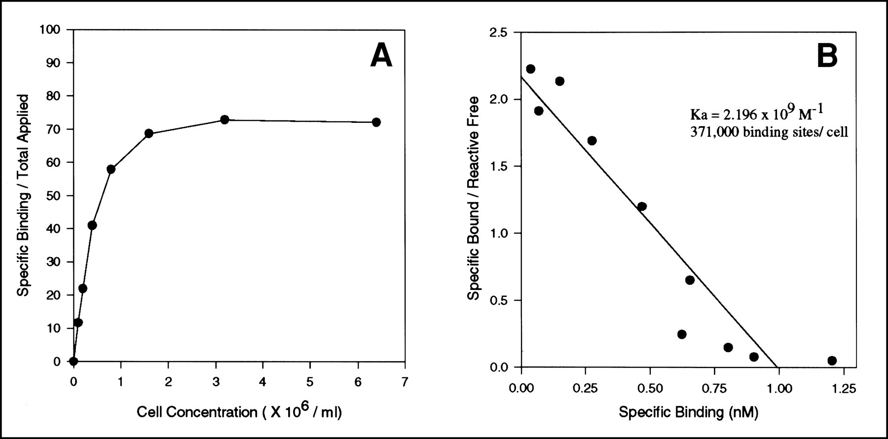
The radiolabeling efficiency of huA33 using 124I and chloramine-T was 40%. After chromatographic purification, >98% of 124I was bound to protein as determined by thin-layer chromatography. The specific activity of 124I-huA33 obtained was 7.4 MBq/mg. Labeled antibody retained the ability to bind A33 antigen–expressing cells as shown using the assay of Lindmo et al. (20) (Fig. 1A). Double-reciprocal plotting of values from the same data (Fig. 1A) and extrapolation to the ordinate provided a quantitative measure of immunoreactivity in the presence of infinite antigen excess. This immunoreactive fraction of the labeled antibody was found to be 0.78. Alternatively, the immunoreactivity is expressed as 78% binding. Scatchard analysis of binding of 124I-huA33 to SW1222 colorectal cancer cells is shown in Figure 1B. The intercept at the abscissa represents the binding capacity of the cell. This value was determined to be 371,000 antibody molecules bound per cell. The apparent association constant can be derived from the slope of the line (Fig. 1B), giving a value of 2.2 × 10⁹ M−1.

Binding assay for determination of immunoreactive fraction of anti–colorectal cancer 124I-huA33. (A) Plot of specific binding over total applied radioactivity against increasing SW1222 cell concentration. (B) Scatchard plot of binding of 124I-huA33 mAb to colorectal cancer cells. From intercept value at abscissa and slope, binding capacity and apparent association constant can be determined. Ka = association constant.
Serum Stability
The stability of radioconjugate was also evaluated by analyzing serum taken from mice injected with antibodies on days 0, 3, and 7 after injection. Thin-layer chromatographic analysis indicated that 98.7% and 85.3%, respectively, of 124I was bound to protein on days 0 and 3. By day 7, the radioactivity in serum was too low to be analyzed by this method. The immunoreactivity of 124I-huA33 in mouse serum at these times after injection was 72%, 69.8%, and 51.3%, respectively. From these results, the radioconjugate was shown to remain stable and to retain an ability to bind to target A33–expressing cells for several days while remaining in the circulation.
Biodistribution and Pharmacokinetics
The relative levels of 124I-huA33 in colon xenografts and blood at times after injection are shown in Figure 2. Assuming a two-compartment model with a four-parameter fit of mean blood levels, the T1/2α was 1.5 h and the T1/2β was 38.2 h.

Radioactivity concentration of 124I-huA33 in blood (•), SW1222 human colon tumor (▴), and LIM 2537 control tumor (▪) in BALB/c nude mice bearing xenografts, as function of time after infusion. %ID = percentage injected dose.
The mean uptake of 124I-huA33 by antigen-expressing cells SW1222 reached a maximum percentage injected dose (%ID) per gram of 50 ± 7.0 by 4 d after injection, with a gradual reduction over the next 6 d. In contrast, the maximal uptake in the control colon xenograft LIM 2537 was 6.27 ± 0.80 %ID/g at 4 h. The tumor-to-blood ratio for SW1222 tumor increased from 5:1 to 35:1 from day 2 to day 7 after injection.
The tissue distribution of 124I-huA33 in xenografted mice is shown in Table 1. Radioactivity was found mainly in blood and several organs by 4 h after injection as a result of blood-pool activity. Activity in the tissues continued to decrease with time, whereas tumor (SW1222) uptake of radioconjugate was substantial and specific. Uptake of radioconjugate in control tumor (LIM 2537) was low.
Biodistribution of 124I-HuA33 mAb in BALB/c Nude Mice Bearing SW1222 and Control (LIM 2537) Colon Carcinoma Xenografts
PET Imaging
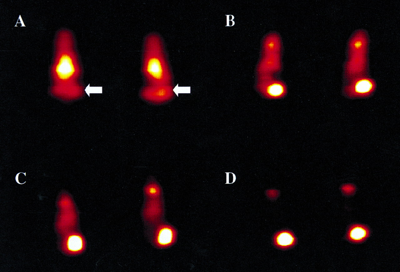
At 4 h after injection (Fig. 3A), blood-pool activity was evident in the heart, with some tumor visualization. Tumor uptake of 124I-huA33 was distinct by 24 h after injection (Fig. 3B) in SW1222 tumors. No radioconjugate uptake was observed in LIM 2537 control tumor located in the contralateral flank. Blood-pool activity in the heart continued to decrease by 48 h after injection (Fig. 3C), and delayed imaging up to 10 d after injection was possible (Fig. 3D). At that late time, other than radioactivity in the tumor and thyroid, the whole-body activity is essentially clear. Tumor sizes ranged from 0.5 to 0.81 g in mass and from 530 to 730 mm3 in volume by 7 d, with growth of tumor up to 1.0 g by day 10.

Immuno-PET images of 124I-huA33 in groups of two BALB/c nude mice bearing SW1222 colorectal cancer xenografts of masses ranging from 0.2 to 0.7 g. Images were acquired 4 h (A), 24 h (B), 48 h (C), and 10 d (D) after injection of 124I-huA33. Location of SW1222 tumors in left flank is indicated in (A) by arrow. Control LIM 2537 tumors are in contralateral flank.
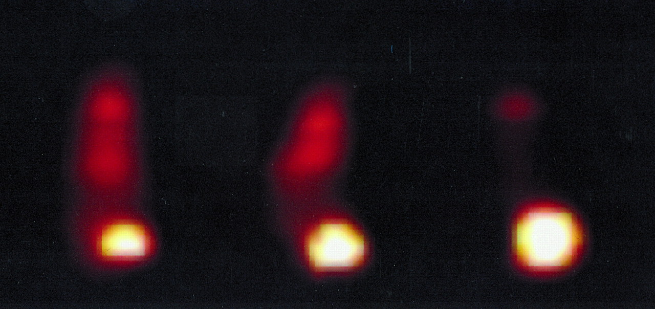
In another experiment, the effect of tumor size on PET imaging was examined with mice bearing tumors weighing 0.21 g (292 mm3), 0.68 g (615 mm3), and 1.2 g (1,079 mm3) (Fig. 4). The mice were imaged 96 h after injection. All tumors could be imaged clearly, and control tumors again did not have any detectable radioconjugate. Larger tumors showed reduced blood-pool activity, consistent with the increased uptake of radioconjugate in tumor mass (Fig. 4).

Effect of tumor mass on immuno-PET detection of 124I-huA33 uptake in SW1222 colorectal cancer xenografts in BALB/c nude mice 96 h after infusion. SW1222 tumors are in left flank, as in Figure 3. Tumor masses were 0.21, 0.68, and 1.2 g.
DISCUSSION
18F-FDG PET has been shown useful for diagnosing and staging colorectal cancer and predicting the response of patients to chemotherapy (22). The superior sensitivity and resolution of PET, and its quantitative nature, have been combined with the selectivity and specificity of mAbs for targeting colorectal tumors in patients (23). Metastatic colorectal tumors as small as 1–2 cm found in the abdomen or pelvis were detectable using 64Cu-1A3 murine mAb despite the shorter, 12.7-h, T1/2 of this positron emitter. Because FDG is readily available, more research is required to determine whether immuno-PET has a role in detecting colorectal carcinoma, especially when CT or metabolic PET imaging is less satisfactory.
In this context, immuno-PET is likely to be applicable for detecting and quantitating biodistribution of radiolabeled antibodies using this noninvasive technique. With reduced tissue attenuation, this approach will permit more exact calculations of dosimetry for normal organ exposure and more accurate quantitation of the radioactivity required to kill enough tumor cells for a clinical response or cure. Targeting the A33 antigen with mAb is ideally suited to this approach, in view of the high uptake of murine and humanized A33 mAb in human colorectal tumors and the promising results in early radioimmunotherapy studies (1,4,5,9,24). The ability to administer repeated doses of antibody is critical to this approach, and A33 has been humanized to address this issue (6). In recent years, antibody protein reengineering has achieved considerable success in allowing new therapeutics to be developed, as shown by the Food and Drug Administration approval of cancer therapeutic products of a chimeric anti-CD20 mAb and a humanized anti-HER-2/neu mAb.
The preparation of radiolabeled antibody for immuno-PET is a critical factor in the evaluation of this technique. In our experiments, the activity concentration of prepared 124I was 104 MBq/mL in dilute NaOH. Despite this low radionuclide concentration, a 40.7% labeling efficiency was obtainable, providing a 124I-huA33 conjugate of 6.7 MBq/mg in specific activity. This mAb has been labeled in our laboratory to specific activities in excess of 370 MBq/mg protein using concentrated 131I (14.8 GBq/mL) with retention of immunoreactivity under similar conditions (7). Preparation of more concentrated and high-specific-activity 124I, on the order of 1,850 MBq/mL, may improve labeling efficiency of antibodies provided the level of oxidants is sufficiently low (17). In this study, 111 kBq labeled antibody bound to 16 μg protein were injected into mice. Previous clinical studies used approximately 74 MBq 124I-3F8 mAb to image neuroblastoma. On the basis of the currently achievable labeling efficiency, this amount will involve only 5–10 mg antibody if a 37- to 74-MBq dose is required for imaging. Preparation of a higher 124I activity concentration will reduce the amount of antibody protein needed. However, the optimal protein and 124I doses need to be determined during clinical studies.
Analysis of radiolabeled antibody taken from mouse serum showed the 124I-huA33 to be stable in the circulation. The analysis consisted of determining the extent of 124I associated with antibody protein and the immunoreactivity of 124I-huA33 mAb in the circulation. Another indication of the quality of radioconjugate is the high tumor uptake of 124I-huA33, reaching a maximum of 50 ± 7.0 %ID/g tumor by 2–4 d after injection. Tumor-to-blood ratios increased from 5:1 to 35:1 over time. The time course, extent, and duration of uptake were comparable with 125I-huA33 (6–7) and 125I-murine A33 (8). These characteristics of tumor uptake and tumor-to-blood ratios indicate efficient targeting of xenografts. The values for 124I-huA33 localization to tumors are greater when compared with 124I-labeled antibodies targeting placental alkaline phosphatase (11) and carcinoembryonic antigen–expressing xenografts (12), as well as breast (13) and ovarian cancer xenografts (14). Indeed, high-resolution PET images of colon xenografts were obtainable as early as 24 h after injection, and 0.21-g tumors could readily be imaged. These results portend well for the currently proposed use of huA33 for targeting colorectal tumors using such radioconjugates.
CONCLUSION
HuA33 could be labeled with the positron emitter 124I with retention of immunoreactivity and avidity. The resulting radioconjugate specifically targeted antigen-positive colorectal xenografts with substantial uptake, permitting the acquisition of high-resolution images. Immuno-PET is expected to permit dosimetric determinations in clinical trials involving the use of radioiodine-labeled huA33 for radioimmunotherapy.
Acknowledgments
The authors thank Anthony Hannah, J. Gordon Chan, and Antony Burgess for their contributions to the experiments and analysis.
Footnotes
Received May 30, 2000; revision accepted Sep. 18, 2000.
For correspondence or reprints contact: Fook T. Lee, PhD, Tumor Targeting Program, Ludwig Institute for Cancer Research, Austin & Repatriation Medical Centre, Studley Rd., Heidelberg, Victoria 3084, Australia.
REFERENCES
In this issue


Jump to section
Related Articles
Cited By...
- Targeted Chemoradiation in Metastatic Colorectal Cancer: A Phase I Trial of 131I-huA33 with Concurrent Capecitabine
- Immuno-PET Quantitation of de2-7 Epidermal Growth Factor Receptor Expression in Glioma Using 124I-IMP-R4-Labeled Antibody ch806
- Immuno-PET: A Navigator in Monoclonal Antibody Development and Applications
- Radioimaging of Light Chain Amyloid with a Fibril-Reactive Monoclonal Antibody
- Phase I Trial of 131I-huA33 in Patients with Advanced Colorectal Carcinoma
- A Phase I Trial of Humanized Monoclonal Antibody A33 in Patients with Colorectal Carcinoma: Biodistribution, Pharmacokinetics, and Quantitative Tumor Uptake
- Quantitative Immuno-Positron Emission Tomography Imaging of HER2-Positive Tumor Xenografts with an Iodine-124 Labeled Anti-HER2 Diabody
- The Promise of Immuno-PET in Radioimmunotherapy
- 124I-Labeled Engineered Anti-CEA Minibodies and Diabodies Allow High-Contrast, Antigen-Specific Small-Animal PET Imaging of Xenografts in Athymic Mice
- Genetically Engineered Antibody Fragments and PET Imaging: A New Era of Radioimmunodiagnosis
- Immuno-PET for Tumor Targeting
- 89Zr Immuno-PET: Comprehensive Procedures for the Production of 89Zr-Labeled Monoclonal Antibodies
- Expression and Targeting of Human Fibroblast Activation Protein in a Human Skin/Severe Combined Immunodeficient Mouse Breast Cancer Xenograft Model
- A Phase I Dose-Escalation Study of Sibrotuzumab in Patients with Advanced or Metastatic Fibroblast Activation Protein-positive Cancer
- PET Imaging for Planning Cancer Therapy