


Abstract
Prolonged clearance kinetics have hampered the development of intact antibodies as imaging agents, despite their ability to effectively deliver radionuclides to tumor targets in vivo. Genetically engineered antibody fragments display rapid, high-level tumor uptake coupled with rapid clearance from the circulation in the athymic mouse/LS174T xenograft model. The anticarcinoembryonic antigen (CEA) T84.66 minibody (single-chain Fv fragment [scFv]-CH3 dimer, 80 kDa) and T84.66 diabody (noncovalent dimer of scFv, 55 kDa) exhibit pharmacokinetics favorable for radioimmunoimaging. The present work evaluated the minibody or diabody labeled with 124I, for imaging tumor-bearing mice using a high-resolution small-animal PET system. Methods: Labeling was conducted with 0.2–0.3 mg of protein and 65–98 MBq (1.7–2.6 mCi) of 124I using an iodination reagent. Radiolabeling efficiencies ranged from 33% to 88%, and immunoreactivity was 42% (diabody) or >90% (minibody). In vivo distribution was evaluated in athymic mice bearing paired LS174T human colon carcinoma (CEA-positive) and C6 rat glioma (CEA-negative) xenografts. Mice were injected via the tail vein with 1.9–3.1 MBq (53–85 μCi) of 124I-minibody or with 3.1 MBq (85 μCi) of 124I-diabody and imaged at 4 and 18 h by PET. Some mice were also imaged using 18F-FDG 2 d before imaging with 124I-minibody. Results: PET images using 124I-labeled minibody or diabody showed specific localization to the CEA-positive xenografts and relatively low activity elsewhere in the mice, particularly by 18 h. Target-to-background ratios for the LS174T tumors versus soft tissues using 124I-minibody were 3.05 at 4 h and 11.03 at 18 h. Similar values were obtained for the 124I-diabody (3.95 at 4 h and 10.93 at 18 h). These results were confirmed by direct counting of tissues after the final imaging. Marked reduction of normal tissue activity, especially in the abdominal region, resulted in high-contrast images at 18 h for the 124I-anti-CEA diabody. CEA-positive tumors as small as 11 mg (<3 mm in diameter) could be imaged, and 124I-anti-CEA minibodies, compared with 18F-FDG, demonstrated highly specific localization. Conclusion: 124I labeling of engineered antibody fragments provides a promising new class of tumor-specific probes for PET imaging of tumors and metastases.
Radioimmunopharmaceuticals represent a promising class of agents for cancer diagnosis, offering the possibility of discrimination between normal and malignant tissues based on specific antigen expression. Recombinant approaches can be used to modify many characteristics of monoclonal antibodies to optimize them for in vivo targeting (1,2). In particular, engineered fragments can be produced that demonstrate rapid uptake into tumors and fast clearance from blood and nontarget tissues, key characteristics required of an imaging agent (2).
Single-chain Fv fragments (scFv), consisting of the variable regions of the immunoglobulin light and heavy chains tethered via a short peptide linker, have been derived from an array of antitumor antibodies and evaluated for in vivo targeting in tumor xenograft-bearing mice (3). Because scFvs are smaller (25–30 kDa) than intact IgG (150 kDa), they show improved tissue penetration (4) and rapid clearance from circulation, resulting in improved tumor-to-blood ratios. However, because of their monovalent nature and rapid elimination from the circulation, scFvs demonstrate substantially lower tumor uptakes than does the corresponding intact IgG (3).
Antitumor diabodies (noncovalent dimers of scFv fragments, 55 kDa), formed by using short linker peptides to promote cross-pairing, show tumor uptakes superior to those of their scFv counterparts and retain rapid blood clearance (2). This leads to high tumor-to-blood ratios at early times after administration. Larger fragments such as the minibody (scFv-CH3; 80 kDa) (5) and scFv-Fc (110 kDa) (6) fusion proteins can demonstrate even higher tumor uptakes; localization is improved because of the longer serum half-lives of these species, allowing longer exposure of the target tissue to the antibody fragment. The trade-off, however, is that longer intervals are required between administration and image acquisition for optimal contrast.
PET is well established as an imaging modality, offering high sensitivity and resolution; furthermore, tissue uptakes can be quantitated using PET. 18F (half-life, 109 min) has been the favored radionuclide for labeling of small molecule tracers. However, the slower targeting and clearance kinetics of biomolecules, including antibodies and fragments, has prompted the evaluation of intermediate-half-life positron emitters including 64Cu (half-life, 12.7 h) and 124I (half-life, 4.18 d) for PET imaging. In particular, targeting and imaging of 124I-labeled intact antibodies have been evaluated preclinically in animal models of breast or ovarian cancer (7–9) and in humans (10) using clinical PET scanners. However, PET imaging applications using 124I may be hampered by the relatively low positron yield (23%), low inherent resolution due to high energy and longer range of the positron emission, and quantitation issues arising from γ-rays emitted in coincidence with positrons or with other γ-emissions (11). The high γ-emissions also raise issues of high radiation doses and additional shielding requirements. Furthermore, the availability of 124I is currently limited to research purposes only. Nonetheless, development of tracers labeled with positron emitters of longer half-life remains an important goal in PET.
In our previous work, diabodies and minibodies were generated from the anticarcinoembryonic antibody (CEA) T84.66. These were radiolabeled with 123I/131I or conjugated with 1,4,7,10-tetraazacyclododecane N,N′,N″,N‴-tetraacetic acid (DOTA) for radiolabeling with 111In. The T84.66 diabody (55 kDa) showed rapid tumor localization in athymic mice bearing CEA-positive LS174T human colon carcinoma xenografts, reaching 13.68 ± 1.49 percentage injected dose per gram of tissue (%ID/g) (123I) or 12.5 ± 0.4 %ID/g (111In) 2 h after administration (5,12,13). The larger T84.66/GS18 Flex minibody (80 kDa; above the threshold for renal filtration) reached tumor accumulations of 26.23 ± 4.57 %ID/g (131I) or 24.47 ± 6.1 %ID/g (111In) at 12 h after injection (13). However, in radiometal-labeled forms, these fragments exhibited normal tissue retention as seen for other radiometal-labeled antibody fragments (1,14). In particular, the 111In-minibody showed elevated retention of activity in the liver (25.97 ± 0.99 %ID/g at 24 h), and the 111In-diabody reached very high levels of activity in the kidney (>180 %ID/g at 6 h) (13). PET imaging studies confirmed the hepatic accumulation of 64Cu-DOTA-T84.66 (15). Because neither renal nor hepatic accumulation was significant for radioiodine-conjugated fragments, evaluation using a positron-emitting isotope of iodine was warranted. The combination of 124I-labeled anti-CEA diabody or anti-CEA minibody and PET imaging should take advantage of the favorable biologic properties of these radioiodinated proteins, including rapid reduction of activity in nonspecific tissues and organs.
MATERIALS AND METHODS
Production and Purification of T84.66 Minibody and Diabody
The anti-CEA scFv-CH3 minibody and diabody (shown schematically in Fig. 1) used in these studies were produced and purified as previously described in detail (16). Briefly, the T84.66/GS18 minibody and diabody were expressed in NS0 murine myeloma cells using the pEE12 expression vector and a cytomegalovirus promoter and glutamine synthetase as the selectable marker (Lonza Biologics) (17). Protein was purified by hydrophobic interaction chromatography (Source ISO; Amersham Pharmacia Biotech) followed by anion exchange chromatography (HQ50; PE Biosystems) using a BioCAD 700E perfusion chromatography system (PE Biosystems). Activity was determined by enzyme-linked immunosorbent assay using microtiter wells coated with a recombinant CEA protein (N and A3 domains) (18) and antihuman Fc (minibody) or antimurine Fab (diabody) (Jackson ImmunoResearch) for detection.
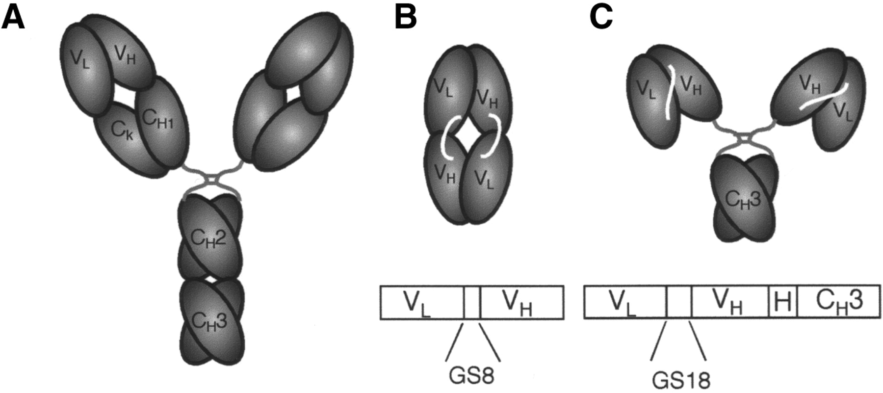
Schematic drawing shows domain structure of parental intact T84.66 antibody (A), T84.66 diabody (B), and T84.66 minibody (C). Anti-CEA minibody (80 kDa) and diabody (55 kDa) were derived from T84.66, a high-affinity and highly specific antibody that recognizes an epitope on A3 domain of CEA. Minibody (C) has glycine-serine-rich 18-amino-acid linker (GS18) between variable light (VL) and variable heavy chains (VH) and human IgG1 hinge (H) joining them to CH3 domain, also derived from human IgG1. Gene encoding diabody (B) encodes glycine-serine-rich 8-amino-acid linker (GS8) between VL and VH regions. Amino acid sequence is GSTSGGGSGGGSGGGGSS for GS18 and GGGSGGGG for GS8.
Radioiodination of T84.66 Minibody and Diabody
Radioiodination of the engineered antibody fragments was accomplished by the IODO-GEN method (Pierce Biotechnology) as previously described (12). Carrier-free 124I was produced at the Cyclotron Facility of Memorial Sloan-Kettering Cancer Center as previously described (19). Labeling reactions (0.1–0.2 mL) typically contained 0.2–0.3 mg of purified protein and 65–98 MBq (1.75–2.65 mCi) of 124I. Radioiodinated proteins were purified by size-exclusion high-performance liquid chromatography (HPLC) on either a TSK2000 (Tosoh Biosep) or Superdex 75 (Amersham Biosciences) column, with phosphate-buffered saline (PBS)/1% human serum albumin (HSA) as the running buffer. The radiolabeling efficiency was determined by integrating areas on the HPLC trace and determining the radioactivity associated with the 80-kDa protein peak for minibody or 55-kDa peak for diabody as a percentage of total radioactivity eluted. Immunoreactivity was determined by incubation of the labeled protein with a 20-fold excess (w/w) of either purified CEA or the recombinant N-A3 fragment of CEA in 0.15 mL of PBS/1% HSA as previously described. Samples were analyzed by size-exclusion HPLC on tandem Superose 6 HR 10/30 columns (Amersham Biosciences) to assess formation of Ab:Ag complexes.
Cell Lines and Xenografts
The LS174T human colorectal carcinoma cell line (CL 188) and C6 rat glioma cell line (CCL 107) were obtained from the American Type Culture Collection and maintained under standard conditions. Xenografts were established in 8-wk-old CD1 female nude mice (Charles River Laboratories) by subcutaneous inoculation of 1–2 × 10⁶ cells 10–14 d before the imaging studies. All animal handling and studies were performed in accordance with the guidelines of the University of California, Los Angeles (UCLA), Animal Research Committee.
PET Scanning
Mice were imaged using the prototype small-animal PET scanner developed at the Crump Institute for Molecular Imaging, UCLA (20). Thyroid uptake of radioiodine was blocked by pretreatment using 10 drops of saturated KI per 100 mL of drinking water for 24 h before injection of radioiodinated protein. Stomach uptake was blocked by administration of 1.5 mg of potassium perchlorate in 0.2 mL of PBS by gastric lavage 30 min before injection. The mice were injected in the tail vein either with 1.9–3.1 MBq (53–85 μCi) of 124I-minibody or with 3.1 MBq (85 μCi) of 124I-diabody in saline/1% HSA. After the appropriate uptake time had elapsed, the mice were anesthetized by intraperitoneal injection of a mixture of ketamine (80 mg/kg final dose) and xylazine (10 mg/kg final dose), placed prone, and imaged using the PET scanner with the long axis of the mouse parallel to the long axis of the scanner. Acquisition time was 28 min (4 min per bed position; 7 bed positions). Images were reconstructed using a 3-dimensional filtered backprojection reconstruction algorithm (21) for quantitation (see below) or using a maximum a posteriori reconstruction protocol (22) for presentation of images. Some mice were also imaged using 18F-FDG 2 d before the 124I-minibody scan. These were administered 5.6–7.4 MBq (150–200 μCi) of 18F-FDG via the tail vein, and scans were acquired as described above, beginning 1 h after tracer injection. In some experiments, after scanning, the animals were euthanized; the tumors were excised, weighed, and counted in a well counter (Cobra II Auto-Gamma; Packard); and the %ID/g was calculated.
Digital Whole-Body Autoradiography (DWBA)
The mice were sacrificed and frozen in carboxymethyl cellulose (Aldrich) in preparation for sectioning using a Cryostat (PMV). Coronal cross-sections were obtained with a thickness of 45 μm. DWBA was performed using a Fujifilm BAS 5000 PhosphorImager (Fujifilm Medical Systems U.S.A., Inc.) and digital plates with a final resolution of ∼100 μm. DWBA data were analyzed using Mac BAS software, version 2.4 (Fujifilm Medical Systems U.S.A., Inc.).
Data Analysis
Preliminary quantitation of the small-animal PET images was performed using the Crump Institute Integrated Imaging Software Package, or CRIIISP (Crump Institute for Molecular Imaging, UCLA). From the 3-dimensional filtered backprojection reconstruction, several planes (of a total of 64) encompassing the tumors were selected in the coronal orientation and averaged. Regions of interest (ROIs) were drawn for both the control and LS174T CEA-positive tumors centered on the peak of the activity profile. A soft-tissue region in the neck, as well as the area of maximum activity in the abdominal region, was also included for ROI analysis. Approximately equal-sized ROIs were drawn. ROI counts per pixel per minute were converted to counts per cubic centimeter per minute using a calibration factor obtained from scanning a cylinder containing a known amount of 124I activity. After decay correction, these data were converted to %ID/g by dividing by the known amount of injected activity. Target-to-background ratios were then determined for individual mice and averaged. The effects of ROI positioning were determined by averaging at least 3 ROIs and assessing the variability across regions. Statistical analysis was performed using the Excel 2000 (Microsoft) software package.
RESULTS
124I-Labeling of Anti-CEA Minibody and Diabody
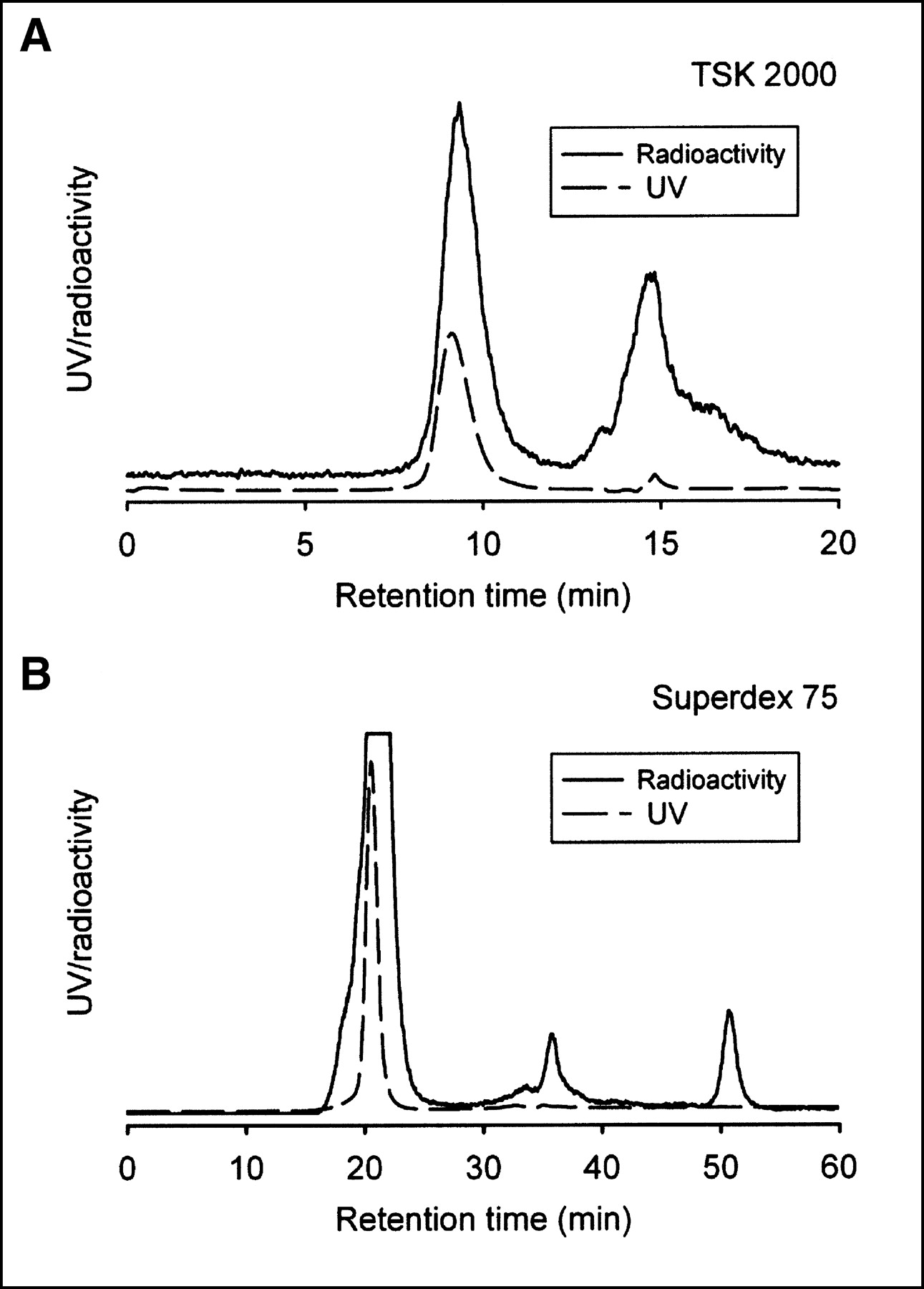
As shown in Figure 2, 124I was readily conjugated to the T84.66 minibody and T84.66 diabody after short incubations (3–5 min). Specific activities after labeling (n = 3) ranged from 81 to 133 kBq/μg (2.2–3.6 μCi/μg). The immunoreactivity of the labeled protein, as determined by size-exclusion HPLC analysis after incubation with excess antigen, was >90% in the case of minibodies and 42% for diabodies.

Size-exclusion HPLC analysis of 124I-radiolabeled anti-CEA minibody (A) and diabody (B). 124I was conjugated to T84.66 minibody and T84.66 diabody as described in Materials and Methods. Radiolabeling efficiency was determined by integrating areas on HPLC trace and determining radioactivity associated with 80-kDa protein peak for minibody or 55-kDa peak for diabody as percentage of total radioactivity eluted. Labeling efficiencies were 33% (not shown) or 46% for minibody (A) and 88% for diabody (B). Peak fractions (based on protein absorbance) were pooled for animal studies. Smaller peaks represent unincorporated label and low-molecular-weight components.
PET Imaging of Xenografts Using 124I-Anti-CEA Minibody
To evaluate tumor targeting of the 124I-minibody, antigen-positive (LS174T colorectal carcinoma) and antigen-negative control (C6 glioma) xenografts were established by subcutaneous inoculation in athymic mice. PET imaging studies were conducted on animals bearing CEA-positive tumors averaging 142 mg (range, 11–487 mg). In one of the study phases, each of 4 mice was injected in the tail vein with ∼2 MBq (53 μCi) of 124I-minibody, and whole-body PET scans were obtained beginning 4 and 18 h after administration. The image in Figure 3C clearly demonstrates high uptake into the positive (LS174T) tumor and low activity in the control (C6) tumor. The positive tumor-to-control tumor uptake ratio for the 124I-anti-CEA minibody was 3.46:1 at 4 h and 10.85:1 at 18 h, demonstrating high specificity (n = 8; Table 1).

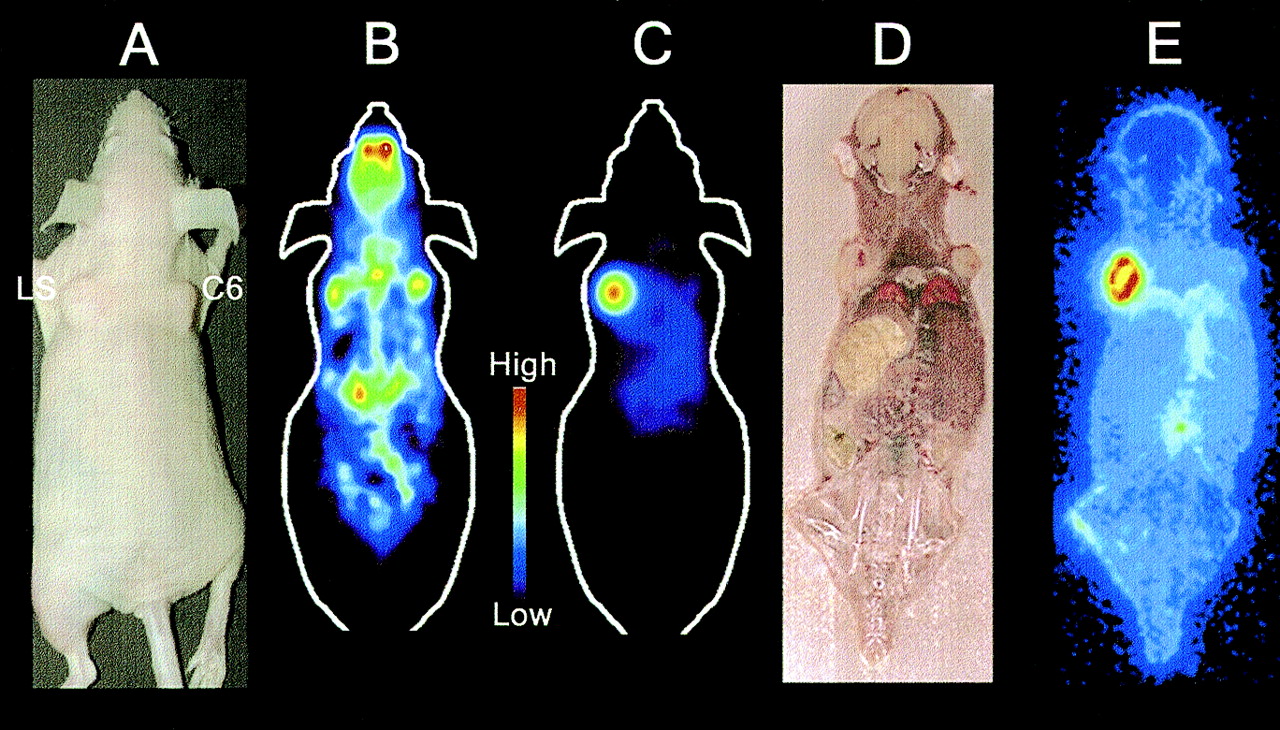
(A) Subcutaneous LS174T (left shoulder) and C6 glioma (right shoulder) xenografts were established in nude mouse. (B) PET imaging with 18F-FDG was done 2 d before injection of 124I (10/64 planes shown). (C) Mouse was injected with 3.1 MBq (85 μCi) 124I-minibody and imaged at 18 h by PET (10/64 planes shown). Both these images (B and C) are on same color scale. (D) After 18-h scan, mouse was euthanized and frozen, and whole-body coronal sections were cut in cryostat and processed for DWBA. (E) DWBA confirms specific localization of 124I minibody to CEA-positive tumor and low levels of background activity.
Ratios Derived from PET Data Comparing Target-to-Background Levels of 124I Activity
Comparison of the PET image encompassing the C6 and LS174T xenografts, and a corresponding DWBA section (Fig. 3E), clearly demonstrates that the region of high uptake observed in the scan correlates with autoradiographically detected tumor-associated radioactivity. Furthermore, an 18F-FDG PET scan of the same animal, performed before the 124I-minibody scan, allowed visualization of both tumors (Fig. 3B). The accumulation of 18F-FDG activity in the control and CEA-positive tumors was not significantly different. These studies confirm that the observed high localization of the 124I-minibody was antigen specific.
Visualization of CEA-Positive Xenografts Using 124I-Anti-CEA Diabody
Experiments were conducted similarly using the 124I-anti-CEA diabody in animals (n = 4) bearing CEA-positive tumors with an average weight of 183 mg (range, 40–420 mg). PET imaging performed 4 h after administration of 124I-anti-CEA diabody revealed rapid and specific uptake in the CEA-positive tumors (Fig. 4 C), with a tumor-to-background uptake ratio of 3.99:1 (Table 1). The contrast between the CEA-positive LS174T and CEA-negative C6 xenografts was even more striking at 18 h (Figs. 4D and 4E); a ratio could not be calculated because the C6 xenografts were not distinguishable.
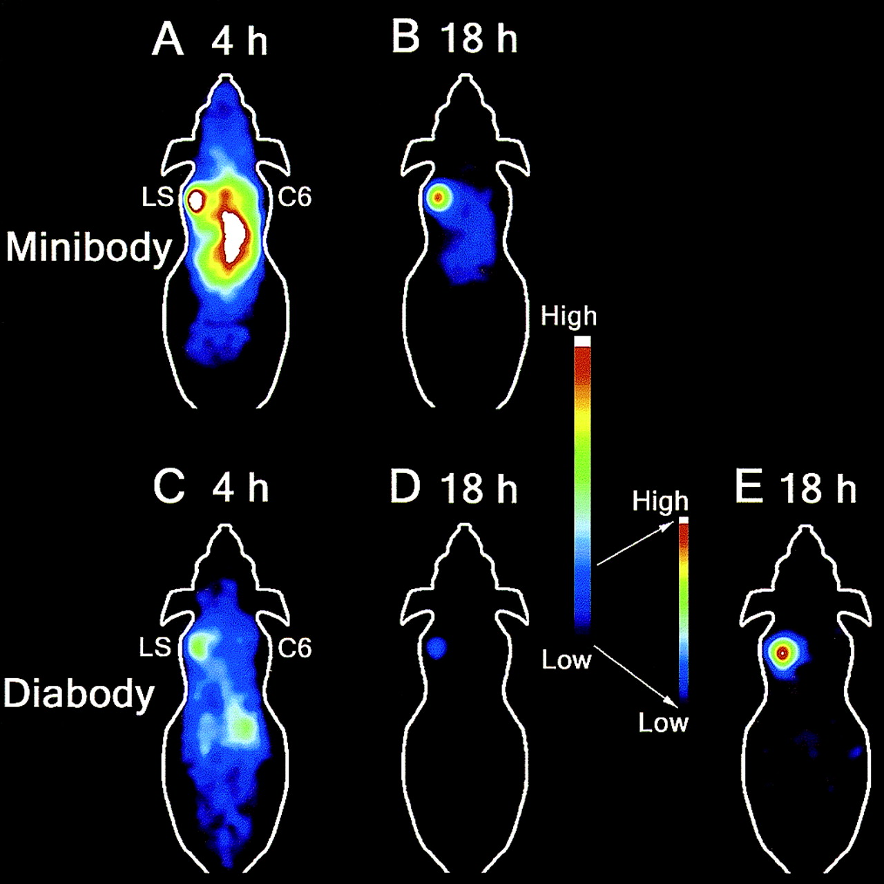
Comparison of 124I minibody and 124I diabody by serial PET imaging at 4 and 18 h. Mice bearing LS174T (LS) and C6 rat glioma (C6) xenografts were injected via tail vein with 1.9–3.1 MBq (65–85 μCi) 124I minibody (A and B) or diabody (C–E) and imaged at 4 and 18 h. At 18 h, background activity in central region was minimal in 124I diabody (D and E)–injected animal, resulting in high contrast, which can be seen in these maximum a posteriori reconstructed images (10/64 planes shown). A–D are on common scale. D was rescaled to E to illustrate excellent contrast achieved with 124I diabody at 18 h.
Tumor-to-Background Ratios from Serial Imaging
Imaging by PET allowed evaluation of tracer localization and distribution over time in the same animal. All animals were imaged 4 and 18 h after injection of minibody or diabody. In animals that were injected with 124I-anti-CEA minibody, there was substantial activity in the blood pool at the 4-h time point and hence the tumor-to-background ratio was only about 0.8:1 (Fig. 4A; Table 1). Subsequently, this ratio improved to 3.5:1 at the 18-h time point as most of the activity in the nonspecific areas cleared, whereas the activity in the CEA-positive tumors remained high (Fig. 4B). Background ratios of tumor to soft tissue averaged 3.05:1 at 4 h and improved to 11.03:1 at 18 h (Table 1).
Higher tumor-to-background ratios were achieved in animals administered 124I-labeled anti-CEA diabody. In these animals (n = 4), the tumor-to-background (background in abdominal region) ratios improved from 1.8:1 at 4 h after injection (Fig. 4C) to 6.1:1 at 18 h after injection (Figs. 4D and 4E). Background ratios of tumor to soft tissue were even higher, increasing from 4:1 at 4 h to 11:1 by the 18-h scans. This improvement in the tumor-to-background ratios achieved with 124I-labeled anti-CEA diabody, especially regarding the central activity in the abdominal region, provides greater contrast and renders a clear image of the CEA-positive tumor (Fig. 4E).
Tissue Counting
After the 18-h scans, animals not used in DWBA studies were sacrificed, and activity in various tissues was quantitated using a γ-counter. The results in Table 2 confirm high tumor uptake in the LS174T model, with a mean uptake of 20.55 ± 2.74 %ID/g for the 124I-T84.66 minibody (n = 5), consistent with previous work using 123I/131I-radiolabeled T84.66 minibody (5,13). Uptake of 124I-minibody by the control C6 xenografts and activities in heart, liver, and kidney were significantly lower than uptake in the positive tumors, and the tumor-to-control ratios determined by counting exceeded 13:1 for the indicated tissues and were consistent with ratios determined from the PET scans.
%ID/g Values and Target-to-Background (LS174T/Organ) Ratios Derived from Ex Vivo Counting of Weighed Tissues in γ-Counter After 18-Hour Uptake Time
Similarly, biodistribution data for the anti-CEA diabody (Table 2) yielded activity levels of 4.53 ± 0.50 %ID/g in the LS174T xenografts (n = 4) at 18 h, comparable to the previously published 4.56 ± 0.5 %ID/g at 24 h using 123I-T84.66 diabody (12). Control C6 tumor activity was only 0.23 ± 0.03 %ID/g, resulting in a tumor-to-control tumor activity ratio of 21.3:1 (Table 2). The highest observed tumor-to-normal ratio was 61.6:1 for LS174T to heart. Again, these results validate the high contrast observed in the PET images using 124I-diabody as the tracer.
DISCUSSION
The present intermediate-molecular-weight engineered anti-CEA antibody fragments, T84.66 minibody and diabody, demonstrated excellent targeting and imaging properties when labeled with 124I and evaluated in an athymic mouse/tumor xenograft model by PET. Confirming previous work, mean tumor uptake levels of 20.55 %ID/g (minibody) or 4.5 %ID/g (diabody) were achieved at 18 h after administration. Fast blood clearance, combined with metabolism and clearance of radioiodine activity from peripheral tissues, leads to high ratios of tumor to normal activity and excellent visualization using either fragment by 18 h. Tumors as small as 11 mg were detected by small-animal PET. High ratios of tumor (LS174T) to control tumor (C6) localization were achieved, demonstrating specificity, although this conclusion may be somewhat tempered by the use of a different tumor type as control. It will be important to confirm these results in paired xenografts that differ only in CEA expression.
Development of tracers for radioimmunoimaging requires attention to at least 3 main components: target antigen, antibody (including format), and radionuclide. Carcinoembryonic antigen, a classic tumor-associated antigen in colorectal carcinoma as well as other adenocarcinomas, remains a prime target for antibody-based detection and therapeutic applications (23). The anti-CEA minibody and diabody were derived from T84.66, a high-affinity (KA = 2 × 10¹¹ L/mol), highly specific antibody that recognizes an epitope on the A3 domain of CEA (24). Previous studies demonstrated the favorable tumor-targeting properties of these fragments labeled with radioiodine or 111In in LS174T-xenografted mice (5,12,13). However, radiometal-labeled fragments showed retention of activity in liver (minibody) or kidney (diabody). The T84.66 minibody was radiolabeled with 64Cu, a positron-emitting radiometal with a 12.7-h half-life, to explore its utility as a PET tracer (15). Excellent tumor localization was observed, but as expected, liver activity was elevated, limiting imaging applications to extrahepatic sites.
The present work demonstrated that radiolabeling of the anti-CEA minibody and diabody using 124I for PET imaging preserves the excellent tumor targeting of these fragments while enhancing the overall images because of lower background activity in normal tissues. The question arises as to which of these fragments is preferable as an imaging agent. The T84.66 minibody reaches higher absolute activity levels in tumor, but background activity takes longer to clear and is still apparent at 18 h in the murine model. By contrast, radioactivity delivered to the CEA-positive xenografts by diabody is substantially lower; however, this apparent disadvantage is ameliorated by the much lower background activity seen in the mouse. Further optimization of diabody radioiodination to retain high immunoreactivity should also enhance performance. In this animal imaging model, the diabody appears to have the advantage over the minibody in achieving higher overall contrast; however, performance still needs to be evaluated in the clinic.
Guidance on the selection of an appropriate fragment can be provided by analysis such as calculation of the imaging figure of merit (IFOM) (25), an approach based on determining statistically the required counting time to distinguish tumor from blood background. We calculated IFOM values for 124I-labeled fragments from previously published biodistribution data on radioiodinated minibody and diabody. Results (not shown) suggested that the minibody should be the superior agent, with an optimal imaging time of 20–24 h after administration, and the optimal imaging interval for the diabody would be 8–12 h. However, the current IFOM calculations are dependent on tumor size and do not take into account normal tissue activity. Further PET imaging studies at additional time points will clarify some of these issues, as will human tumor targeting and pharmacokinetic data from ongoing clinical imaging studies of 123I-T84.66 minibody. Either of these fragments labeled with 124I may prove especially useful for imaging hepatic metastases.
Several groups have explored the use of 124I for PET imaging with clinical scanners, including antibody applications. Bakir et al. (8) demonstrated detection of c-erbB2/Her2-overexpressing tumor xenografts in athymic mouse using 124I-ICR12, a rat monoclonal antibody. Tumors as small as 6 mm in diameter were imaged at 24, 48, and 120 h. More recently, Lee and associates (7) imaged human colon carcinoma xenografts using humanized 124I-A33 monoclonal antibody. This intact antibody reached 50 %ID/g 4 d after injection, and clear tumor images (tumor sizes, 0.2–0.7 g) were obtained when mice were imaged at 24, 48, or 240 h after injection. Clinical investigations have included PET imaging of breast cancer patients using 124I-anti-mucin antibodies (10) and evaluation of 124I-3F9 antibody administration for dose estimation in a patient with neuroblastoma (26). Use of a positron-emitting radionuclide with a long physical half-life, such as 124I, has been necessary for applications using intact antibodies because of their extended biologic half-lives. As noted in the above literature, optimal imaging was obtained days after administration. Results presented here indicate that 124I is also well suited for imaging using engineered fragments, with the added advantage of shorter intervals to optimal imaging times (same day or next day).
The use of 124I for radiolabeling the anti-CEA fragments, in particular the minibody, provides advantages over previous work. As expected, the high, persistent liver activity observed in our previous PET studies using 64Cu-anti-CEA minibody was dramatically reduced because of the different metabolism and clearance of activity after administration of radioiodinated protein compared with radiometal-labeled protein. The fact that CEA is not internalized on binding of antibody probably contributes to retention of activity in the LS174T xenografts, enhancing the tumor-to-liver and tumor-to-kidney ratios of the 124I-diabodies and -minibodies. In addition to the favorable biologic properties of these radioiodinated proteins, 124I-minibody and -diabody should also offer the advantage of high avidity compared with 99mTc-labeled Fab′ fragment (arcitumomab [CEA-Scan]; Immunomedics, Inc.), as these represent high-affinity bivalent, rather than monovalent, CEA-binding proteins.
The present work suggests that qualitative PET imaging using 124I-engineered fragments may provide a sensitive method for visualization of tumor masses. Relative quantitation (determination of target-to-background ratios) is straightforward. However, absolute quantitation of 124I activity will require correction of the imaging data to compensate for the unfavorable spectrum of emissions generated during the decay of 124I. In particular, 124I decay results in only a 23% yield of positrons, compared with the near 100% positron yield of 18F (27). The average energy of the 124I positrons is higher than that for 18F, and the increased positron range would result in degradation of resolution. 124I also has a complex decay scheme, resulting in a high γ-component, with many high-energy γ-emissions occurring in coincidence with each other and with the annihilation photons. Despite these shortcomings, PET imaging studies using phantoms or xenografted mouse models have been conducted under realistic conditions. Results indicate that suitable images could be obtained and that relative quantitation was feasible using a variety of clinical PET scanners (11,27). Recently, a Monte Carlo simulation scheme has been developed for correction of 124I images for scatter, γ-coincidences, and attenuation and should be applicable to the small-animal PET data generated in the current studies (C. Holdsworth, oral communication, May 2003). Correction of the current datasets is in progress. A final challenge in the current work arises from the small size of some of the tumors used—close to the resolution of the prototype small-animal PET instrument (1.8–2.0 mm). As a result, correction for the partial-volume effect will also be required for accurate quantitation (28).
The present approach can be highly complementary to existing PET oncology imaging, which primarily uses 18F-FDG as a tracer for glucose uptake and use by cancer cells. Direct comparison of quantitative image analysis was not attempted here because of the complexity of the 124I emissions as noted above. However, it is apparent from Figure 3 (which presents raw images of the same tumor-bearing mouse imaged using both tracers on the same color scale) that antibody-fragment imaging resulted in highly specific tumor localization in this xenograft model, when compared with 18F-FDG. Uptake levels for the engineered fragments equal (diabody) or substantially exceed (minibody) the %ID/g levels attained by 18F-FDG (which maximizes at approximately 3–8 %ID/g depending on the tumor model). Imaging modes such as antibody-based imaging would be especially useful as an alternative to 18F-FDG PET in cancers that exhibit low metabolic activity such as low-grade lymphomas, many prostate cancers, and other indolent tumors. There are many well-characterized tumor-associated antigens currently under evaluation for antibody-targeted imaging and therapy in lymphomas and solid tumors, such as CD20, Her2, and TAG-72, that could similarly be targeted using 124I-radiolabeled recombinant fragments.
CONCLUSION
The combination of 124I and an appropriate genetically engineered antibody fragment can provide a new class of PET tracers for imaging tumors on the basis of cell-surface antigen expression. Evaluation of 123I-T84.66 minibody for γ-camera/SPECT imaging in patients is in progress. Results from these studies and 123I-diabody studies will guide selection of appropriate 124I-labeled recombinant fragment for clinical PET studies. Furthermore, current progress in the genomics and proteomics of cancer is rapidly generating new molecular targets that can potentially be accessed using antibody technology. Reformatting of antibodies into diabodies or minibodies for 124I radiolabeling can provide a rapid route for generation of new PET tracers for cancer imaging.
Acknowledgments
We are thankful to Dr. David Stout, Judy Edwards, and Waldemar Ladno for providing expert technical help with PET imaging, and to Khoi Nguyen and Xiaoman Zhou Lewis for their help with animal studies. We are also grateful to Dr. Clay Holdsworth and Andreas Loening for their valuable suggestions on image reconstruction and analysis. Appreciations are due to Chia-Wei Cheung and to Anne-Line Anderson for conducting the protein purification and radioiodination. This study was supported by the National Institutes of Health/National Cancer Institute (SAIRP-R24-CA-92845, NCI-CA-43904, NCI-CA-33572, NCI-CA-08748, and NCI-CA-86438), the Department of Defense (DAMD-17-00-1-0150), and the Department of Energy (DE-F02-86-E60407 and DE-FC03-87ER60615).
Footnotes
Received Feb. 26, 2003; revision accepted Jun. 13, 2003.
For correspondence or reprints contact: Anna M. Wu, PhD, Crump Institute for Molecular Imaging, David Geffen School of Medicine at UCLA, 700 Westwood Plaza, Crump A342B, P.O. Box 951770, Los Angeles, CA 90095-1770.
E-mail: awu{at}mednet.ucla.edu
REFERENCES
In this issue


Jump to section
Related Articles
Cited By...
- Preclinical Evaluation of an Engineered Single-Chain Fragment Variable-Fragment Crystallizable Targeting Human CD44
- In Vivo Imaging of the Programmed Death Ligand 1 by 18F PET
- Anti-MET ImmunoPET for Non-Small Cell Lung Cancer Using Novel Fully Human Antibody Fragments
- Development of 124I Immuno-PET Targeting Tumor Vascular TEM1/Endosialin
- A Novel Engineered Anti-CD20 Tracer Enables Early Time PET Imaging in a Humanized Transgenic Mouse Model of B-cell Non-Hodgkins Lymphoma
- Advances in Immuno-Positron Emission Tomography: Antibodies for Molecular Imaging in Oncology
- Anti-carcinoembryonic Antigen Single-chain Variable Fragment Antibody Variants Bind Mouse and Human Neonatal Fc Receptor with Different Affinities That Reveal Distinct Cross-species Differences in Serum Half-life
- Evaluation of the Anti-HER2 C6.5 Diabody as a PET Radiotracer to Monitor HER2 status and Predict Response to Trastuzumab Treatment
- Cytotoxic Enhancement of a Bispecific Diabody by Format Conversion to Tandem Single-chain Variable Fragment (taFv): THE CASE OF THE hEx3 DIABODY
- Identification of Internalizing Human Single-Chain Antibodies Targeting Brain Tumor Sphere Cells
- Monodispersed DOTA-PEG-Conjugated Anti-TAG-72 Diabody Has Low Kidney Uptake and High Tumor-to-Blood Ratios Resulting in Improved 64Cu PET
- Recombinant Anti-CD20 Antibody Fragments for Small-Animal PET Imaging of B-Cell Lymphomas
- PET Imaging of Prostate Cancer Xenografts with a Highly Specific Antibody against the Prostate-Specific Membrane Antigen
- Antibodies and Antimatter: The Resurgence of Immuno-PET
- Humanized Radioiodinated Minibody For Imaging of Prostate Stem Cell Antigen-Expressing Tumors
- Comprehensive Analysis of the Factors Contributing to the Stability and Solubility of Autonomous Human VH Domains
- Immuno-PET: A Navigator in Monoclonal Antibody Development and Applications
- Imaging of Weak-Source Distributions in LSO-Based Small-Animal PET Scanners
- Bispecific Antibody Pretargeting of Radionuclides for Immuno-Single-Photon Emission Computed Tomography and Immuno-Positron Emission Tomography Molecular Imaging: An Update
- PET Imaging of Colorectal Cancer in Xenograft-Bearing Mice by Use of an 18F-Labeled T84.66 Anti-Carcinoembryonic Antigen Diabody
- Radioiodinated versus Radiometal-Labeled Anti-Carcinoembryonic Antigen Single-Chain Fv-Fc Antibody Fragments: Optimal Pharmacokinetics for Therapy
- Radioimaging of Light Chain Amyloid with a Fibril-Reactive Monoclonal Antibody
- Bispecific Antibody Pretargeting PET (ImmunoPET) with an 124I-Labeled Hapten-Peptide
- The Progress and Promise of Molecular Imaging Probes in Oncologic Drug Development
- Optimizing Radiolabeled Engineered Anti-p185HER2 Antibody Fragments for In vivo Imaging
- Radiolabeled Small-Molecule Ligands for Prostate-Specific Membrane Antigen: In vivo Imaging in Experimental Models of Prostate Cancer
- Quantitative Immuno-Positron Emission Tomography Imaging of HER2-Positive Tumor Xenografts with an Iodine-124 Labeled Anti-HER2 Diabody
- Tailoring the Pharmacokinetics and Positron Emission Tomography Imaging Properties of Anti-Carcinoembryonic Antigen Single-Chain Fv-Fc Antibody Fragments
- The Promise of Immuno-PET in Radioimmunotherapy
- Tumor imaging by means of proteolytic activation of cell-penetrating peptides
- Genetically Engineered Antibody Fragments and PET Imaging: A New Era of Radioimmunodiagnosis