


Abstract
Radioimmunotherapy (RIT) is currently being considered for the treatment of solid tumors. Although results have been encouraging for pretargeted 131I RIT with the affinity enhancement system (AES), the radionuclide used is not optimal because of its long half-life, strong γ emission, poor specific activity, and low β particle energy. 188Re, though unsuitable for direct antibody labeling, could be used with the AES two-step targeting technique. The purpose of this study was to compare the distribution and dosimetry of a bivalent hapten labeled with 188Re or 125I. For dosimetry calculations and biodistribution data, 125I was substituted for 131I. Methods: After preliminary injection of a bispecific anticarcinoembryonic antigen (CEA) or antihapten antibody (Bs-mAb F6-679), AG 8.1 or AG 8.0 hapten radiolabeled with 188Re or 125I was injected into a nude mouse model grafted subcutaneously with a human colon carcinoma cell line (LS-174-T) expressing CEA. A dosimetry study was performed for each animal from the concentration of radioactivity in tumor and different tissues. Results: Radiolabeling of AG 8.1 with 125I afforded a 40% yield with a specific activity of 11.1 MBq/nmol after purification. Radiolabeling of AG 8.0 with 188Re afforded a 72% yield with a specific activity of 31.82 MBq/nmol. In all experiments, the percentage of tumor uptake of 125I-AG 8.1 was always significantly greater than that of 188Re-AG 8.0. The corresponding tumor-to-tissue ratios reflected uptake values. The least favorable tumor-to-normal tissue ratios in the dosimetry study were 8.1 and 8.5 for 131I (tumor-to-blood ratio and tumor-to-kidney ratio, respectively) and 2.3 for 188Re (tumor-to-intestine ratio). Conclusion: This study indicates that 188Re can be used for radiolabeling of hapten in two-step radioimmunotherapy protocols with the AES technique. 188Re has a greater range than 131I, which should allow the treatment of solid tumors around 1 cm in diameter. Although the method used for hapten radiolabeling did not provide optimal tumor uptake, the use of a bifunctional chelating agent associated with AG 8.1 should solve this problem.
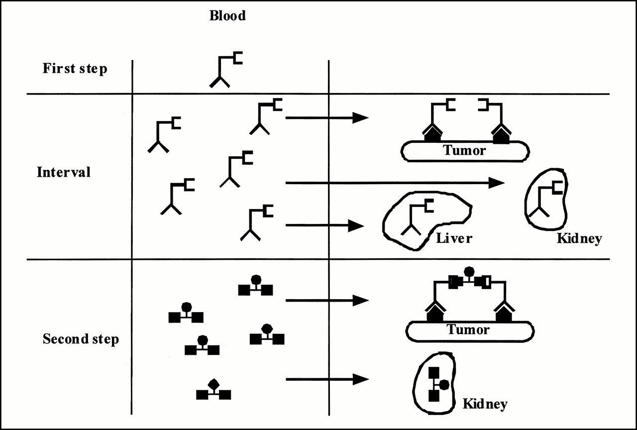
Radioimmunotherapy (RIT) has proven to be efficient for the treatment of low-grade lymphomas, but results for solid (generally bulky) tumors have been disappointing (1–4) because of the low percentage of tumor uptake achieved (<0.01%) and the relatively high nonspecific uptake with the directly labeled monoclonal antibodies (in whole immunoglobulin G or fragment form) used in most studies. Two-step targeting techniques, which substantially reduce the uptake of activity in normal tissues, should help overcome these disadvantages (5). Several preclinical and clinical studies have shown the benefit of using the affinity enhancement system (AES), which associates a bispecific antibody with a bivalent hapten (Fig. 1). The haptens used have been radiolabeled first with 111In for diagnostic studies and then with 131I for RIT. The mean energy of β− particles emitted by 131I limits the efficacy of this radionuclide to small tumor targets. For larger targets, radionuclides emitting more energetic β− particles seem to be preferable (6). Among these isotopes, 188Re is a good candidate because of its physical characteristics (half-life, 16.98 h; β−, 2.118 and 1.962 MeV) and production mode (188W/188Re generator) (7–10). However, the possibility of using this radionuclide with a directly radiolabeled antibody for RIT is limited by its short physical half-life and the specific activities obtained during direct radiolabeling of antibodies (70–80 MBq/nmol) (7–9) as well as slow tumor uptake (11,12). The AES two-step targeting technique provides rapid tumor uptake because of the small size and hydrophilic properties of the bivalent hapten and its long retention time at the tumor site attributed to specific binding to prelocalized antibodies (5,13–15).

AES.
The purpose of this study was to compare the distribution and dosimetry of a bivalent hapten labeled with 188Re or 125I after preliminary injection of a bispecific anticarcinoembryonic antigen (CEA) or antihapten antibody into a nude mouse model grafted subcutaneously with a human colon carcinoma cell line (LS-174-T) expressing CEA.
MATERIALS AND METHODS
The bispecific antibody (Bs-mAb F6–679) used in this study was composed of an F(ab′) fragment of an antiCEA antibody (F6) coupled chemically to an F(ab′) fragment of an antiglycylsuccinimidylhistamine antibody (679). The bispecific antibody was supplied by Immunotech (Marseille, France) as a 3 mg/mL solution in 0.1 mol/L phosphate buffer and 0.05 mol/L EDTA, pH 7.
The hapten used, AG 8.1, was an octapeptide grafted with two glycylsuccinimidylhistamine residues in ε of lysines 1 and 3. Its semideveloped formula is shown in Figure 2. AG 8.1 may be readily radiolabeled with radioactive iodine.
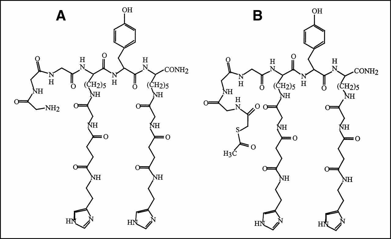
Plane semideveloped formula of AG 8.1 (A) and AG 8.0 (B).
For 188Re labeling, the peptide was derivatized with an S-acetylthioacetyl residue at the N-terminal position. The new derivative thus obtained was designated as AG 8.0 (16). The two haptens (AG 8.1 and AG 8.0) were supplied by Immunotech as an aqueous solution at 1 mg/mL.
Hapten Radiolabeling
125I-AG 8.1.
AG 8.1 was labeled with 125I using the chloramine-T technique (17). After labeling, the hapten was purified using a SepPak C18 cartridge (Millipore, Saint Quentin Yvelines, France) by successive injections of the labeling solution (5 mL 0.05% trifluoroacetic acid in water [Aldrich, Saint Quentin Fallavier, France] and then 5 mL of a 3:2 mixture of 0.1 mol/L phosphate buffer solution, pH 7, and ethanol). Under these conditions, free 125I was eluted in the acid aqueous phase and the radiolabeled hapten was eluted in the first 2 mL of the ethanol phase. The radiochemical purity of the purified solution was measured by thin-layer silica gel chromatography (Kieselgel 60 F254; Merck, Nogent sur Marne, France) using methanol as the migration solvent. The immunoreactive fraction was measured by determining the percentage of activity binding to tubes coated with the antihistamine antibody 679. The chromatograms were analyzed after exposure on a phosphorus screen (Molecular Dynamics, Sunnyvale, CA) using IPLab-Gel software (Analytics Corp., Atlanta, GA).
188Re-AG 8.0.
AG 8.0 was labeled with 188Re by ligand exchange according to a method derived from that of Joiris et al. (18). Briefly, 188Re-glucoheptonate was obtained quantitatively by addition under a nitrogen atmosphere of 740 MBq (20 mCi; 3 mL) sodium perrhenate in a glass flask containing 150 mg sodium glucoheptonate (Aldrich), 8.4 mg sodium hydrogenocarbonate (Aldrich), and 1.25 mg stannous chloride (Aldrich). The solution was incubated for 1 h at room temperature and controlled by instant thin-layer chromatography on silica gel (ITLC-SG; Gelman-OSI, Elancourt, France) using 0.9% sodium chloride and acetone as migration solvents. During the incubation period, AG 8.0 was deacetylated by addition of excess sodium hydroxide (1N). A spot test with dithionitrobenzoic acid was performed on an aliquot of the deprotected solution to check for the presence of free thiol (19). Twenty-one nanomoles of deprotected AG 8.0 were added to the 188Re-glucoheptonate solution, which was then incubated for 15 min at 100°C before purification, and the control solution under the same conditions as those during radiolabeling of AG 8.1 by 125I.
Animal Studies
For each radionuclide, 15 nude mice (Swiss/nu/nu; Iffa-Credo, L’Arbresle, France) were used (30 animals total). Each animal was xenografted subcutaneously in the right flank with 1 million cells of the LS-174-T colon cancer cell line (American Type Culture Collection, Rockville, MD), which strongly expresses CEA. Two weeks after grafting, the tumors measured 50–500 mm3. Each mouse was injected intravenously in a tail vein with 50 μg (0.5 nmol/100 μL) Bs-mAb F6-679. On the basis of data obtained from other studies (20–25), a second injection was performed (24 h after this first injection) with either 0.25 nmol 125I-AG 8.1 (11.1 MBq/nmol) or 0.25 nmol 188Re-AG 8.0 (31.8 MBq/nmol).
Biodistribution Study
For each radionuclide, a group of 3 animals was killed at 5 min and at 1, 5, 24, and 48 h after injection of the radiolabeled hapten. For each time point, the main tissues and tumors were removed, dried, weighed, and counted in a γ scintillator (Compugamma; LKB-Pharmacia, Uppsala, Sweden) in parallel with a calibrated radioactive decay standard. The results are expressed as a percentage of the injected dose per gram (%ID/g) of tissue and as tumor-to-tissue ratios.
188Re-AG 8.0 Stability
An analysis of the immunoreactive fraction of blood and urine was also performed using the same procedure as that used for the radiolabeling control. Statistical analysis of the results was done by ANOVA using Statview II software (Abacus Concepts, Berkeley, CA).
Measurements of immunoreactive fractions were performed at 5 min and at 1, 5, 24, and 48 h in blood, urine, and the 188Re-labeled hapten solution after purification. This control solution was kept at room temperature throughout the experiment, with a starting activity of 185 MBq/mL (5 mCi/mL). Measurements were performed to study the instability of the radiolabeled hapten.
Dosimetry Study
For each mouse, the concentration of radioactivity (kBq/g) was calculated in different tissues or tumors. 131I was considered for dosimetry instead of 125I for several reasons: cost, radioprotection, and identical biodistributions. The counting and calculation of the percentage of the injected dose per animal tissue was determined simultaneously with that of a weighed standard aliquot of the injected dose. To minimize counting errors associated with radiation self-absorption by tissues, the same weight of each organ sample was measured. The mean value of this concentration at each time point allowed curves to be plotted according to a theoretic one-compartment extravascular model. The results (initial concentration, C0) were used, after correction of the physical half-life, to calculate the mean dose delivered by a theoretic injection of 3.7 MBq 131I or 188Re according to a MIRD formula (26):
 where D̄(target ← source) is the mean absorbed dose (in Gy) delivered to the target by the source, A�source is the cumulated activity in the source (in Bq × s), Δ (in J/(Bq × s)) is the sum of energies emitted by desintegration, φ(target ← source) is the fraction of energy emitted by the source that is absorbed by the target, and mcible is the mass of the target.
To simplify the calculations, it was considered in a first approximation that the sources and targets were identical, and the emissions were regarded as nonpenetrating. In this case,
 becomes:
 which assumes that the emitted particles deposit their energy locally:

Calculations were performed with the tabulated Δ (27) for 131I and 188Re, which gave respectively:
 and
and

RESULTS
Hapten Radiolabeling
125I-AG 8.1.
During radiolabeling of AG 8.1 with 125I, a 40% yield was obtained after addition of 259 MBq 125I to 11 nmol AG 8.1. The specific activity of 125I-AG 8.1 after purification was 11.1 MBq/nmol, with a radiochemical purity of 100% by chromatography. The immunoreactive fraction was measured to 96.4%.
188Re-AG 8.0.
The radiochemical purity of the 188Re-glucoheptonate solution used in the radiolabeling of AG 8.0 was 80% by chromatography. The impurities in the preparation represented 12% reduced and hydrolyzed rhenium and 8% perrhenate. Because these impurities could be removed easily during the purification step for the radiolabeled hapten, the solution was used in this state. Under these conditions, the uptake yield after addition of 1 GBq 188Re-glucoheptonate to 21 nmol AG 8.0 was 72%. The specific activity of 188Re-AG 8.0 after purification was estimated at 90% by chromatography. The immunoreactive fraction was measured to 91%. The major impurities detected by chromatography could not be identified. The purification step allowed elimination of perrhenate and 188Re hydrolysates.
Animal Study
Biodistribution Study.
The results obtained (Table 1) show that the maximum injected dose in all tissues appeared at the same time point for both radionuclide-associated haptens: 5 min for blood, liver, kidney, spleen, and bone; 1 h for intestine; and 5 h for tumor. The percentage of tumor uptake of 125I-AG 8.1 was always statistically greater than that of 188Re-AG 8.0 (P < 0.01 for all time points). The distribution of radioactivity was comparable for blood, spleen, and bone up to 1 h after injection and then became statistically greater for 125I-AG 8.1 than for 188Re-AG 8.0—except for bone, in which uptake of 188Re-AG 8.0 was greater than that of 125I-AG 8.1 at 48 h (P < 0.01).
Comparative F6–679/125I-AG 8.1 Versus F6–679/188Re-AG 8.0 Biodistribution After Injection in Nude Mouse Model Grafted Subcutaneously with Human Colon Carcinoma Cell Line (LS-174-T)
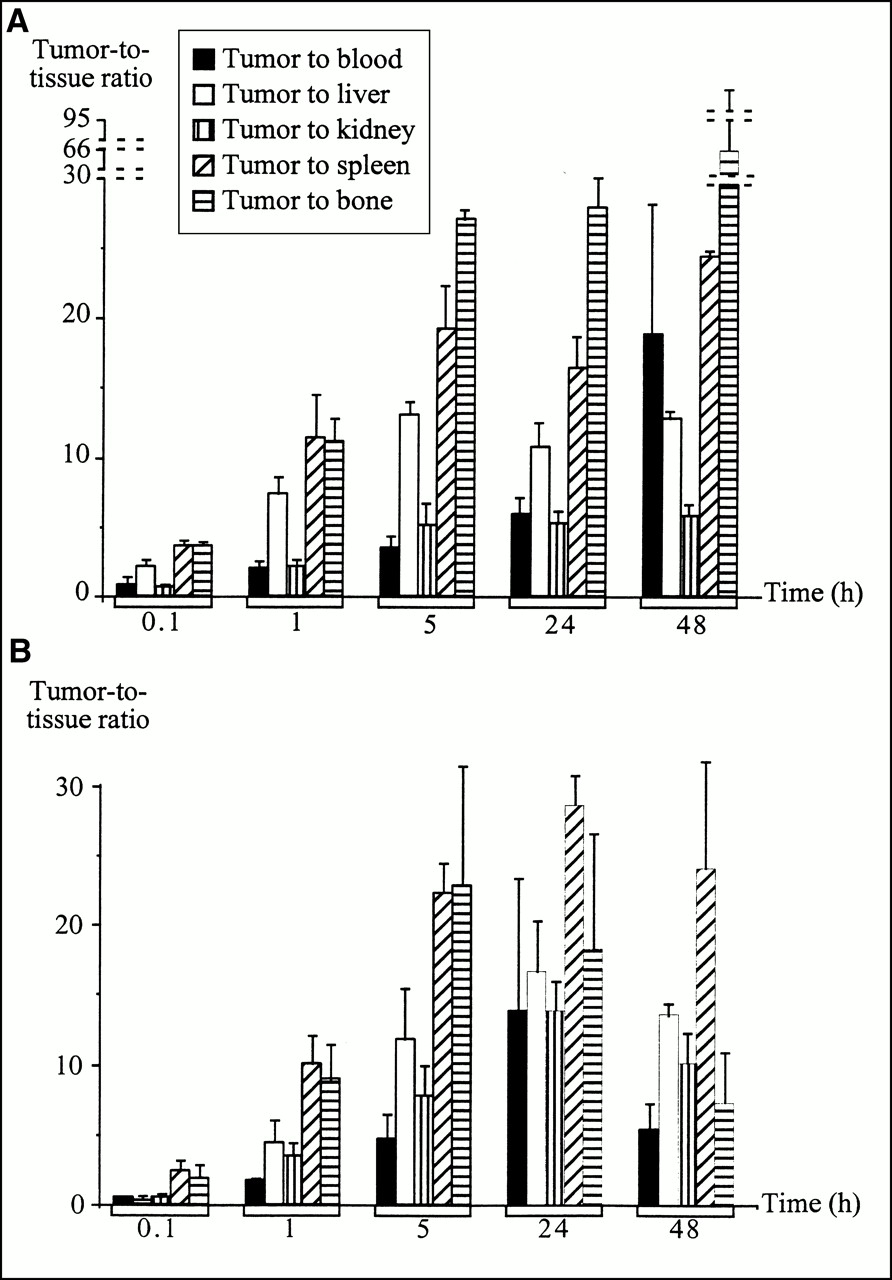
The corresponding tumor-to-tissue ratios (Fig. 3) reflected these uptake values. They were higher with 125I-AG 8.1 (Fig. 3A) at 5 min for blood, spleen, and bone (P < 0.05) and were comparable with the ratios of 188Re-AG 8.0 (Fig. 3B) for these same tissues at 1, 5, and 24 h. At late times (48 h), 188Re-AG 8.0 showed ratios that were lower than those for 125I-AG 8.1, except in liver and kidney (P < 10−4), which were indicative of higher tumor uptake and lower bone uptake for 125I-AG 8.1. At 5 min, liver uptake of 188Re-AG 8.0 was higher than that of 125I-AG 8.1, resulting in very low tumor-to-liver ratios (P < 0.01), whereas at 5, 24, and 48 h uptake was greater for 125I-AG 8.1 (P < 0.05), with comparable tumor-to-liver ratios for both substances (P > 0.05). In the intestine, uptake of 125I-AG 8.1 and 188Re-AG 8.0 was not significantly different at any time point, except at 1 h when uptake was markedly greater for 188Re-AG 8.0 (P < 0.01), with a very large SD indicative of considerable interindividual variation. In kidney, regardless of analysis time, uptake percentages for 125I-AG 8.1 were greater than those for 188Re-AG 8.0 (P < 0.05). Despite high tumor uptake of 125I-AG 8.1, except at 5 min, the tumor-to-kidney ratios obtained with 188Re-AG 8.0 were greater than those for 125I-AG 8.1 (P < 0.05).

Comparison of tumor-to-tissue ratios obtained with 125I-AG 8.1 (A) and 188Re-AG 8.0 (B) after injection in nude mouse model grafted subcutaneously with human colon carcinoma cell line (LS-174-T). Data are expressed as mean ± SD.
In summary, tumor-to-tissue ratios were highest for 188Re-AG 8.0 at 24 h, except for the tumor-to-bone ratio, which was highest at 5 h. For 125I-AG 8.1, the tumor-to-blood ratio was highest at 48 h, whereas the other ratios were highest between 5 and 48 h.
188Re-AG 8.0 Stability.
The results for immunoreactive measurements are shown in Figure 4. Analysis of the stability of the purified 188Re-AG 8.0 solution over time showed that the 188Re-labeled peptide, used as a control, lost a large part of its immunoreactivity at early times. It decreased from 90.8% at 5 min to 17.7% at 5 h but remained quite constant after that time point: 14.2% at 24 h and 8% at 48 h.

Analysis of stability of 188Re-AG 8.0 by measurement of serum and urinary immunoreactive fractions after injection in nude mouse model grafted subcutaneously with human colon carcinoma cell line (LS-174-T).
The immunoreactive fraction in urine was around 38% at 5 min and then became very low (7.1%) between 1 and 5 h before rising to 44% at 24 h and dropping slightly to 36% at 48 h. Most of the radioactivity was eliminated rapidly. The immunoreactive fractions were quite stable in blood, ranging from 84% at 5 min to 26.7% at 48 h. This phenomenon could have been caused by radiolysis resulting from the high activity used or by instability of the radiolabeled hapten (or both).
The study of immunoreactive fractions relating to 188Re-labeled hapten showed that urinary radioactivity was eliminated mainly in the form of nonimmunoreactive metabolites, whereas circulating forms in the blood were composed mainly of immunoreactive hapten until 5 h after injection and were then considerably reduced at late times.
Dosimetry Study.
The results of the dosimetric study are shown in Table 2. For each tissue and tumor, the cumulative concentration is indicated in kBq/s/kg, as extrapolated by exponential adjustment of kinetic values and the constant of the dose corresponding to each radionuclide studied. Calculations for 131I were performed on the basis of results for 125I, assuming that the kinetics of AG 8.1 kinetics was identical for the two iodine radioisotopes.
Dosimetry Estimations Obtained from Biodistribution Results for 125I-AG 8.1 and 188Re-AG 8.0 for Theoretic Injection of 3.7 MBq 131I or 188Re in Nude Mouse Model Grafted Subcutaneously with Human Colon Carcinoma Cell Line (LS-174-T)
With 188Re, the highest doses were obtained for tumor, intestine, blood, and kidney, whereas with 125I the most irradiated tissues were tumor, kidney, blood, and liver. The least favorable tumor-to-normal tissue ratios were 8.1 and 8.5 for 131I (tumor-to-blood ratio and tumor-to-kidney ratio, respectively) and 2.3 for 188Re (tumor-to-intestine ratio).
DISCUSSION
The use of mercaptoacetyltriglycine (MAG3) derivatives for the radiolabeling of proteins with 99mTc or 188Re is one of the most common techniques (28–32). With 188Re, the usual approach is to radiolabel MAG3 first and then purify the 188Re-MAG3 thus obtained before activating an acid residue (generally butyric acid). The radiolabeling of proteins is then achieved by incubating the activated, radiolabeled MAG3 with a slightly alkaline protein solution (28,29). Although this procedure provides radiolabeling with high radiochemical purity, it is time-consuming and thus unsuitable in terms of the short physical half-life of 188Re. Radiolabeling of antibodies with 188Re can be performed directly (7–9,33), but the radioantibodies obtained generally lack stability in vivo (8,29). To avoid these drawbacks, the modified derivative of AG 8.1 used in this study had a structure derived from MAG3 but could be radiolabeled directly with 188Re. Moreover, to facilitate the radiolabeling of this AG 8.0 peptide, a ligand-exchange technique was developed, which allowed 188Re-AG 8.0 to be obtained in 15 min with good yields. As a reference, we used another peptide closely related to AG 8.0, which was labeled with a low specific activity of 125I to minimize radiolysis (11.1 MBq/nmol). AG 8.0 was labeled with a higher specific activity of 188Re (31.82 MBq/nmol), which should make 188Re-AG 8.0 suitable for future RIT protocols. The same purification technique used for labeling both radionuclides afforded excellent results for 125I-AG 8.1 but was less efficient in removing impurities from the 188Re-AG 8.0 solution. However, the latter was used because radiochemical purity was better than or equal to that reported for direct radiolabeling of antibodies with 188Re (60%–80% as measured by the immunoreactive fraction) (7–9).
The results obtained with 125I-AG 8.1 were comparable with those reported in the same animal model (34) for a radiolabeled peptide that was only slightly different from the one used in this study. The results for tumor uptake of 188Re-AG 8.0 were poorer than those with 125I labeling but were comparable with values in the literature for antibodies radiolabeled directly with 188Re (16,29). Kinetic analysis of biodistribution showed that the blood clearance of 125I-AG 8.1 was lower than that of 188Re-AG 8.0 (1.23 mL/h vs. 2.1 mL/h). The higher blood clearance of AG 8.0 was apparently one of the reasons for lower tumor uptake with this peptide than with AG 8.1. However, the radiochemical purity of 188Re-AG 8.0 decreased very rapidly over time when it was concentrated in the purification medium, which suggests that impurities formed during the period required for preparation and injection of 188Re-AG 8.0 into the animal. These impurities associated with radiolabeling complexation instability or radiolysis (or both) were apparently responsible for part of the increase in the renal clearance of radioactivity and for reduced tumor uptake. This was confirmed by the study of urinary immunoreactive fractions, which showed that only 8% of the radioactivity consisted of hapten that was recognized by antibody 679 at 1 h, whereas this fraction was >30% at all other time points. This difference may be attributed to rapid urinary elimination of impurities caused by the release of noncomplexed 188Re from 188Re-AG 8.0 before administration. At later time points, the metabolism of this radiolabeled peptide was a contributing factor, although a highly significant fraction of peptide circulating in the blood remained immunoreactive over time. Moreover, the results of the biodistribution study show that 188Re-AG 8.0 had very high hepatic and intestinal uptake at early time points compared with that of 125I-AG 8.1, apparently because of biliary elimination of 188Re-AG 8.0 radioactivity. This mode of elimination could have caused increased clearance of the radiolabeled peptide and thus decreased tumor uptake at later times. It appears that 125I-AG 8.1 was eliminated preferentially by the renal route. In fact, the percentages of renal uptake were always greater for 125I-AG 8.1 than for 188Re-AG 8.0 at all time points. Finally, bone uptake was greater at 48 h with 188Re-AG 8.0 than with 125I-AG 8.1, possibly because 188Re-AG 8.0 lacked labeling stability in vivo. This could have caused local uptake by exchange between the 188Re-MAG3 complex of the peptide and the phosphocalcic structures of the bone network.
For all normal tissues studied, the %ID/g with 125I-AG 8.1 was lower than that for antibody radiolabeled directly with 125I (5,16,35,36). This phenomenon, which has been reported for other bispecific antibody or hapten pairs (14,15), resulted in tumor-to-tissue ratios that were greater than those observed with antibodies that were radiolabeled directly (16,28,37). Comparison of the biodistribution obtained with AG 8.1 labeled with 125I and 188Re showed differences in the in vivo distribution of radioactivity depending on the radioelement used. In fact, this phenomenon could have been attributed to the structural difference between the two haptens. The elimination kinetics of 188Re-AG 8.0 appeared to be faster than that of 125I-AG 8.1, partly because 188Re-AG 8.0 has significant hepatobiliary clearance.
Two assumptions were made in this study relative to dosimetry calculations. First, the effect of γ emissions was disregarded at the doses delivered because of the low proportion of photon emissions by 188Re (9.18 × 10⁻¹⁵ kg × Gy/Bq × s) compared with particle emissions (1.24 × 10⁻¹³ kg × Gy/Bq × s) and the fact that this energy was deposited at greater distances than with β− emissions. Second, particle emissions were considered nonpenetrating in the geometry of our experimental conditions. This assumption, though frequently made for dosimetry in vivo in humans, cannot be verified in mice. Thus, our results need to be interpreted with caution, given the size of murine organs and tissues, which are generally smaller than the maximal range of β− particles of 188Re (6). Nevertheless, it is necessary to make these calculations to compare the results obtained with both radionuclides with those published using the same animal model (21–24).
On the whole, our results are encouraging for the development of RIT protocols with this technique, even if improvement of 188Re-AG 8.0 stability is still necessary. Under these conditions, the injection of quantities of radioactivity three to four times higher than the quantities we used may allow the delivery of doses approximating those reported in other studies relating to this method of human tumor treatment (33,38,39).
CONCLUSION
This study indicates that 188Re can be used for the radiolabeling of hapten in two-step RIT protocols with the AES. The main advantage of this radionuclide over 131I is its range, which should allow the treatment of solid tumors around 1 cm in diameter. However, the method used for hapten radiolabeling did not provide optimal tumor uptake. Other rhenium-chelating agents easily substituted for the N-terminal end of AG 8.1 could offer improved labeling efficiency and in vivo stability.
Acknowledgments
The authors thank James Gray for technical assistance. This study was supported by a grant from the Comité Départemental de la Ligue Nationale Contre le Cancer, Département de la Vendée, and by a grant from the Comité Départemental de la Ligue Nationale Contre le Cancer, Département des Deux-Sèvres.
Footnotes
Received Mar. 15, 2000; revision accepted Jun. 20, 2000.
For correspondence or reprints contact: Jean F. Gestin, PhD, Institut de Biologie, Unit 463, Institut National de la Santé et de la Recherche Médicale, 9 Quai Moncousu, 44093 Nantes cedex, France.
REFERENCES
In this issue


Jump to section
Related Articles
Cited By...
- Bispecific Antibody Pretargeting PET (ImmunoPET) with an 124I-Labeled Hapten-Peptide
- Successful Radiotherapy of Tumor in Pretargeted Mice by 188Re-Radiolabeled Phosphorodiamidate Morpholino Oligomer, a Synthetic DNA Analogue.
- Pretargeting of Carcinoembryonic Antigen-Expressing Tumors with a Biologically Produced Bispecific Anticarcinoembryonic Antigen x Anti-Indium-Labeled Diethylenetriaminepentaacetic Acid Antibody
- Pretargeting with Bispecific Anti-Renal Cell Carcinoma x Anti-DTPA(In) Antibody in 3 RCC Models
- Pretargeting with Labeled Bivalent Peptides Allowing the Use of Four Radionuclides: 111In, 131I, 99mTc, and 188Re
- Murine S Factors for Liver, Spleen, and Kidney
- Pretargeted Radioimmunotherapy of Cancer: Progress Step by Step
- A Universal Pretargeting System for Cancer Detection and Therapy Using Bispecific Antibody
- Molecular Advances in Pretargeting Radioimunotherapy with Bispecific Antibodies
- Tumor Pretargeting in Mice Using 99mTc-Labeled Morpholino, a DNA Analog