


Abstract
Short-lived α-emitting isotopes individually conjugated to monoclonal antibodies have now reached human use, but little is still known about their toxicity. Use of antibody targetable 225Ac nanogenerators is a new approach in the field of α-immunotherapy offering the advantage of a 10-d half-life (t1/2) and increased potency due to generation of 3 new atoms, yielding a total of 4 α-particles. However, the 3 α-emitting daughter elements generated have the potential for significant toxicity as these nuclides are no longer bound to the carrier IgG. Methods: Cynomolgus monkeys were used to evaluate the toxicity of prototype 225Ac nanogenerators. Monoclonal antibody HuM195 (anti-CD33) is the carrier for planned human clinical trials of 225Ac; there are no CD33 sites in cynomolgus monkeys. In one experiment, 2 monkeys received a single intravenous dose of 225Ac-HuM195 at 28 kBq/kg. This dose level is approximately the planned initial human dose. In another experiment, 2 animals received a dose escalation schedule of 3 increasing 225Ac-HuM195 doses with a cumulative activity of 377 kBq/kg. The whole-blood t1/2 of 225Ac, ratios of 225Ac to its ultimate α-emitting daughter nuclide 213Bi, generation of monkey anti-HuM195 antibodies (MAHA), hematologic indices, serum biochemistries, and clinical parameters were measured. Monkeys were euthanized and examined histopathologically when the dose escalation reached toxicity. Results: The blood t1/2 of 225Ac-HuM195 was 12 d, and 45% of generated 213Bi daughters were cleared from the blood. MAHA production was not detected. Approximately 28 kBq/kg of 225Ac caused no toxicity at 6 mo, whereas a cumulative dose of ∼377 kBq/kg caused severe toxicity. In the cumulative dosing schedule, single doses of ∼37 kBq/kg resulted in no toxicity at 6 wk. After ∼130 kBq/kg were administered, no toxicity was observed for 13 wk. However, 28 wk after this second dose administration, mild anemia and increases of blood urea nitrogen and creatinine were detected. After administration of an additional 185 kBq/kg, toxicity became clinically apparent. Monkeys were euthanized 13 and 19 wk after the third dose administration (cumulative dose was 377 kBq/kg). Histopathologic evaluation revealed mainly renal tubular damage associated with interstitial fibrosis. Conclusion:225Ac nanogenerators may result in renal toxicity and anemia at high doses. The longer blood t1/2 and the lack of target cell antigens in cynomolgus monkeys may increase toxicity compared with human application. Therefore, a dose level of at least 28 kBq/kg may be a safe starting dose in humans. Hematologic and renal function will require close surveillance during clinical trials.
Radioimmunotherapy is a promising approach to selectively treat a variety of cancers (1,2). It takes advantage of a growing number of monoclonal antibodies to target tumor cells preferentially while sparing normal, healthy tissue. This potentially increases the therapeutic index compared with other treatment modalities. Recent advances in chemistry have led to methods for stable conjugation of a variety of radionuclides, including both halogens and metals to proteins and peptides (3–5).
Radiation as a cytotoxic agent has several advantages over therapeutics that interfere chemically with their target cells. Most importantly, radiation is able to kill tumor tissue without the need for binding or internalization into every target cell. Second, combinations of radiotherapy with some chemotherapeutic approaches results in a more than additive therapeutic effect (6). Due to optimizations in external beam therapy, local control of many tumors is possible, whereas micrometastatic disease is currently amenable only to treatment with systemic chemotherapeutic agents. One important step toward feasible radiotherapeutic treatment of micrometastatic disease is to target radiation via monoclonal antibodies directly to tumor throughout the body. Commonly used isotopes for this are the β-emitting isotopes 131I and 90Y (7). Recently, several α-emitting isotopes such as 213Bi and 211At have been studied clinically as radioisotopes in cancer therapy (3,8). α-Particles are characterized by 400-fold greater linear energy transfer than β-particles. Whereas the β-particle’s range is typically between 0.5 and 8 mm, α-particles have a tissue pathlength of only 40–90 μm. Therefore, α-radiation is suitable for killing single-cell diseases and small tumor cell clusters consisting only of a few thousand cells. One of the most potent, yet challenging, therapeutic α-emitting approaches is the use of an α-generator: 225Ac. 225Ac has the convenient half-life (t1/2) of 10 d and decays via a chain, yielding a total of 4 α-particles. The α-emitting daughter nuclides are short-lived (221Fr, t1/2 = 4.8 min; 217At, t1/2 = 32.3 ms; 213Bi, t1/2 = 45.6 min), leading to an extremely high cytotoxicity when retained in or near tumor cells or tumor cell clusters. A key drawback of this same property is the release of the daughters from 225Ac immunoconjugates; such daughters may not be retained in tumor tissue (9). Though used successfully in small animal systems, 225Ac generators have not been used in humans (10).
In this study, we describe initial monkey studies of a new radioimmunoconjugate consisting of the α-emitting isotope 225Ac conjugated to the anti-CD33 antibody HuM195. HuM195 is a humanized antibody that targets CD33 on acute myeloid leukemia cells. HuM195 has been used as the targeting moiety in a variety of clinical trials as a native antibody and conjugated to β-emitting and α-emitting isotopes (8).
The longest lived α-emitting daughter nuclide of 225Ac is 213Bi. Bismuth accumulates in renal tissue and, therefore, nephrotoxicity was hypothesized to be the dose-limiting toxicity in 225Ac immunotherapy due to constant kidney accumulation of 213Bi arising from systemic 225Ac (11). Therefore, prior to human trials, we evaluated 225Ac-HuM195 toxicity in a nonhuman primate model.
MATERIALS AND METHODS
Preparation of 225Ac-HuM195
225Ac was conjugated to HuM195 antibody using a 2-step labeling method as described previously (12). Briefly, labeling and preparation of the doses were done at the Memorial Sloan-Kettering Cancer Center (MSKCC) Central Isotope Laboratory to ensure consistency and to standardize each step for human clinical use. 225Ac (37 MBq in 25 μL 0.2 mol/L HCl; Oak Ridge National Laboratory) was incubated with l-ascorbic acid (150 g/L, 20 μL), 2-(p-isothiocyanatobenzyl)-1,4,7,10-tetraazacyclododecane-1,4,7,10-tetraacetic acid (DOTA-NCS) (10 g/L, 50 μL; Macrocyclics), and tetramethylammonium acetate (2 mol/L, 50 μL; Aldrich Chemical Co.) to facilitate incorporation of 225Ac into DOTA. The reaction was allowed to continue for 30 min at 60°C (pH 5.0). For conjugation of 225Ac-DOTA to HuM195 (the second-step reaction), another 20 μL of ascorbic acid were added before adding 1 mg of HuM195 (200 μL; Protein Design Laboratories). The pH was adjusted with carbonate/bicarbonate buffer (1 mol/L, 100 μL) to 9.0 and incubation was for 30 min at 37°C. Afterward, free 225Ac along with other metals was absorbed with 20 μL 10 mmol/L diethylenetriaminepentaacetic acid (DTPA) and the unconjugated 225Ac was separated from 225Ac-HuM195 by PD10 size exclusion (Bio-Rad) using 1% human serum albumin in 0.9% saline as eluent. Quality control of the final product included thin-layer chromatography to determine radiopurity, a cell-based binding assay to measure immunoreactivity of the antibody vehicle, Limulus amebocyte lysate testing to determine pyrogen content, and microbiologic culture in fluid thioglycollate of soybean-casein digest medium to verify sterility.
Nonhuman Primate Study Design
Four male cynomolgus monkeys, 2–4 y old, were used in this study. Housing and care were in accordance with the Animal Welfare Act and the Guide for the Care and Use of Laboratory Animals (13). Animal protocols were approved by the Institutional Animal Care and Use Committee at MSKCC. All procedures were performed under short-term intramuscular ketamine anesthesia.
In one experiment, 2 animals received a single intravenous dose of 225Ac-HuM195 at 28 kBq/kg via femoral venipuncture. This dose level was scaled from 1/20 of the maximum tolerated dose values determined in rodents (10). With regard to the total number of α-particles emitted until total decay of the 225Ac, this dose was similar to a 213Bi dose that has proven safe in patients previously (8).
Animals were observed twice daily for levels of brightness, alertness, and reactivity and weighed weekly. Biochemical and hematologic indices of the blood (IDEXX Laboratory) as well as blood radioactivity values were determined on days 1, 5, 8, 14, and 18 and monthly thereafter.
In another experiment, 2 animals received a dose escalation schedule of 3 increasing 225Ac-HuM195 doses (at 0, 6, and 34 wk) with a cumulative activity of ∼370 kBq/kg, also administered by femoral venipuncture. Animals were followed similarly for laboratory values and for clinical toxicity.
Determination of Monkey Antihuman Antibodies
Monkey antihuman antibody production was assessed on days 6 and 12 after the first injection and on day 6 after the third injection in monkeys 3 and 4. Serum samples (100 μL) were incubated with 250 ng/mL 111In-HuM195 for 10 min at room temperature and then the molecular weight of the radioactive fraction was determined using Beckman Coulter System Gold high-performance liquid chromatography (HPLC) equipped with a γ-RAM Radio HPLC detector (IN/US Systems, Inc.). Therefore, 50-μL samples were injected onto a size-exclusion column (300 × 7.8 mm TSK 3000SWXL; Supelco) and an elution profile was measured over 16 min. The mobile phase consisted of 0.15 mol/L sodium chloride/0.02 mol/L sodium acetate, pH 6.5. The sensitivity of the HPLC radiodetector and the 111In-HuM195 antibody concentration allows accurate detection of monkey anti-HuM195 antibody (MAHA) titer above 50 ng/mL.
Histopathologic Evaluation
Necropsy was performed after euthanasia by barbiturate overdose. Tissues from the cerebrum, testis, salivary gland, skeletal muscle, esophagus, trachea, stomach, small intestine, spleen, lung, liver, adrenal grand, large intestine, and kidneys were stained with hematoxylin–eosin and examined by 2 veterinary pathologists.
Pharmacokinetic Analysis and Absorbed-Dose Estimation
Due to the concern of iatrogenic anemia, blood samples were kept to a minimum and only blood samples of ∼1 mL were analyzed for blood activity level by γ-counting (Packard Cobra γ-Counter; Packard Instrument Co.). The blood pharmacokinetic data obtained in the 221Fr and 213Bi photon windows were used to estimate blood and renal cortex absorbed dose. Starting at 30 min after each blood drawing, blood activity was measured separately for 213Bi and 221Fr and measured several times thereafter until equilibration of the daughters. The steady-state ratio of 213Bi to 221Fr was back-extrapolated by using exponential growth of 213Bi and constant activity of 221Fr. Due to the 4.5-min t1/2 of 221Fr, its activity approximates 225Ac after 25 min. In this way, time–activity curves for 225Ac and 213Bi were obtained separately.
The blood dose was obtained by fitting the 225Ac time–activity curve to a 1-component (monkey 2) or 2-component (monkey 1) exponential expression. The resulting fitted expressions were analytically integrated over time to yield the total number of 225Ac disintegrations occurring in the blood. Fitting was performed using the Simulation Analysis and Modeling software package, SAAM II (SAAM Institute, Inc.). Two different assumptions were then applied to estimate the number of 221Fr and 217At disintegrations in blood. In the first assumption, these 2 daughters were assumed to decay at the site of 225Ac decay—that is, in the blood. In the second assumption, these daughters were assumed to decay at the site of 213Bi decay. 213Bi decays not occurring in the blood were assumed to occur in the kidneys, specifically the renal cortex portion of the kidneys. Once the decays of each radionuclide were apportioned to blood or the renal cortex of the kidneys, the absorbed dose was obtained assuming a uniform deposition of α-particle energy in the tissue mass. The energy emitted per disintegration was, therefore, multiplied by the number of disintegrations and by the α-particle energy emitted per disintegration; the result was then divided by the blood or renal cortex mass. The electron and photon energy contributions to the tissue absorbed dose were considered negligible relative to the α-particle energy and were, therefore, not included.
The number of 213Bi disintegrations occurring in the blood was obtained separately by integrating, over time, the back-extrapolated estimate of 213Bi radioactivity concentration in blood at each time. This provided an estimate of 213Bi disintegrations occurring in the blood. The difference between the total number of 225Ac disintegrations and the 213Bi disintegrations—that is, those disintegrations resulting from 213Bi that was no longer in the blood—was assigned to the renal cortex portion of the kidneys (14). The decay of an equal number of 221Fr and 217At was assigned to the blood (assumption A) or to the renal cortex of the kidneys (assumption B).
The mass of blood in cynomolgus monkeys was obtained as the product of 80 mL/kg and the total body weight of the monkeys (15). The mass of the renal cortex was obtained by assuming that the kidney mass of a 5-kg cynomolgus monkey is the same as that of a 5-kg (2–3 y old) human (16). The adult renal cortex-to-total kidney mass ratio (17) was then used to obtain the renal cortex mass.
RESULTS
Toxicity of Single-Dose 225Ac-HuM195
At a dose level of 28 kBq/kg administered once intravenously, no clinical or laboratory toxicity was observed for >6 mo. Serum tests including hepatic enzymes (serum glutamic-oxalocetic transaminase, serum glutamic-pyruvic transaminase, alkaline phosphatase, γ-glutamyl transpeptidase, and bilirubin), renal values (blood urea nitrogen [BUN] and creatinine), hematology values (hemoglobin, red blood cell count, and white blood cell [WBC] count), and electrolytes (sodium, potassium, chloride, carbon dioxide, calcium, and phosphorus) were normal at all times. A nonspecific rise in serum creatine phosphokinase (CPK) was observed after intramuscular anesthesia. Independence of this finding from radioimmunoconjugate administration was shown by elevated CPK values after each handling, even if no drug was administered. The peak of CPK elevation was reproducibly observed 24 h after intramuscular ketamine anesthesia with significant elevation over 4–5 d.
Blood t1/2 of 225Ac-HuM195
The blood t1/2 of 225Ac-HuM195 in both monkeys was ∼12 d. In monkey 1, biphasic pharmacokinetics were observed comprising a rapid clearing component with a t1/2 of 2.3 ± 0.2 d and a slowly clearing component with a t1/2 of 12 ± 1 d. In monkey 2, a single-component exponential fit demonstrated a t1/2 of 12 ± 2 d (Fig. 1).

Blood time–activity curves of 225Ac. Activity concentration for 225Ac was estimated from 221Fr counting after equilibration. Dashed line (monkey 1, ⋄) corresponds to 2-component exponential fit to data. α-Component (42%) cleared with t1/2 of 2.3 ± 0.2 d and slow β-component (58%) cleared with t1/2 of 12 ± 1 d. Solid line (monkey 2, ▵) is single-component exponential fit with intercept and t1/2 of 60.3 percentage injected dose per kilogram (%ID/kg) and 12 ± 2 d, respectively.
MAHA Detection
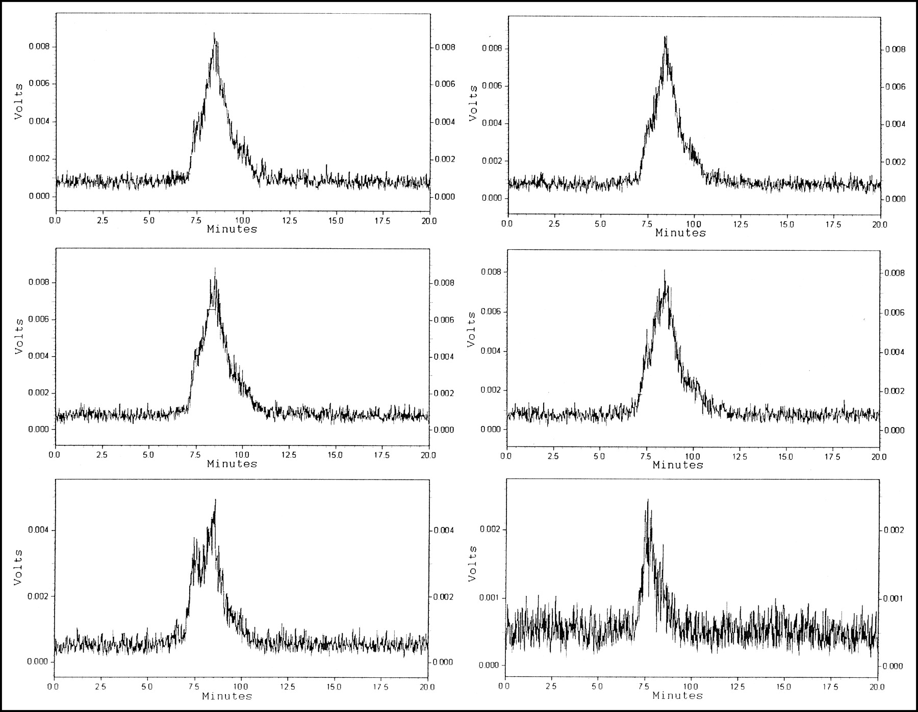
The engineered HuM195 is a potential immunogen in cynomolgus monkeys. Monkey antihuman antibody response (MAHA) could result in a major change in biodistribution of the second or third injection of 225Ac-HuM195 compared with the first administration. In particular, it would be expected that if complexed by MAHA an increased percentage of 225Ac-HuM195 would be cleared rapidly through the liver or spleen. HPLC of monkey sera incubated with 111In-HuM195 showed no radioactive fraction at ≥300 kDa, indicating that the 2 monkeys injected with 3 doses of HuM195 did not generate neutralizing antibody against the human protein throughout the course of the study (Fig. 2).

HPLC assay for monkey antihuman antibody response (MAHA). 111In-HuM195 was incubated with monkey serum obtained 6 d after injection 1 (top row), 12 d after injection 1 (middle row), and 6 d after injection 3 (bottom row). Antibody dimers (300 kDa) or multimers (>450 kDa) suggestive of autoantibodies against HuM195 were not detected. Retention time of ∼150-kDa HuM195 is 8.4 min. Higher molecular weight 300 kDa would have markedly shorter retention time (<6.4 min). (Left) Data for monkey 4. (Right) Data for monkey 3.
Clinical Toxicity of 225Ac-HuM195
Animals in the dose-escalating study showed no signs of toxicity 6 wk after administration of doses of 30 and 59 kBq/kg. The second dose at 133 and 130 kBq/kg (cumulative doses,163–189 kBq/kg) also did not result in any clinical toxicity for up to 3 additional months after administration. However, a trend toward decrease of hemoglobin levels was observed (Fig. 3). Anemia could have arisen from toxicity, such as decreased production of erythropoietin, hemolysis, or the repeated bleedings.

Hemoglobin levels in monkeys receiving dose escalation of 225Ac-HuM195. Doses were given at weeks 0, 6, and 28. Arrows indicate time of intravenous injection of 0.15, 0.67, and 1.07 MBq in monkey 3 (○) and 0.3, 0.67, and 0.93 MBq in monkey 4 (♦).
After a 4-mo hiatus, animals were reevaluated at the time of an additional injection. At time of administration of this third dose (215 and 189 kBq/kg; cumulative dose, 377 kBq/kg), 28 wk after administration of the second dose, evidence of renal failure with elevated creatinine levels (monkey 3, 1.6 mg/dL; monkey 4, 2.6 mg/dL) and worsening anemia (monkey 3, 7.3 mg/dL; monkey 4, 6.8 mg/dL) were observed. Therefore, the third dose was likely to be cleared slowly from the kidney and would likely compound the toxicity already present. Monkey 4 suffered from increasing anorexia, weight loss, and lethargy during the next 13 wk and was euthanized 47 wk after the first dose. Monkey 3 deteriorated clinically at a slower pace. He was euthanized at 1 y after the first dose, for histopathologic examination (Figs. 4 and 5). Marrow toxicity as measured by reduced WBC counts was not found to be a toxicity of 225Ac-HuM195 (Fig. 6).

Serum creatinine levels in monkeys receiving dose escalation of 225Ac-HuM195. Dose and schedule were as in Figure 3. Arrows indicate intravenous injection times. ○, Monkey 3; ♦, monkey 4.

Serum BUN levels in monkeys receiving dose escalation of 225Ac-HuM195. Dose and schedule were as in Figure 3. Arrows indicate intravenous injection times. ○, Monkey 3; ♦, monkey 4.

WBC counts in monkeys receiving dose escalation of 225Ac-HuM195. Dose and schedule were as in Figure 3. Arrows indicate intravenous injection times. ○, Monkey 3; ♦, monkey 4.
Histopathologic Examination
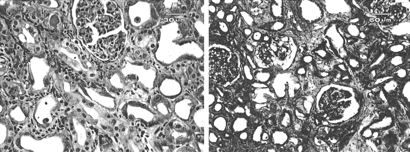
Monkey 4 showed histopathologic damage in renal tissue, but not elsewhere. This damage was characterized by multifocal proximal dilated convoluted tubules due to the loss of tubular epithelial cells. These tubules were also associated with interstitial fibrosis and, rarely, small aggregates of mononuclear cells in the interstitium (Fig. 7). Bone marrow was mildly hypocellular in the sternum and femur.

Histopathologic examination of kidneys taken after euthanasia of monkeys. Kidney damage was observed in different stages. (Left) Monkey 3 shows tubular necrosis and regeneration mixed with some interstitial fibrosis, indicative of more chronic damage. (Right) Monkey 4 also displays tubular damage and marked interstitial fibrosis in more acute state.
Monkey 3 showed histopathologic damage in a variety of other organs. Changes in renal lesions were less overt than in monkey 4 but were similar in nature. Some tubular epithelial necrosis and marked regeneration, corresponding to more chronic lesions, could be seen. Some interstitial fibrosis and hyaline casts were also noted (Fig. 7). Other organs showed histopathologic damage consistent either with radiation damage or with severe stress. Myocardial tissue showed areas of interstitial fibrosis and aggregates of mononuclear cells with degeneration of surrounding myocytes; the gastric mucosa showed many well-demarcated erosions; liver showed areas of mild hepatocellular degeneration and testis showed seminiferous tubular lumens containing multinucleated giant cells. Cellularity of bone marrow was moderately hypocellular in sternum and severely hypocellular in femur.
Pharmacokinetic Analysis and Absorbed Dose Estimation
Dose estimates were made based on the time–activity curves for 225Ac (Fig. 1) and 213Bi (Fig. 8). Steady-state in vivo ratios of 213Bi to its parent nuclide 225Ac was 0.57:1 (±0.11) in monkey 1 and 0.54:1 in monkey 2 (±0.13) (Figs. 8A and 8B). Tables 1 and 2 summarize the dosimetry results obtained under the 2 different assumptions described in the Materials and Methods. Blood and renal cortex absorbed doses were obtained by an extrapolation of kinetics measured for monkey 1 to monkey 3 and monkey 4, both of whom received a total of 1.89 MBq 225Ac-HuM195. Under the assumption that 221Fr and 217At decay with the 225Ac, the blood dose is 4 Gy and the renal cortex dose is 5 Gy. Assuming that the kinetics on monkey 2 apply, the blood dose is 3 Gy and the renal cortex dose is 3 Gy. Under the second assumption, that 221Fr and 217At decay with the 213Bi, using kinetics of monkey 1, the blood dose is 3 Gy and the renal cortex dose is 13 Gy; using kinetics of monkey 2, the blood dose is 2 Gy and the renal cortex dose is 9 Gy.

(A and B) Relationship between 213Bi and 221Fr activity in whole blood. Recorded activities of 213Bi (○) and 221Fr (▪) in monkey 1 (A) and monkey 2 (B) are shown. Each individual small curve represents data from single day (days 1, 5, 8, 14, and 18, respectively, after 225Ac-HuM195 administration). For each curve, x-axis is linear with respect to time during which isotope equilibrium is reached ex vivo. (C) Blood time–activity curves of 213Bi. Activity concentration for 213Bi was estimated by back-extrapolating 213Bi counts to time of blood collection. Dashed line (monkey 1) corresponds to 1-component exponential fit with intercept and t1/2 of 49 percentage injected dose per kilogram (%ID/kg) and 9 ± 2 d, respectively. Corresponding values for monkey 2 (solid line) are 34 %ID/kg and 11 ± 3 d, respectively.
Dosimetry of Single Dose of 225Ac-HuM195 Following Pharmacokinetics in Monkey 1
Dosimetry of Single Dose of 225Ac-HuM195 Following Pharmacokinetics in Monkey 2
DISCUSSION
Targeted 225Ac represents a new class of drugs, which have not yet been used in humans (10). 225Ac-HuM195 was developed as a highly cytotoxic α-radiation emitter targeted by specific binding to CD33 and internalization of the monoclonal antibody-generator construct. While several forms of radioimmunotherapy have been developed and used in humans, the unique features of targeted nanogenerators required that potential side effects be evaluated in primate studies before application in humans. 225Ac has the unique property of decaying via an atomic chain containing 3 additional α-emitting isotopes, each with different chemical properties. We have shown that clinical grade 225Ac-HuM195 production, quality control, and administration are feasible. In the future, the 10-d t1/2 of 225Ac will allow central production of 225Ac immunoconjugates under controlled reproducible conditions and distribution to multiple sites of clinical application.
225Ac labeling of HuM195 did not affect its immunoreactivity nor did labeling change the blood t1/2 when compared with previous studies in monkeys using HuM195 (18). In addition, the chelated 225Ac is stable in vivo. This suggests that metabolism will be determined primarily by the carrier IgG and the physical properties of the radioisotopes. The blood t1/2 of a monoclonal antibody is affected by specific recognition by the immune system or by binding to Fc receptors, thereby increasing clearance, and by specific binding to target cells. Monkeys do not express CD33. Therefore, HuM195 blood t1/2 in monkeys is long (∼12 d), whereas in patients displaying CD33 binding sites on both normal monocytes and leukemia cells it is far shorter (2–3 d) (19,20). In humans, after binding sites become saturated or leukemia cells are cytoreduced, blood t1/2 increased (18). The biologic t1/2 of monoclonal antibody and the physical half-live of the 225Ac radioisotope are matched well. In contrast, the more commonly used short-lived α-emitting isotopes 213Bi (t1/2 = 46 min) and 211At (t1/2 = 7 h) demand more injections to maintain high blood activity concentrations. This results in a more complicated and expensive regimen and as well in accumulation of unlabeled, potentially blocking monoclonal antibodies over the course of the treatment.
Due to the short pathlength of α-radiation, unbound α-immunoconjugates circulating in blood are unlikely to cause significant toxicity. After the initial decay of 225Ac to 221Fr, however, this daughter α-emitting element, and the subsequent α-emitting daughters, 217At and 213Bi, would be released. The distributions of these 3 α-emitting daughter nuclides of 225Ac are no longer determined by the carrier molecule and thus may increase toxicity as compared with drugs carrying a single α-particle–emitting isotope. Therefore, estimating appropriate doses of 225Ac from chelated 213Bi or chemically conjugated 211At data (both of which have been used in humans) might not be accurate. Difficulties may arise when estimating the impact of free 221Fr, 217At, and 213Bi for therapeutic efficacy or for toxicity, as there are no previous data available for the first 2 elements, and minimal data for bismuth. Studies with longer-lived bismuth isotopes show that a considerable fraction of bismuth will be accumulated and excreted in the kidneys (21). Thus, renal toxicity in this study was an expected finding. The data also suggest that toxicity will be determined to a major extent by the redistribution of 225Ac daughters. Nearly half of the last α-emitting daughter nuclide (213Bi) was cleared from the blood, indicating that redistribution of daughter nuclides occurred. Excretion of heavy metals can be enhanced by administration of chelators such as 2,3-dimercapto-1-propanesulfonic acid, ethylenediaminetetraacetic acid, DTPA, and dimercaptosuccinic acid (22). Therefore, similar methods might be beneficial to reduce toxicity.
In the 2 monkeys showing acute radiation nephritis, the estimated absorbed dose to the renal cortex (assuming localization of 221Fr and subsequent daughters to the renal cortex) was 9–13 Gy. The renal cortex was chosen as the primary target tissue since there is evidence that heavy metals localize to this portion of the kidney (14). It is important to note that the absorbed-dose estimates are based on assuming a uniform deposition of α-particle energy throughout the renal cortex mass. As with almost all absorbed-dose calculations, this is a necessary oversimplification in the absence of detailed microbiodistribution data. The implications and possible errors associated with such an oversimplification, however, are profound for α-particle emitters due to their high potency and short range. Depending on the actual microbiodistribution, the biologic effects could be substantially greater or smaller than one might expect from an estimated absorbed dose of 13 Gy. In humans, the tolerated dose of external beam radiation yielding radiation nephritis in 50% of humans within 5 y (TD50/5) is 28 Gy (17). Assuming that a relative biologic effectiveness (RBE) factor of 5 applies, the RBE-adjusted absorbed-dose range of 45–65 Gy to the monkey renal cortex is consistent with the toxicity observed in the monkeys.
CONCLUSION
In this study we showed that 225Ac-HuM195 can safely be administered at a dose level of 28 kBq/kg, whereas dose escalation caused significant toxicity at cumulative doses of 215–370 kBq/kg. For repeated treatment cycles, pharmacokinetics did not appear to be influenced by an immune response against HuM195. However, antibody production against xenogeneic monoclonal antibodies can occur at any time during the treatment. Significant human antibody response to HuM195 has not been reported. In humans, the biologic t1/2 of HuM195 is 4–5 times shorter than that in monkeys because of rapid targeting of the antibody-based drug and subsequent cellular internalization of the immune complex. Therefore, we assume that a dose level of at least 28 kBq/kg may be administered with tolerable acute side effects.
Acknowledgments
We thank Krista LaPerle, VMD, PhD, and Hai Nguyen, VMD, for careful review and valuable discussion of histopathology. We also thank Laike Stewart, VMD, and Felix Homberger, VMD, PhD, for helpful advice. The help from Ron Finn, PhD, Jing Qiao, Michael Curcio, and Catalina Cabassa for preparing the radiopharmaceuticals and George Gonzales and Donna Ortiz for excellent care of the animals is acknowledged. This work was supported by National Institutes of Health grants P01 33049 and R01 55349; the Steps for Breath Fund; William H. Goodwin and Alice Goodwin and the Commonwealth Cancer Foundation for Research and the Experimental Therapeutics Center of MSKCC; and the Doris Duke Charitable Foundation.
Footnotes
Received May 28, 2003; revision accepted Sep. 25, 2003.
For correspondence or reprints contact: David A. Scheinberg, MD, PhD, Department of Molecular Pharmacology and Chemistry, Memorial Sloan- Kettering Cancer Center, 1275 York Ave., New York, NY 10021.
E-mail: d-scheinberg{at}ski.mskcc.org
REFERENCES
In this issue


Jump to section
Related Articles
Cited By...
- Treatment of prostate cancer with CD46 targeted 225Ac alpha particle radioimmunotherapy
- [18F]-Labeled PARP-1 PET Imaging of PSMA Targeted Alpha Particle Radiotherapy Response
- Preclinical Evaluation of 213Bi- and 225Ac-Labeled Low-Molecular-Weight Compounds for Radiopharmaceutical Therapy of Prostate Cancer
- A Self-Assembling and Disassembling (SADA) Bispecific Antibody (BsAb) Platform for Curative Two-step Pretargeted Radioimmunotherapy
- Preclinical Evaluation of 203/212Pb-Labeled Low-Molecular-Weight Compounds for Targeted Radiopharmaceutical Therapy of Prostate Cancer
- Therapeutic Radiometals Beyond 177Lu and 90Y: Production and Application of Promising {alpha}-Particle, {beta}--Particle, and Auger Electron Emitters
- The Future of Radioligand Therapy: {alpha}, {beta}, or Both?
- Preclinical Evaluation of Multistep Targeting of Diasialoganglioside GD2 Using an IgG-scFv Bispecific Antibody with High Affinity for GD2 and DOTA Metal Complex
- Molecular Pathways: Targeted {alpha}-Particle Radiation Therapy
- Anti-CD45 pretargeted radioimmunotherapy using bismuth-213: high rates of complete remission and long-term survival in a mouse myeloid leukemia xenograft model
- MIRD Pamphlet No. 22 (Abridged): Radiobiology and Dosimetry of {alpha}-Particle Emitters for Targeted Radionuclide Therapy
- Radioimmunotherapy of Breast Cancer Metastases with {alpha}-Particle Emitter 225Ac: Comparing Efficacy with 213Bi and 90Y
- A Pharmacokinetic Model for Radioimmunotherapy Delivered Through Cerebrospinal Fluid for the Treatment of Leptomeningeal Metastases
- Multidrug Resistance in Acute Myeloid Leukemia: Potential New Therapeutics
- Preclinical Evaluation of the {alpha}-Particle Generator Nuclide 225Ac for Somatostatin Receptor Radiotherapy of Neuroendocrine Tumors
- Phase I Study of Targeted Radioimmunotherapy for Leptomeningeal Cancers Using Intra-Ommaya 131-I-3F8
- 111In-Labeled Trastuzumab (Herceptin) Modified with Nuclear Localization Sequences (NLS): An Auger Electron-Emitting Radiotherapeutic Agent for HER2/neu-Amplified Breast Cancer
- Anti-CD45 Monoclonal Antibody YAML568: A Promising Radioimmunoconjugate for Targeted Therapy of Acute Leukemia
- Nuclear Localizing Sequences Promote Nuclear Translocation and Enhance the Radiotoxicity of the Anti-CD33 Monoclonal Antibody HuM195 Labeled with 111In in Human Myeloid Leukemia Cells
- Renal Tubulointerstitial Changes after Internal Irradiation with {alpha}-Particle-Emitting Actinium Daughters
- Melanoma Therapy via Peptide-Targeted {alpha}-Radiation
- Treatment of Neuroblastoma Meningeal Carcinomatosis with Intrathecal Application of {alpha}-Emitting Atomic Nanogenerators Targeting Disialo-Ganglioside GD2
- Alpha-Particle Emitting Atomic Generator (Actinium-225)-Labeled Trastuzumab (Herceptin) Targeting of Breast Cancer Spheroids: Efficacy versus HER2/neu Expression