


Abstract
18F-FDG PET is a promising tool for detecting vulnerable plaques, depending on the extent of macrophage infiltration; however, it is still not clear which stage of the lesion can be detected by 18F-FDG PET. Methods: In this study, we investigated the effect of foam cell formation on 18F-FDG uptake using cultured mouse peritoneal macrophages. Results: 18F-FDG accumulation was increased by foam cell formation, but the uptake was decreased to the control level after complete differentiation to foam cells. Changes in hexokinase activity tended to accompany changes in 18F-FDG uptake. In contrast, changes in glucose-6-phosphatase activity and glucose transporter 1 expression did not parallel 18F-FDG uptake. Conclusion: Our results suggest that 18F-FDG PET detects the early stage of foam cell formation in atherosclerosis.
The rupture of atherosclerotic plaques is responsible for myocardial and cerebral infarctions. Macrophage infiltration and vulnerable plaque formation play an essential role in plaque rupture. Infiltrated macrophages increase their expression of scavenger receptors during a maturation process. The receptors bind and internalize modified low-density lipoprotein (LDL) such as oxidized LDL and acetylated LDL, thereby causing foam cell formation. Foam cells release several proteases and cytokines, which lead to plaque rupture (1,2). We and other groups have reported that 18F-FDG PET is a promising tool for detecting vulnerable atherosclerotic plaques, depending on the extent of macrophage infiltration into the atherosclerotic lesion (3–5). To date, some clinical trials have been done at several institutes, and the utility of 18F-FDG PET has been established (3,6). However, it is still not clear which stage of the lesion can be detected by 18F-FDG—that is, whether foam cell formation affects 18F-FDG uptake. Earlier work has shown that 18F-FDG accumulates in cultured macrophages in vitro, according to the incubation time and glucose concentration (7); however, the molecules that are responsible for delivering 18F-FDG uptake to the vulnerable plaque are unknown (8). To establish the molecular evidence that can validate the use of 18F-FDG PET in making clinical diagnoses, it is necessary to know at what stage of plaque formation 18F-FDG shows up in atherosclerosis imaging.
In this study, we evaluated the effects of foam cell formation on 18F-FDG uptake using mouse peritoneum–derived macrophages. In addition, uptake to arterial smooth muscle cells (SMCs) was investigated to evaluate the effects of 18F-FDG uptake by the aortic wall in atherosclerosis imaging. Also, changes in hexokinase and glucose-6-phosphatase (G6Pase) activities, as well as glucose transporter (GLUT) expression, were examined in order to elucidate the molecular mechanism of 18F-FDG uptake by macrophages in vulnerable plaques.
MATERIALS AND METHODS
Preparation of Peritoneal Macrophages and Foam Cell Formation
Mouse peritoneal macrophages were isolated as previously described, with slight modifications (9). Four days after the 10% thioglycolate injection (intraperitoneally) to female ddY mice (Japan SLC Co. Ltd.), the mice were sacrificed and macrophages were isolated from the peritoneal cavity by phosphate-buffered saline (PBS) lavage. The macrophages were plated into 6-well plates and cultured in Dulbecco modified Eagle medium (DMEM, 4,000 mg/L of glucose; Sigma–Aldrich Co.) supplemented with 10% fetal bovine serum and a 100 U/mL concentration of penicillin and streptomycin at 37°C in 95% air–5% CO2. After 24 h of incubation, macrophages were treated with acetylated LDL (50 μg/mL, BTI Inc.) for 12, 24, or 48 h to promote formation of foam cells. Isolated macrophages from each mouse were plated in separate wells, which were individually investigated (n = 4–5 for each experiment). To check the foam cell formation, at least 1 well from each condition was used for oil red O staining after the cells had been fixed with 4% paraformaldehyde. The animal study was approved by the Animal Care and Use Committee of the Hamamatsu University School of Medicine.
Preparation of Aortic SMCs
Mouse aortic SMCs were isolated as previously described (10). Briefly, male C57BL/6J mice (8–12 wk old; Japan SLC Co. Ltd.) were anesthetized with pentobarbital and the aorta was dissected. The endothelial cells were removed through incubation of the dissected aorta in collagenase type II solution (2 mg/mL), and the cells were flushed out with DMEM (20% fetal bovine serum). The aorta was cut and placed on a gelatin-coated dish with the cut side facing the gelatin (Asahi Glass Co., Ltd.) and was cultured in DMEM (10% fetal bovine serum) for 10 d. The dishes were analyzed separately (n = 5).
18F-FDG Uptake Studies
The macrophages or SMCs were preincubated with 1.5 mL of low-glucose medium (DMEM, 1,000 mg/L of glucose) at 37°C for 1 h. Then, 37 kBq of 18F-FDG (Nihon Medi-Physics Co., Ltd.) were added to each well, and the cells were incubated at 37°C for 3 h in 5% CO2. After incubation, the medium was removed and the cells were rinsed twice with 1 mL of PBS. Macrophages were removed from the wells mechanically in 0.5 mL of PBS, and the wells were rinsed with an additional 0.5 mL of PBS, which was combined with the cell suspension. SMCs were peeled from the wells by trypsin treatment and washed with PBS by centrifugation to avoid contamination with coated gelatin in the protein concentration measurement. The radioactivity of each medium and the cell suspension was measured with an automated γ-counter. Protein concentration was measured by the method of Lowry et al. (11). The 18F-FDG uptake was expressed as the ratio of radioactivity in the cell to the initial dose per milligram of protein (percentage injected dose).
Hexokinase Activity
Hexokinase activity was measured by the method of Vinuela et al. (12). In brief, the reaction solution (40 mM Tris-HCl buffer [pH 8.0], 10 mM glucose, 4 mM adenosine triphosphate, 10 U of glucose 6-phosphate dehydrogenase II per milliliter, 1 mM nicotinamide adenine dinucleotide phosphate, 10 mM MgCl2) was preincubated at 37°C. The cultured macrophages were washed with PBS and collected in 0.3 mL of 0.1 M KH2PO4-NaOH buffer. Then, 50 μL of the cell suspension were mixed with 0.5 mL of prewarmed reaction buffer. The absorbance was measured at 340 nm every 1 min to measure the produced nicotinamide adenine dinucleotide phosphate (NADP+) by the hexokinase reaction.
G6Pase Activity
G6Pase activity was measured by the method of Koide et al. (13). In brief, after being washed with 0.1 M citrate buffer (pH 6.5), the cultured macrophages were mechanically collected in 0.4 mL of citrate buffer. Then, 100 μL of 0.2 M glucose-6-phosphate were added to the cell suspension, and the cells were incubated at 37°C for 30 min. The reaction was terminated by the addition of 500 μL of 10% trichloroacetic acid. The generated phosphate was measured by the malachite green molybdate assay.
GLUT Expression
Macrophages were collected in lysis buffer (50 mM Tris-HCl [pH 7.5], 1 mM ethylenediamine tetraacetic acid, 150 mM NaCl, 1% NP-40, 0.25% sodium deoxycholate, inhibitor cocktail). Lysates containing 30 μg of protein samples were subjected to sodium dodecylsulfate polyacrylamide gel electrophoresis and transferred to a polyvinylidene fluoride membrane (Millipore). The membrane was blocked for 1 h in Blotto A (Santa Cruz Biotechnology Inc.) and then probed with anti-GLUT-1 (Abcam) or anti-actin antibodies at room temperature for 2 h. The membrane was washed in wash buffer (0.05% polysorbate-20 in Tris-buffered saline [25 mM Tris-HCl (pH 7.5), 2.7 mM KCl, 137 mM NaCl]). Primary antibodies were detected using horseradish peroxidase–conjugated secondary antibody and visualized with a western blotting detection system (ECL Plus; GE Healthcare). Bands were visualized by an LAS 3000 Mini system (Fujifilm), and densitometry measurements were performed.
Statistical Analysis
Data are presented as the mean ± SD. Statistical analysis was performed using the Mann–Whitney U test for comparison between and within the groups. Statistical significance was established at a P value of less than 0.05.
RESULTS
Macrophage Foam Cell Formation
Isolated macrophages were moderately stained with oil red O after 24 h of acetylated LDL treatment, strongly stained at 48 h, but not stained at 12 h (Fig. 1). That is, macrophages formed foam cells with a 24-h acetylated LDL treatment and then completely differentiated to foam cells during a 48-h treatment.

Results of oil red O staining. Cells were treated with acetylated LDL for 0 h (A), 12 h (B), 24 h (C), or 48 h (D). Moderate staining of macrophages at 24 h (pale red) and strong staining at 48 h (deep red) indicates foam cell formation.
18F-FDG Uptake to Macrophages and SMCs
18F-FDG uptake by macrophages and SMCs is summarized in Figure 2.
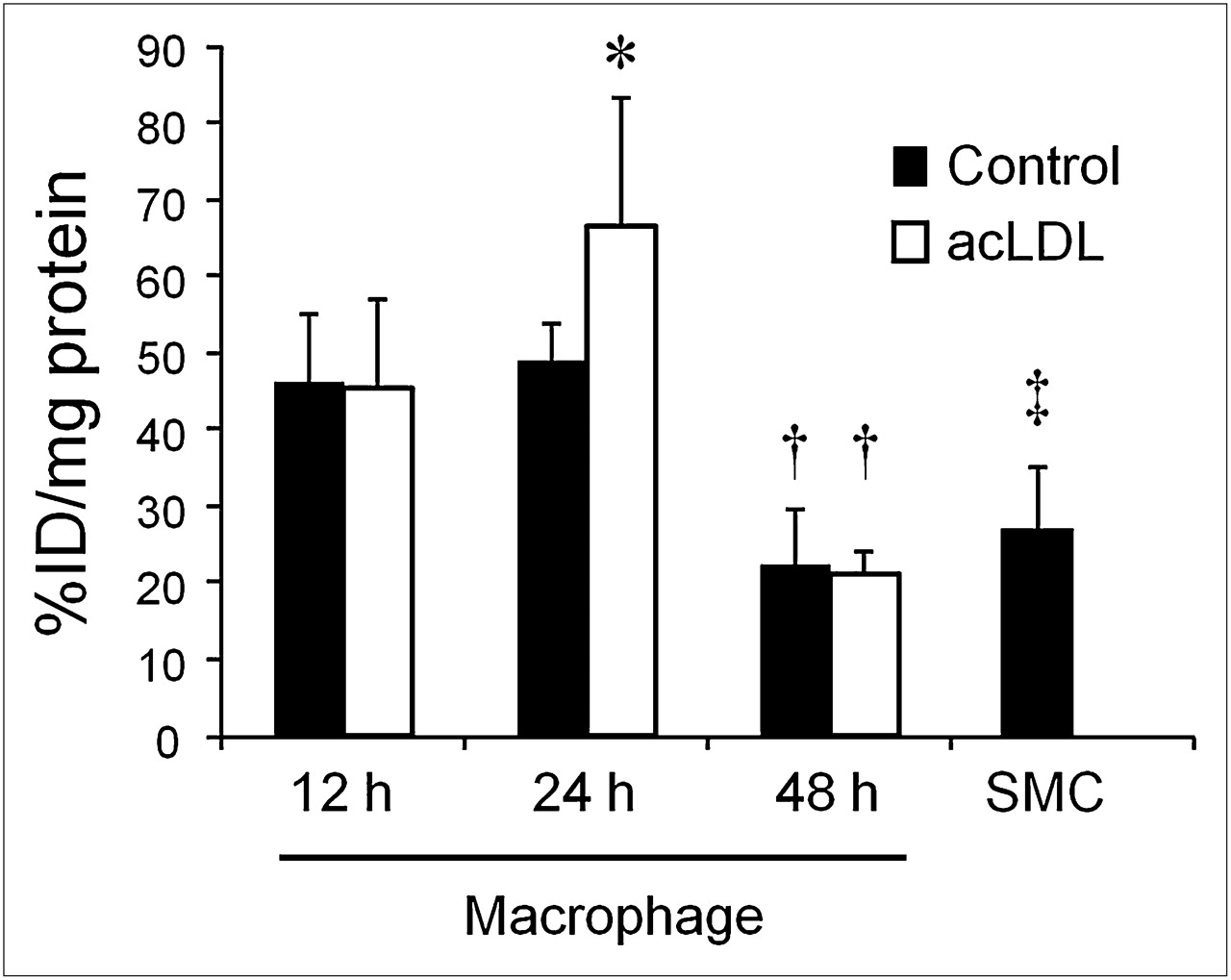
18F-FDG uptake by macrophages and SMCs. Macrophages were treated with acetylated LDL for 12, 24, or 48 h. As control, PBS was added instead of acetylated LDL. Significantly higher uptake was observed under 24-h acetylated LDL–treated condition than under 24-h control condition (*P < 0.05). Uptake was significantly lower in 48-h culture than in 12- and 24-h culture under both acetylated LDL–treated and control conditions (†P < 0.05). Uptake by SMCs was significantly lower than uptake by 12- or 24-h PBS- or acetylated LDL–treated macrophages (‡P < 0.05) but was almost at same level as that of 48-h treated groups. acLDL = acetylated low-density lipoprotein; %ID = percentage injected dose.
The uptake was significantly higher under the acetylated LDL-treated condition than under the control condition in the 24-h group. There was no significant difference between acetylated LDL–treated and control conditions in either the 12-h or the 48-h group. Decreased 18F-FDG uptake was observed in the 48-h culture under the control condition, compared with the 12- and 24-h cultures. Uptake by SMCs was lower than that by the 12- and 24-h PBS- or acetylated LDL–treated macrophages but was almost at the same level as that of the 48-h treated groups.
Changes in Hexokinase, G6Pase Activities, and GLUT Expression by Foam Cell Formation
No significant difference was observed in hexokinase activity among any of the tested conditions, but a tendency toward higher activity was observed after 24 h of acetylated LDL loading than under the control condition (Fig. 3A) (P = 0.08). In contrast, the changes in G6Pase activity and GLUT-1 expression did not parallel 18F-FDG uptake (Figs. 3B–4D). G6Pase activity remained stable with acetylated LDL treatment for 12 h and 24 h but was lower than under the control condition after a 48-h treatment. GLUT-1 expression was decreased after a 24-h treatment under the control condition, but in the acetylated LDL–loaded condition, expression had already decreased after a 12-h treatment.

Changes in hexokinase activity (A), G6Pase activity (B), and GLUT-1 expression (C and D) by acetylated LDL treatments. For western blot analysis, MCF7 cell lysate was used as molecular-weight marker for GLUT-1 and actin was used as loading control (D). Elevated hexokinase activity was observed after 24 h of acetylated LDL treatment, compared with control condition, and changes in hexokinase activity tended to follow changes in 18F-FDG uptake. G6Pase activity and GLUT-1 expression were independent of 18F-FDG uptake. acLDL = acetylated low-density lipoprotein; Ctr = control.
DISCUSSION
During the course of vulnerable plaque development, macrophages differentiate to foam cells by acetylated LDL loading, and foam cell formation leads to plaque rupture (2). In this study, 18F-FDG accumulation was increased during foam cell formation, but the uptake was decreased to the control level after the cells had differentiated completely to foam cells. These findings suggest that 18F-FDG PET detects the early stage of foam cell formation in atherosclerosis. The decrease in 18F-FDG uptake at 48 h under the control condition may be due to the decrease in cell viability because of their long time in culture. A somewhat large SD under the 24-h acetylated LDL condition could be due to the variations in each cell condition, since the cells were in the course of forming foam cells.
Physiologic accumulation of 18F-FDG in the healthy aortic vessel interferes with visualization of the adjacent vulnerable plaque in in vivo imaging. Previously, Maschauer et al. performed a 18F-FDG uptake study using cultured human endothelial cells (14). Their results showed that 18F-FDG uptake was higher in these cells than in human macrophages. However, as Buck et al. pointed out in their comments on that article, endothelial cells are not appropriate alternatives to arterial SMCs (15). Moreover, Maschauer et al. conducted the study at 4°C, and most enzymes, including hexokinase and G6Pase, cannot work at that temperature. In our study, 18F-FDG uptake by cultured aortic SMCs was almost at the same level as that of 3-d cultured macrophages (48 h after the PBS or acetylated LDL treatment). Therefore, it would be difficult to visualize the later stages of macrophage-infiltrated plaques. A limitation to our study is that we used 10-d cultured SMCs, since it takes time to obtain enough cells from the aortic segments. This situation might not reflect the physiologic conditions in vivo, and further investigation is needed to predict the capability of 18F-FDG PET to distinguish SMCs in the arterial wall from macrophages in plaque, especially in advanced lesions. Another limitation is that mouse-derived macrophages and SMCs were used in this study. The use of these cells might create a physiologic situation somewhat different from that with human cells.
The changes in hexokinase activity modestly corresponded to the changes in 18F-FDG uptake. In contrast, the changes in G6Pase activity and GLUT-1 expression did not parallel the changes in 18F-FDG uptake. These results suggest that hexokinase activity is responsible for 18F-FDG uptake by atherosclerotic vulnerable plaques. The major GLUT isoforms in macrophages and monocytes are GLUT-1, -3, and -5. GLUT-3 expression decreases during differentiation of monocytes to macrophages, and GLUT-3 is present only in monocytes (16). GLUT-5 works as a fructose transporter. On this basis, we checked the GLUT-1 expression, and it was independent of the changes in 18F-FDG uptake.
CONCLUSION
In atherosclerosis imaging, 18F-FDG PET appears to visualize the early stage of foam cell formation in vulnerable plaques. Changes in hexokinase activity tended to accompany changes in 18F-FDG uptake.
DISCLOSURE STATEMENT
The costs of publication of this article were defrayed in part by the payment of page charges. Therefore, and solely to indicate this fact, this article is hereby marked “advertisement” in accordance with 18 USC section 1734.
Acknowledgments
This work was supported by Grant-in-Aid for Scientific Research from the Japan Society for the Promotion of Science (JSPS). No other potential conflict of interest relevant to this article was reported.
Footnotes
Published online Nov. 29, 2011.
- © 2012 by the Society of Nuclear Medicine, Inc.
REFERENCES
- Received for publication May 9, 2011.
- Accepted for publication September 6, 2011.


Jump to section
Related Articles
Cited By...
- Low-dose interleukin 2 for the reduction of vascular inflammation in acute coronary syndromes (IVORY): protocol and study rationale for a randomised, double-blind, placebo-controlled, phase II clinical trial
- Therapeutic Antibody Against Phosphorylcholine Preserves Coronary Function and Attenuates Vascular 18F-FDG Uptake in Atherosclerotic Mice
- Vascular Inflammation in Subclinical Atherosclerosis Detected by Hybrid PET/MRI
- Characterization of Macrophage Polarization States Using Combined Measurement of 2-Deoxyglucose and Glutamine Accumulation: Implications for Imaging of Atherosclerosis
- Arterial Stiffness Is Positively Associated With 18F-fluorodeoxyglucose Positron Emission Tomography-Assessed Subclinical Vascular Inflammation in People With Early Type 2 Diabetes
- Imaging Atherosclerosis
- Scintillating Balloon-Enabled Fiber-Optic System for Radionuclide Imaging of Atherosclerotic Plaques
- Oxidized Low-Density Lipoprotein Stimulates Macrophage 18F-FDG Uptake via Hypoxia-Inducible Factor-1{alpha} Activation Through Nox2-Dependent Reactive Oxygen Species Generation
- Development of 111In-Labeled Liposomes for Vulnerable Atherosclerotic Plaque Imaging
- Molecular Imaging of Atherosclerosis for Improving Diagnostic and Therapeutic Development
- Diabetes and Vascular 18F-Fluorodeoxyglucose Positron Emission Tomography Uptake: Another Step Toward Understanding Inflammation in Atherosclerosis