
Main menu

  • Home
  • Content
    • Current
    • Ahead of print
    • Past Issues
    • JNM Supplement
    • SNMMI Annual Meeting Abstracts
    • Continuing Education
    • JNM Podcasts
  • Subscriptions
    • Subscribers
    • Institutional and Non-member
    • Rates
    • Journal Claims
    • Corporate & Special Sales
  • Authors
    • Submit to JNM
    • Information for Authors
    • Assignment of Copyright
    • AQARA requirements
  • Info
    • Reviewers
    • Permissions
    • Advertisers
  • About
    • About Us
    • Editorial Board
    • Contact Information
  • More
    • Alerts
    • Feedback
    • Help
    • SNMMI Journals
  • SNMMI
    • JNM
    • JNMT
    • SNMMI Journals
    • SNMMI

User menu

  • Subscribe
  • My alerts
  • Log in
  • My Cart

Search

  • Advanced search
Journal of Nuclear Medicine
  • SNMMI
    • JNM
    • JNMT
    • SNMMI Journals
    • SNMMI
  • Subscribe
  • My alerts
  • Log in
  • My Cart
Journal of Nuclear Medicine

Advanced Search

  • Home
  • Content
    • Current
    • Ahead of print
    • Past Issues
    • JNM Supplement
    • SNMMI Annual Meeting Abstracts
    • Continuing Education
    • JNM Podcasts
  • Subscriptions
    • Subscribers
    • Institutional and Non-member
    • Rates
    • Journal Claims
    • Corporate & Special Sales
  • Authors
    • Submit to JNM
    • Information for Authors
    • Assignment of Copyright
    • AQARA requirements
  • Info
    • Reviewers
    • Permissions
    • Advertisers
  • About
    • About Us
    • Editorial Board
    • Contact Information
  • More
    • Alerts
    • Feedback
    • Help
    • SNMMI Journals
  • View or Listen to JNM Podcast
  • Visit JNM on Facebook
  • Join JNM on LinkedIn
  • Follow JNM on Twitter
  • Subscribe to our RSS feeds
Research ArticleBrief Communication

What Can Be Seen by 18F-FDG PET in Atherosclerosis Imaging? The Effect of Foam Cell Formation on 18F-FDG Uptake to Macrophages In Vitro

Mikako Ogawa, Satoki Nakamura, Yuriko Saito, Mutsumi Kosugi and Yasuhiro Magata
Journal of Nuclear Medicine January 2012, 53 (1) 55-58; DOI: https://doi.org/10.2967/jnumed.111.092866
Mikako Ogawa
  • Find this author on Google Scholar
  • Find this author on PubMed
  • Search for this author on this site
Satoki Nakamura
  • Find this author on Google Scholar
  • Find this author on PubMed
  • Search for this author on this site
Yuriko Saito
  • Find this author on Google Scholar
  • Find this author on PubMed
  • Search for this author on this site
Mutsumi Kosugi
  • Find this author on Google Scholar
  • Find this author on PubMed
  • Search for this author on this site
Yasuhiro Magata
  • Find this author on Google Scholar
  • Find this author on PubMed
  • Search for this author on this site
  • Article
  • Figures & Data
  • Info & Metrics
  • PDF
Loading

Article Figures & Data

Figures

  • FIGURE 1.
    • Download figure
    • Open in new tab
    • Download powerpoint
    FIGURE 1.

    Results of oil red O staining. Cells were treated with acetylated LDL for 0 h (A), 12 h (B), 24 h (C), or 48 h (D). Moderate staining of macrophages at 24 h (pale red) and strong staining at 48 h (deep red) indicates foam cell formation.
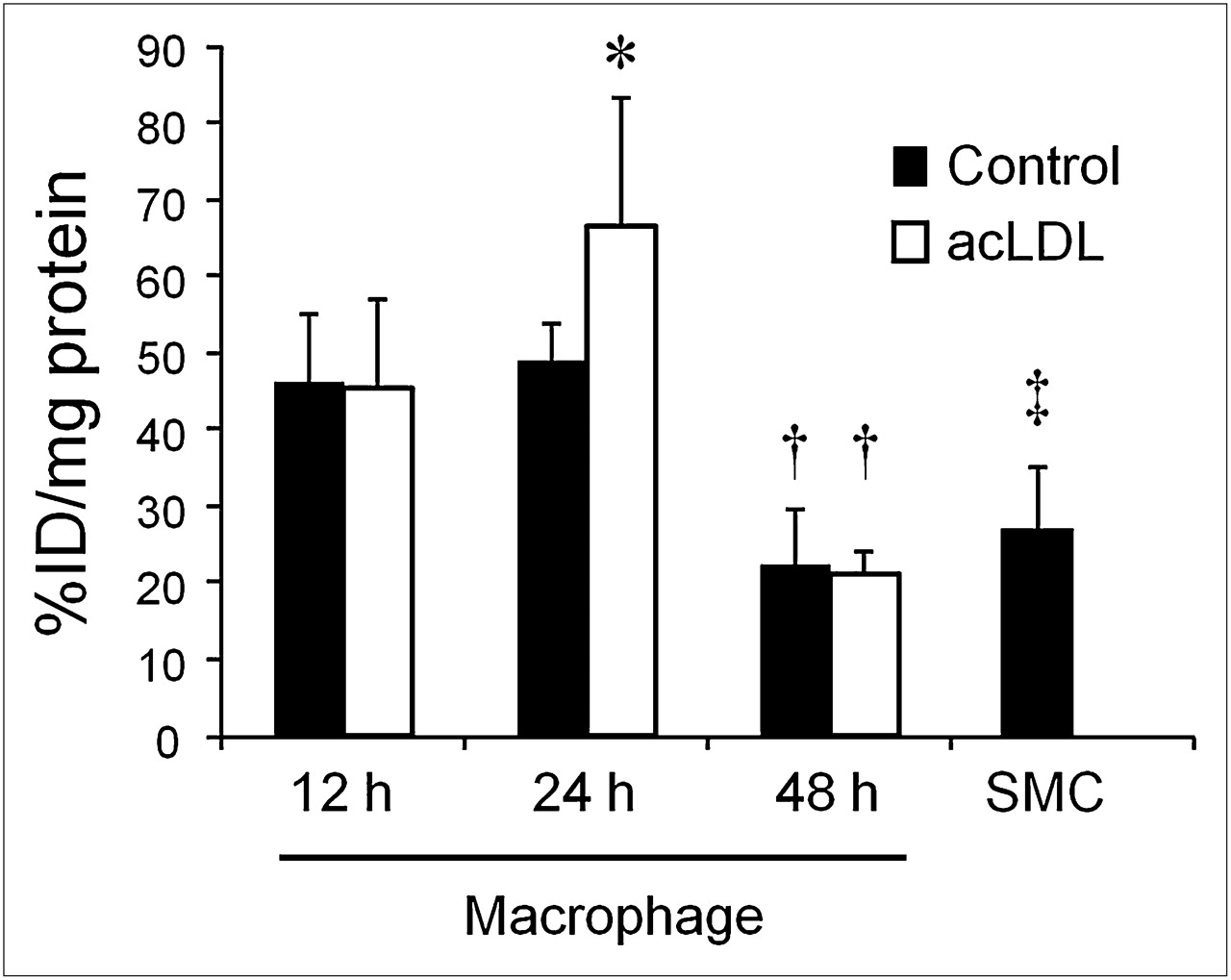
  • FIGURE 2.
    • Download figure
    • Open in new tab
    • Download powerpoint
    FIGURE 2.

    18F-FDG uptake by macrophages and SMCs. Macrophages were treated with acetylated LDL for 12, 24, or 48 h. As control, PBS was added instead of acetylated LDL. Significantly higher uptake was observed under 24-h acetylated LDL–treated condition than under 24-h control condition (*P < 0.05). Uptake was significantly lower in 48-h culture than in 12- and 24-h culture under both acetylated LDL–treated and control conditions (†P < 0.05). Uptake by SMCs was significantly lower than uptake by 12- or 24-h PBS- or acetylated LDL–treated macrophages (‡P < 0.05) but was almost at same level as that of 48-h treated groups. acLDL = acetylated low-density lipoprotein; %ID = percentage injected dose.

  • FIGURE 3.
    • Download figure
    • Open in new tab
    • Download powerpoint
    FIGURE 3.

    Changes in hexokinase activity (A), G6Pase activity (B), and GLUT-1 expression (C and D) by acetylated LDL treatments. For western blot analysis, MCF7 cell lysate was used as molecular-weight marker for GLUT-1 and actin was used as loading control (D). Elevated hexokinase activity was observed after 24 h of acetylated LDL treatment, compared with control condition, and changes in hexokinase activity tended to follow changes in 18F-FDG uptake. G6Pase activity and GLUT-1 expression were independent of 18F-FDG uptake. acLDL = acetylated low-density lipoprotein; Ctr = control.

PreviousNext
Back to top

In this issue

Journal of Nuclear Medicine: 53 (1)
Journal of Nuclear Medicine
Vol. 53, Issue 1
January 1, 2012
  • Table of Contents
  • Table of Contents (PDF)
  • About the Cover
  • Index by author
Print
Download PDF
Article Alerts
Sign In to Email Alerts with your Email Address
Email Article

Thank you for your interest in spreading the word on Journal of Nuclear Medicine.

NOTE: We only request your email address so that the person you are recommending the page to knows that you wanted them to see it, and that it is not junk mail. We do not capture any email address.

Enter multiple addresses on separate lines or separate them with commas.
What Can Be Seen by 18F-FDG PET in Atherosclerosis Imaging? The Effect of Foam Cell Formation on 18F-FDG Uptake to Macrophages In Vitro
(Your Name) has sent you a message from Journal of Nuclear Medicine
(Your Name) thought you would like to see the Journal of Nuclear Medicine web site.
Citation Tools
What Can Be Seen by 18F-FDG PET in Atherosclerosis Imaging? The Effect of Foam Cell Formation on 18F-FDG Uptake to Macrophages In Vitro
Mikako Ogawa, Satoki Nakamura, Yuriko Saito, Mutsumi Kosugi, Yasuhiro Magata
Journal of Nuclear Medicine Jan 2012, 53 (1) 55-58; DOI: 10.2967/jnumed.111.092866

Citation Manager Formats

  • BibTeX
  • Bookends
  • EasyBib
  • EndNote (tagged)
  • EndNote 8 (xml)
  • Medlars
  • Mendeley
  • Papers
  • RefWorks Tagged
  • Ref Manager
  • RIS
  • Zotero
Share
What Can Be Seen by 18F-FDG PET in Atherosclerosis Imaging? The Effect of Foam Cell Formation on 18F-FDG Uptake to Macrophages In Vitro
Mikako Ogawa, Satoki Nakamura, Yuriko Saito, Mutsumi Kosugi, Yasuhiro Magata
Journal of Nuclear Medicine Jan 2012, 53 (1) 55-58; DOI: 10.2967/jnumed.111.092866
Twitter logo Facebook logo LinkedIn logo Mendeley logo
  • Tweet Widget
  • Facebook Like
  • Google Plus One
Bookmark this article

Jump to section

  • Article
    • Abstract
    • MATERIALS AND METHODS
    • RESULTS
    • DISCUSSION
    • CONCLUSION
    • DISCLOSURE STATEMENT
    • Acknowledgments
    • Footnotes
    • REFERENCES
  • Figures & Data
  • Info & Metrics
  • PDF

Related Articles

  • This Month in JNM
  • PubMed
  • Google Scholar

Cited By...

  • Low-dose interleukin 2 for the reduction of vascular inflammation in acute coronary syndromes (IVORY): protocol and study rationale for a randomised, double-blind, placebo-controlled, phase II clinical trial
  • Therapeutic Antibody Against Phosphorylcholine Preserves Coronary Function and Attenuates Vascular 18F-FDG Uptake in Atherosclerotic Mice
  • Vascular Inflammation in Subclinical Atherosclerosis Detected by Hybrid PET/MRI
  • Characterization of Macrophage Polarization States Using Combined Measurement of 2-Deoxyglucose and Glutamine Accumulation: Implications for Imaging of Atherosclerosis
  • Arterial Stiffness Is Positively Associated With 18F-fluorodeoxyglucose Positron Emission Tomography-Assessed Subclinical Vascular Inflammation in People With Early Type 2 Diabetes
  • Imaging Atherosclerosis
  • Scintillating Balloon-Enabled Fiber-Optic System for Radionuclide Imaging of Atherosclerotic Plaques
  • Oxidized Low-Density Lipoprotein Stimulates Macrophage 18F-FDG Uptake via Hypoxia-Inducible Factor-1{alpha} Activation Through Nox2-Dependent Reactive Oxygen Species Generation
  • Development of 111In-Labeled Liposomes for Vulnerable Atherosclerotic Plaque Imaging
  • Molecular Imaging of Atherosclerosis for Improving Diagnostic and Therapeutic Development
  • Diabetes and Vascular 18F-Fluorodeoxyglucose Positron Emission Tomography Uptake: Another Step Toward Understanding Inflammation in Atherosclerosis
  • Google Scholar

More in this TOC Section

  • Measuring Total Metabolic Tumor Volume from 18F-FDG PET: A Reality Check
  • Bioanalytic Hybrid System Merging 3-Dimensional Cell Culture and Chromatographic Precision for Unprecedented Preclinical Insights in Molecular Imaging
  • Radiances of Cerenkov-Emitting Radionuclides on the In Vivo Imaging System
Show more BRIEF COMMUNICATION

Similar Articles

SNMMI

© 2025 SNMMI

Powered by HighWire