


Abstract
Annexin V, a human protein with a high affinity for phosphatidylserine, has been labeled with 99mTc to detect apoptosis in vivo. To determine the effectiveness of imaging with this agent as a reflection of the degree of apoptosis after the first dose of chemotherapy, we studied rats with an engrafted hepatoma. Methods: Annexin V was labeled with 99mTc (specific activity, 3.0 MBq/μg protein). Eleven days after being inoculated with allogenic hepatoma cells (KDH-8) in the left calf muscle, the rats were randomized to receive a single dose of cyclophosphamide (150 mg/kg intraperitoneally) or to serve as controls. 99mTc-annexin V was injected 20 h later. Radioactivity in tissues was determined 6 h after injection of 99mTc-annexin V. Tumor uptake of 14C-iodoanitpyrine was determined as a marker of tumor blood flow. Terminal deoxynucleotidyl transferase–mediated deoxyuridine triphosphate nick-end labeling (TUNEL) of tissue harvested at necropsy was performed to detect apoptosis in the tumor. Results: Cyclophosphamide treatment significantly increased the tumor uptake (percentage activity of injected dose per gram of tissue after normalization to the animal’s weight [%ID/g/kg]) of 99mTc-annexin V (0.070 ± 0.007 %ID/g/kg for treated rats and 0.046 ± 0.009 %ID/g/kg for controls, P < 0.001). 14C-iodoantipyrine uptake was similar in the treated and untreated groups. The number of TUNEL-positive cells in the tumor was significantly larger in the treated rats (297.70 ± 50.34 cells/mm2) than in the control rats (168.45 ± 23.60 cells/mm2, P < 0.001). Tumor uptake of 99mTc-annexin V correlated with the number of TUNEL-positive cells in the tumor (r = 0.712; P < 0.001). Conclusion: Tumor uptake of 99mTc-annexin V was significantly increased by a single dose of cyclophosphamide treatment, and the increase was concordant with the number of TUNEL-positive cells in the tumor. The current results are suggestive of the utility of 99mTc-annexin V as a noninvasive means to assess tumor response, although further testing, including clinical evaluation, is required.
Apoptosis plays an important role in both normal physiology and many disease processes (1–5). One of the earliest events in apoptosis is the externalization of phosphatidylserine, a membrane phospholipid normally restricted to the inner leaflet of the lipid bilayer (6,7). Annexin V, a human protein with a high affinity for membrane-bound phosphatidylserine (6,8–14), has been labeled with fluorescent markers for in vitro detection of apoptotic cells (11,14) and with radioactive agents, such as 99mTc, to detect apoptosis in vivo (15–18).
In the case of tumor tissue, successful chemotherapy or radiotherapy induces apoptosis in the neoplastic cells and indicates tumor response to the therapy (15,19–24). Previous studies demonstrated that radiolabeled annexin imaging can detect apoptosis in vivo in experimental models (15–17,25–29) of tumor therapy. This experiment was performed to determine whether radiolabeled annexin imaging could identify this response after the first treatment.
MATERIALS AND METHODS
Animal Studies
All procedures involving animals were performed in accordance with the institutional guidelines of Hokkaido University (Sapporo, Japan). Male Wistar King Aptekman/Hok rats (supplied by the Experimental Animal Institute, Graduate School of Medicine, Hokkaido University) were inoculated with a suspension of KDH-8 rat hepatoma cells (1 × 10⁶ cells per rat) into the left calf muscle (30,31).
Human annexin V was produced by expression in Escherichia coli as previously described (13,14,16,32–34). Annexin V was labeled with 99mTc after derivatization with hydrazinonicotinamide (specific activity, 3.0 MBq/μg protein) (16). Eleven days after intramuscular injection of KDH-8 cells, rats weighing 209–287 g were randomized to receive a single dose of cyclophosphamide (150 mg/kg intraperitoneally; n = 9) or to serve as controls (n = 10). 99mTc-annexin V (3.8 μg protein per rat) was injected intravenously 20 h later. The animals were under light ether anesthesia at the time of injection. Six hours after 99mTc-annexin V injection, the animals were sacrificed and the tumor, blood, and samples of normal tissues were removed. The tissue samples were weighed, and radioactivity was determined with a well-type scintillation counter (1480 WIZARD 3″; Wallac Co. Ltd., Turku, Finland). Aliquots of the tumor tissues were used to prepare formalin-fixed paraffin-embedded specimens for subsequent histologic studies. The accumulation of 99mTc-annexin V in the tissues was expressed as the percentage activity of injected dose per gram of tissue after normalization to the animal’s weight (%ID/g/kg). The tumor-to-muscle ratio (T/M ratio) and the tumor-to-blood ratio (T/B ratio) were calculated from the %ID/g/kg value of each tissue.
To determine whether the chemotherapy altered tumor blood flow (which could alter delivery of radiolabeled annexin to the tumor), a separate group of tumor animals was used to assess tissue blood flow by the uptake of 14C-iodoantipyrine (14C-IAP) (35). Eleven days after being inoculated with KDH-8 cells, rats weighing 240–270 g were randomized to receive a single dose of cyclophosphamide (150 mg/kg intraperitoneally; n = 4) or to serve as controls (n = 4). 14C-IAP (0.37 MBq per rat) purchased from American Radiolabeled Chemicals Inc. (St. Louis, MO) was intravenously injected 20 h later while the rats were under light ether anesthesia. The rats were sacrificed 40 s after injection of 14C-IAP, and the tumor, blood, and samples of normal tissues were removed. The tissue samples were weighed, and radioactivity was determined with a liquid scintillation counter (1414 WinSpectral α/β; Wallac Co. Ltd.). The accumulation of 14C-IAP in the tissues was expressed as %ID/g/kg.
Detection of Apoptosis
Apoptotic nuclei were determined by direct immunoperoxidase detection of digoxigenin-labeled 3′ DNA strand breaks by use of terminal deoxynucleotidyl transferase–mediated deoxyuridine triphosphate nick-end labeling (TUNEL) (36). The formalin-fixed paraffin-embedded tissues were sectioned at 3 μm and stained according to a standard procedure using a commercially available kit (apoptosis in situ detection kit Wako; Wako Pure Chemical Industries, Ltd., Tokyo, Japan). Negative and positive controls were used for the determination of TUNEL-positive cells, instead of using a reference area. The number of TUNEL-positive cells was counted on 10 randomly selected ×200 fields for each section by use of a light microscope.
Statistical Analysis
All values are shown as mean ± SD. Statistical analysis was performed using the unpaired Student t test to evaluate the significance of differences in the values between the control and treated animals. Simple regression analysis was used to compare the uptake of 99mTc-annexin V and the number of TUNEL-positive cells. A 2-tailed value of P < 0.05 was considered significant.
RESULTS
Biodistribution and Tumor Uptake of 99mTc-Annexin V
The tissue distribution of 99mTc-annexin V is shown in Table 1. In control rats, uptake of 99mTc-annexin V was highest in the kidneys, followed in decreasing order by the spleen, liver, and bone marrow. Uptake of 99mTc-annexin V was significantly higher in tumor (0.046 ± 0.009 %ID/g/kg) than in muscle (0.008 ± 0.001 %ID/g/kg, P < 0.01) or in blood (0.031 ± 0.004 %ID/g/kg, P < 0.05). The T/M and T/B ratios were 5.60 ± 0.90 and 1.33 ± 0.20, respectively.
Biodistribution of 99mTc-Annexin V
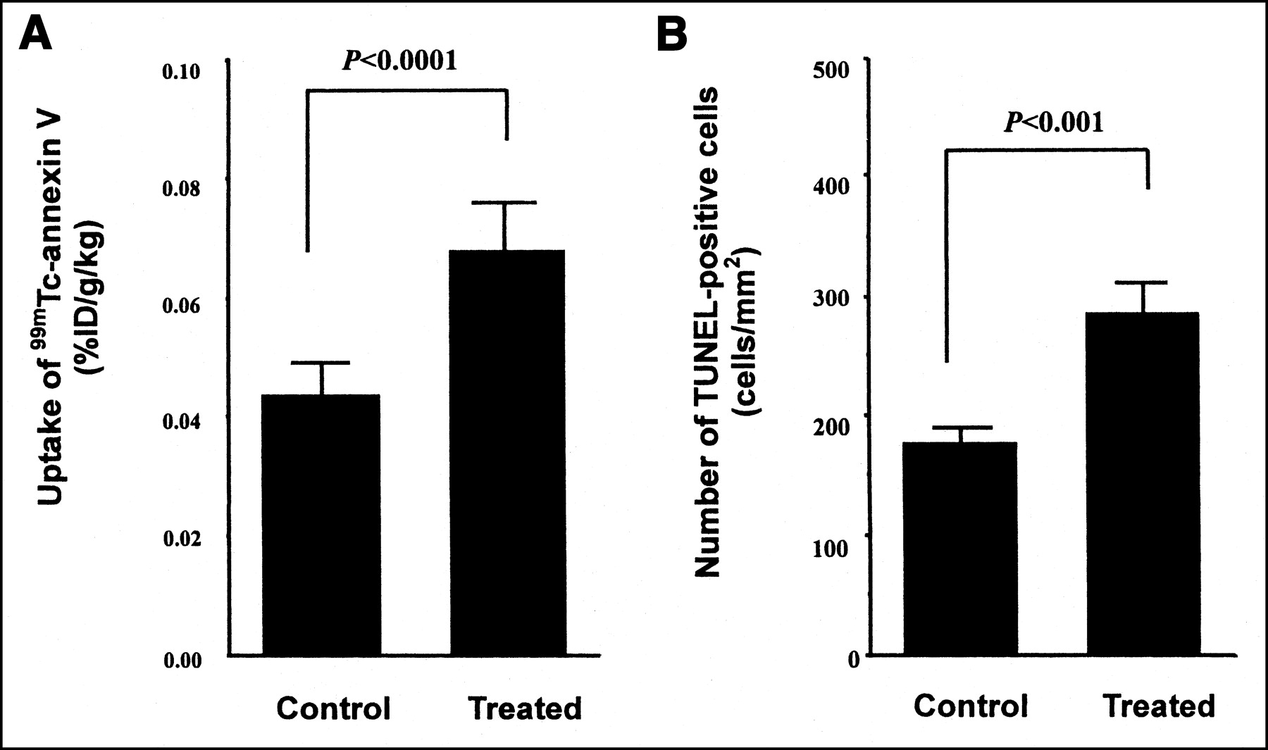
Uptake of 99mTc-annexin V was significantly higher in treated tumors (0.070 ± 0.007 %ID/g/kg) than in control tumors (0.046 ± 0.009 %ID/g/kg, P < 0.001) (Table 1; Fig. 1A). Uptake in the thymus, bone marrow, and spleen was also significantly increased by cyclophosphamide treatment. In contrast, uptake in blood, muscle, the liver, and the kidneys was not significantly affected by the treatment. The T/M and T/B ratios of the treated groups were 7.53 ± 1.06 and 2.14 ± 0.31, respectively, which were significantly higher than those of the control groups (P < 0.05 for T/M and P < 0.001 for T/B).

Uptake of 99mTc-annexin V (A) and number of TUNEL-positive cells (B) in tumor. Uptake of 99mTc-annexin V and number of TUNEL-positive cells were significantly higher in treated tumor than in control tumor. Control = control rats; treated = rats treated with cyclophosphamide (150 mg/kg intraperitoneally).
Biodistribution and Tumor Uptake of 14C-IAP
Tissue uptake of 14C-IAP is shown in Table 2. In control rats, uptake of 14C-IAP was highest in the liver (0.306 ± 0.133 %ID/g/kg), followed in decreasing order by the kidneys, the bone marrow, the spleen, and muscle. Uptake of 14C-IAP in tumor (0.088 ± 0.048 %ID/g/kg) was the lowest among the tissues determined. Uptake of 14C-IAP in tumor was decreased by cyclophosphamide treatment (0.041 ± 0.009 %ID/g/kg, treated rats), although the difference was not significant (P = 0.10). Uptake of 14C-IAP in other tissues did not significantly differ between the treated and control groups.
Biodistribution of 14C-IAP
Apoptotic Cells and Relationship with 99mTc-Annexin V Uptake
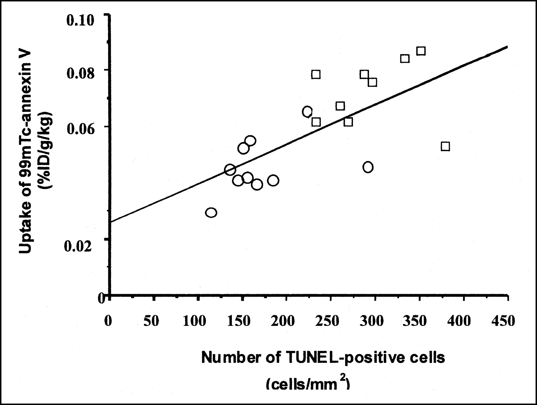
TUNEL-positive cells were seen in the tumor specimens obtained from both the control rats and the treated rats (Fig. 2). The number of TUNEL-positive cells was significantly greater in the treated tumors (297.70 ± 50.34 cells/mm2) than in the control tumors (168.45 ± 23.60 cells/mm2, P < 0.001) (Fig. 1B). Tumor uptake of 99mTc-annexin V correlated with the number of TUNEL-positive cells in the tissue (r = 0.712; P < 0.001) (Fig. 3).
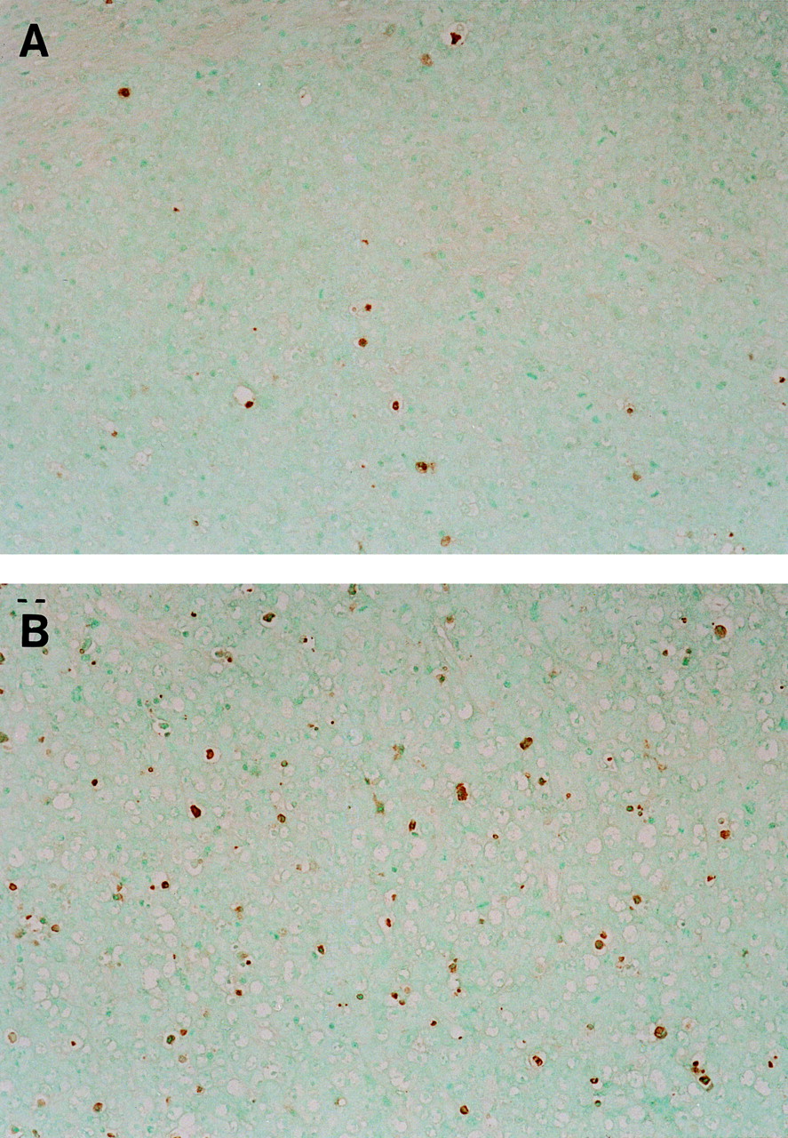
TUNEL staining (×200) of tumor specimens in control rats (A) and treated rats (B). Cells stained brown were considered TUNEL positive. TUNEL-positive cells were observed in tumor specimens obtained from both control and treated rats.

Correlation of uptake of 99mTc-annexin V and number of TUNEL-positive cells in tumor. Tumor uptake of 99mTc-annexin V significantly correlated with number of TUNEL-positive cells in tumor. (Uptake of 99mTc-annexin V) = 0.025 + 1.397 × 10⁻⁴ × (number of TUNEL-positive cells); r = 0.712; P < 0.001. ○ = control rats; □ = treated rats.
DISCUSSION
KDH-8 tumor uptake of 99mTc-annexin V increased significantly after a single dose of cyclophosphamide chemotherapy in a rat malignant tumor model, despite a slight decrease in perfusion. The number of TUNEL-positive cells in the tumor increased after treatment and correlated with radiolabeled annexin uptake.
Chemotherapeutic drugs and irradiation induce apoptosis in both normal tissues and tumors (15,16,19–24). TUNEL staining, which marks the degraded DNA in the cell, has been used as a marker for apoptosis (36). Our results demonstrate a striking relationship between increased 99mTc-annexin V uptake and TUNEL staining after cyclophosphamide treatment. Cyclophosphamide treatment gave a 60% increase in tumor uptake of 99mTc-annexin V and a 77% increase in TUNEL-positive cells, compared with the control value. These results are similar to those previously reported by Blankenberg et al. (16) in a model of murine B cell lymphoma.
Yang et al. (37) reported increased cellular uptake of 99mTc-annexin V after irradiation and paclitaxel treatment in an in vitro cell culture using a breast cancer cell line. They also showed increased tumor-to-nontumor ratios of 99mTc-annexin V after paclitaxel treatment by in vivo imaging experiments. Hofstra et al. (38) observed an enhanced tumor accumulation of 99mTc-annexin V in a patient with an intracardiac tumor.
Our results showed that 99mTc-annexin V uptake significantly correlated with the number of TUNEL-positive cells. Blankenberg et al. (16) observed annexin V accumulation in rodent heart transplants that were undergoing rejection and that occasionally had TUNEL-positive cell nuclei. In contrast, D’Arceuil et al. (29) reported a high uptake of 99mTc-annexin V in hypoxic neurons and glial cells without evidence of apoptotic nuclei by TUNEL staining in a rabbit model of neonatal hypoxic brain injury. This discrepancy appears to largely be ascribed to differences in the tissue insult of each model and the phase of apoptosis detected by annexin V and TUNEL staining (29). TUNEL staining detects DNA degradation that occurs in a late phase of apoptosis, whereas annexin V binds to the phosphatidylserine that is externalized in an earlier phase of apoptosis that immediately follows caspase-3 activation. Further studies, including those on the expression of caspase-3 messenger RNA and protein in relation to 99mTc-annexin V uptake, may be helpful to confirm the present results and provide further evidence for the potential of 99mTc-annexin V as an indicator of apoptotic tumor cells.
In addition to apoptotic cells, annexin V binds to necrotic cells, because of exposure of phosphatidylserine located in the inner leaflet of the highly permeable plasma membrane of necrotic cells (11). Annexin V localization in vivo does not appear to be entirely specific for apoptosis. The present results, however, indicate that 99mTc-annexin V accumulation can largely be ascribed to apoptotic cells in the tumor.
Accumulation of a tracer in a tissue is generally regulated by its delivery to the tissue, uptake by the tissue, and retention in the tissue. Consequently, increased blood flow may cause increased tissue accumulation of the tracer. In our study, cyclophosphamide treatment significantly increased the tumor uptake of 99mTc-annexin V. In contrast, the uptake of 14C-IAP was lower in the treated rats than in the control rats, although the difference was not significant (Table 2). These results indicate that the increased accumulation of 99mTc-annexin V in the tumor is not attributable to changes in blood flow.
The present study was conducted to clarify the potential of 99mTc-annexin V to detect apoptosis early after chemotherapy. Thus, a relatively early time, 20 h after treatment, was selected. 99mTc-annexin V could detect the apoptotic tumor response by cyclophosphamide treatment at this time. However, experiments at various times after treatment are required to further confirm the potential of 99mTc-annexin V.
Regarding the timing after 99mTc-annexin V, Yang et al. (37) reported increased tumor-to-blood, tumor-to-lung, and tumor-to-muscle count density ratios as a function of time during their experimental period of 4 h. Our preliminary study (39) also showed that the tumor-to-blood uptake ratio of 99mTc-annexin V was significantly higher at 6 h than at 1 h. Higher ratios of tumor to blood and tumor to other tissue will contribute to the quality of images from in vivo studies, which are important in the clinical setting. In the present study, we determined the biodistribution and tumor uptake of 99mTc-annexin V at 6 h after injection of 99mTc-annexin V, although the optimum time after 99mTc-annexin V injection remains to be determined. Our results showed significantly higher tumor uptake of 99mTc-annexin V without significant differences in tumor weights between the control and treated animals (3.71 ± 0.56 g for treated rats and 3.62 ± 0.99 g for controls). These results suggest that 99mTc-annexin V imaging can predict treatment efficacy before tumor volumes are reduced by the treatment. However, the potential of 99mTc-annexin V for imaging apoptosis remains to be elucidated. A scintigraphic image would be helpful to demonstrate that apoptosis is visible in the image. Autoradiographic methods are also helpful for the direct comparison of tracer uptake and TUNEL-positive cells.
In our study, a relatively high dose of cyclophosphamide (150 mg/kg intraperitoneally) was used to induce apoptosis in the tumor. Blankenberg et al. (15) used a 100 mg/kg intraperitoneal dose of cyclophosphamide to induce apoptosis in murine B cell lymphoma. They also reported that 99mTc-annexin V preferentially localized to regions of intramedullary and splenic apoptotic injury induced by a 100–150 mg/kg intraperitoneal dose of cyclophosphamide—a finding that is consistent with the present results. Similar increases in tumor apoptosis and radiolabeled annexin uptake were observed by Yang et al. (37) with a 40 mg/kg intravenous dose of paclitaxel.
CONCLUSION
Tumor uptake of 99mTc-annexin V is significantly increased by cyclophosphamide treatment, and the increase was concordant with the number of TUNEL-positive cells in the tumor. The current results are suggestive of the utility of 99mTc-annexin V as a noninvasive means to assess tumor response, although further testing, including clinical evaluation, is required.
Acknowledgments
The authors are grateful to Professors Shinzo Nishi, Kazuo Miyasaka, and Toshiyuki Ohnishi of the Central Institute of Isotope Science, Hokkaido University, for supporting this work. The authors thank Koutaro Suzuki, Hidenori Katsuura, Hidehiko Omote, and Hiroshi Arai for their assistance. This work was financially supported in part by a grant from the Japanese Foundation for Multidisciplinary Treatment of Cancer.
Footnotes
Received Mar. 18, 2002; revision accepted Jul. 26, 2002.
For correspondence or reprints contact: Nagara Tamaki, MD, Department of Nuclear Medicine, Graduate School of Medicine, Hokkaido University, Kita15 Nishi7, Kita-ku, Sapporo 060-8638, Japan.
E-mail: natamaki{at}med.hokudai.ac.jp
REFERENCES
In this issue


Jump to section
Related Articles
Cited By...
- Resolvins suppress tumor growth and enhance cancer therapy
- Quantitative Determination of Apoptosis of Pancreatic {beta}-Cells in a Murine Model of Type 1 Diabetes Mellitus
- Looking Through the Vascular Normalization Window: Timing Antiangiogenic Treatment and Chemotherapy with 99mTc-Annexin A5
- Prolonged High-Fat Feeding Enhances Aortic 18F-FDG and 99mTc-Annexin A5 Uptake in Apolipoprotein E-Deficient and Wild-Type C57BL/6J Mice
- In vivo Targeting of Dead Tumor Cells in a Murine Tumor Model Using a Monoclonal Antibody Specific for the La Autoantigen
- Prognostic Significance of 99mTc Hynic-rh-Annexin V Scintigraphy During Platinum-Based Chemotherapy in Advanced Lung Cancer
- Evaluation of 4'-[Methyl-14C]Thiothymidine for In Vivo DNA Synthesis Imaging
- Counting Heads in the War against Cancer: Defining the Role of Annexin A5 Imaging in Cancer Treatment and Surveillance
- A soluble tissue factor-annexin V chimeric protein has both procoagulant and anticoagulant properties
- Past, Present, and Future of Annexin A5: From Protein Discovery to Clinical Applications
- Imaging {beta}-Cell Death With a Near-Infrared Probe
- A Monoclonal Antibody that Binds Anionic Phospholipids on Tumor Blood Vessels Enhances the Antitumor Effect of Docetaxel on Human Breast Tumors in Mice
- Enhanced Apoptotic Reaction Correlates with Suppressed Tumor Glucose Utilization After Cytotoxic Chemotherapy: Use of 99mTc-Annexin V, 18F-FDG, and Histologic Evaluation
- Time Course of Apoptotic Tumor Response After a Single Dose of Chemotherapy: Comparison with 99mTc-Annexin V Uptake and Histologic Findings in an Experimental Model
- Physiologic Granulocyte Destruction In Vivo by Apoptosis
- Feasibility of 99mTc-Annexin V for Repetitive Detection of Apoptotic Tumor Response to Chemotherapy: An Experimental Study Using a Rat Tumor Model