


Article Figures & Data
Figures
- FIGURE 1.
S value simulation setups for GOT1 and NCI-H69 cells using Geant4. (A) Floating GOT1 clusters during 4-h uptake phase modeled with random close packing, with zoomed-in views on right. (B) Microscopic image of GOT1 cells plated for 7-d follow-up phase (top left) modeled with cluster size probabilities (top right) and hexagonal packing (bottom right) or microscopic image-derived placements (bottom left). (C) Simulated cluster geometry for NCI-H69 cells in suspension at day 0 (compact, N° = 12) and day 7, illustrating exponential growth and random placement after proliferation. Sim. = simulated.
- FIGURE 2.
Time-dependent activity per cell for GOT1 (A) and NCI-H69 (B) cells at various activity concentrations. Asterisks indicate extrapolated data points. Error bars indicate SD.
- FIGURE 3.
Cross-dose variability and absorbed dose rate heterogeneity in GOT1 cells. (A) Cross-dose contribution relative to total for 2 target cells within same setup. (B) Absorbed dose rate maps (excluding medium contribution) for 10 target cells at start of incubation at 0.1 and 1 MBq/mL. Abs. = absorbed.
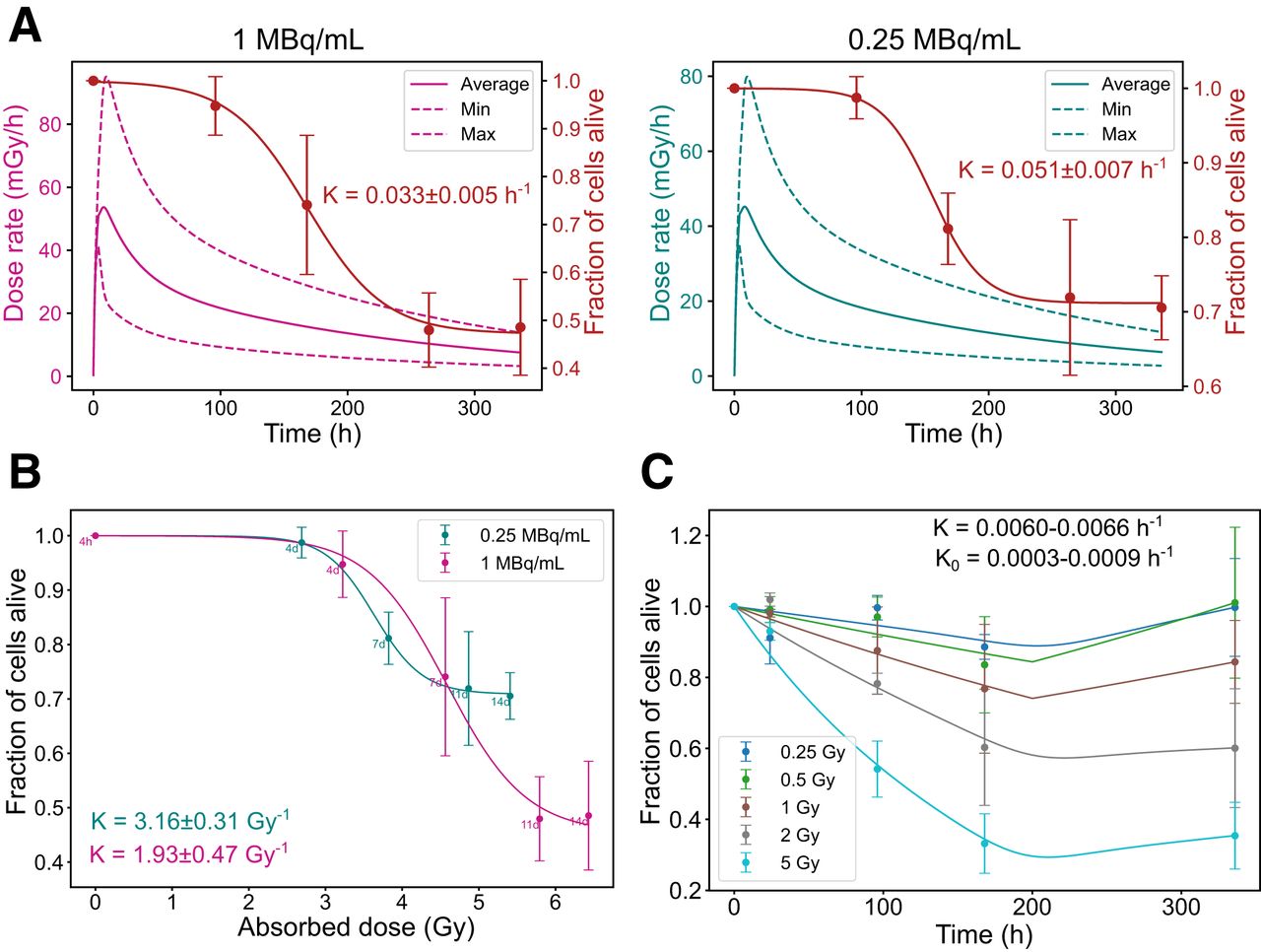
- FIGURE 4.
Radiobiologic correlations for GOT1 cells. (A) Time-dependent dose rate (left, y-axis) and fraction of alive cells (right, y-axis) at 1 and 0.25 MBq/mL. (B) Correlation between absorbed dose and fraction of alive cells for same added activities. (C) Fraction of alive cells over time for EBRT exposure. Error bars indicate SD. Dashed lines indicate dose rate heterogeneity. K = death rate constant; K0 = regrowth rate constant.
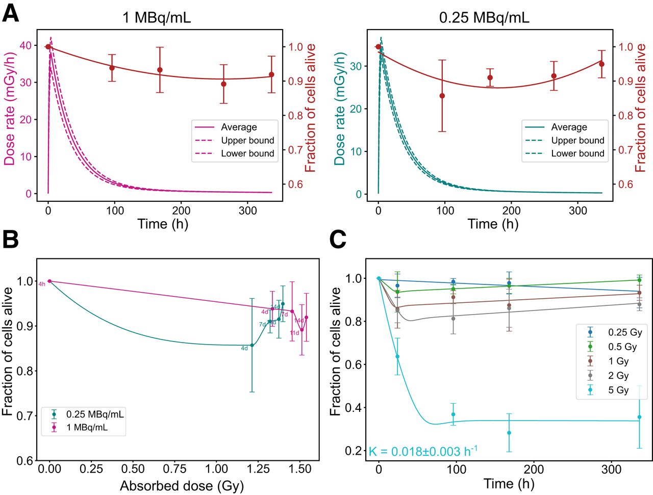
- FIGURE 5.
Radiobiologic correlations for NCI-H69 cells. (A) Time-dependent dose rate (left, y-axis) and fraction of alive cells (right, y-axis) at 1 and 0.25 MBq/mL. (B) Correlation between absorbed dose and fraction of alive cells for same added activities. (C) Fraction of alive cells over time for EBRT exposure. Error bars indicate SD. Dashed lines indicate dose rate heterogeneity. K = death rate constant.
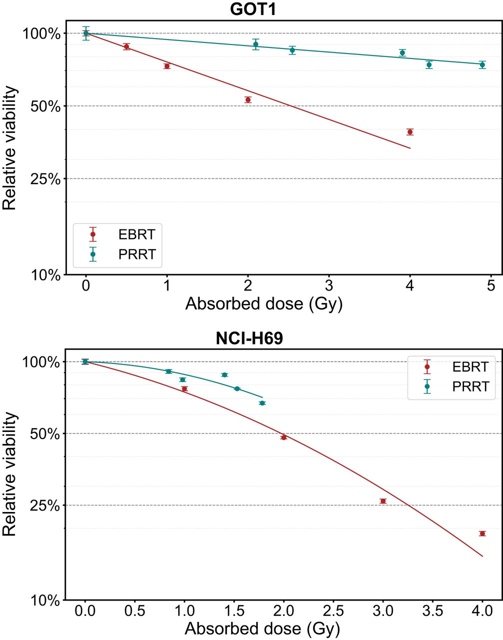
- FIGURE 6.
Dose–response curves showing absorbed dose vs. viability at 7 d for GOT1 and NCI-H69 cells exposed to EBRT or PRRT. Error bars indicate SD.


Tables
- TABLE 1.
Summary of Best Fitting Parameters Used for Dose Responses Shown in Figure 6 After Exposure of GOT1 and NCI-H69 Cells to EBRT and PRRT
NCI-H69 GOT1 LQ L Exposure R2 AIC α (Gy−1) β (Gy−2) Linearized α (Gy−1) R2 AIC α (Gy−1) EBRT 0.99 −32.81 0.24 ± 0.05 0.06 ± 0.02 0.38 ± 0.03 0.97 −31.30 0.27 ± 0.02 177Lu-DOTATATE 0.85 −34.25 0.03 ± 0.10 0.09 ± 0.07 0.16 ± 0.02 0.94 −43.20 0.06 ± 0.02 LQ = linear quadratic; AIC = Akaike information criterion; L = linear.
R2 and AIC are reported to evaluate best fit.
Supplemental Data
Files in this Data Supplement:
In this issue


Jump to section
Related Articles
Cited By...
- No citing articles found.