
Main menu

  • Home
  • Content
    • Current
    • Ahead of print
    • Past Issues
    • JNM Supplement
    • SNMMI Annual Meeting Abstracts
    • Continuing Education
    • JNM Podcasts
  • Subscriptions
    • Subscribers
    • Institutional and Non-member
    • Rates
    • Journal Claims
    • Corporate & Special Sales
  • Authors
    • Submit to JNM
    • Information for Authors
    • Assignment of Copyright
    • AQARA requirements
  • Info
    • Reviewers
    • Permissions
    • Advertisers
  • About
    • About Us
    • Editorial Board
    • Contact Information
  • More
    • Alerts
    • Feedback
    • Help
    • SNMMI Journals
  • SNMMI
    • JNM
    • JNMT
    • SNMMI Journals
    • SNMMI

User menu

  • Subscribe
  • My alerts
  • Log in
  • My Cart

Search

  • Advanced search
Journal of Nuclear Medicine
  • SNMMI
    • JNM
    • JNMT
    • SNMMI Journals
    • SNMMI
  • Subscribe
  • My alerts
  • Log in
  • My Cart
Journal of Nuclear Medicine

Advanced Search

  • Home
  • Content
    • Current
    • Ahead of print
    • Past Issues
    • JNM Supplement
    • SNMMI Annual Meeting Abstracts
    • Continuing Education
    • JNM Podcasts
  • Subscriptions
    • Subscribers
    • Institutional and Non-member
    • Rates
    • Journal Claims
    • Corporate & Special Sales
  • Authors
    • Submit to JNM
    • Information for Authors
    • Assignment of Copyright
    • AQARA requirements
  • Info
    • Reviewers
    • Permissions
    • Advertisers
  • About
    • About Us
    • Editorial Board
    • Contact Information
  • More
    • Alerts
    • Feedback
    • Help
    • SNMMI Journals
  • View or Listen to JNM Podcast
  • Visit JNM on Facebook
  • Join JNM on LinkedIn
  • Follow JNM on Twitter
  • Subscribe to our RSS feeds
Research ArticleClinical Investigation
Open Access

First-in-Human Study of 18F-Labeled PET Tracer for Glutamate AMPA Receptor [18F]K-40: A Derivative of [11C]K-2

Sadamitsu Ichijo, Tetsu Arisawa, Mai Hatano, Waki Nakajima, Tomoyuki Miyazaki, Tsuyoshi Eiro, Yuuki Takada, Ryunosuke Iai, Akane Sano, Masaki Sonoda, Yutaro Takayama, Yuichi Kimura and Takuya Takahashi
Journal of Nuclear Medicine May 2025, jnumed.124.269405; DOI: https://doi.org/10.2967/jnumed.124.269405
Sadamitsu Ichijo
1Department of Physiology, Yokohama City University Graduate School of Medicine, Yokohama, Japan;
  • Find this author on Google Scholar
  • Find this author on PubMed
  • Search for this author on this site
Tetsu Arisawa
1Department of Physiology, Yokohama City University Graduate School of Medicine, Yokohama, Japan;
2Yokohama City University Graduate School of Medicine, Radioisotope Research Center, Yokohama, Japan;
  • Find this author on Google Scholar
  • Find this author on PubMed
  • Search for this author on this site
Mai Hatano
1Department of Physiology, Yokohama City University Graduate School of Medicine, Yokohama, Japan;
  • Find this author on Google Scholar
  • Find this author on PubMed
  • Search for this author on this site
Waki Nakajima
1Department of Physiology, Yokohama City University Graduate School of Medicine, Yokohama, Japan;
  • Find this author on Google Scholar
  • Find this author on PubMed
  • Search for this author on this site
Tomoyuki Miyazaki
1Department of Physiology, Yokohama City University Graduate School of Medicine, Yokohama, Japan;
3Center for Promotion of Research and Industry–Academic Collaboration, Yokohama City University, Yokohama, Japan;
  • Find this author on Google Scholar
  • Find this author on PubMed
  • Search for this author on this site
Tsuyoshi Eiro
1Department of Physiology, Yokohama City University Graduate School of Medicine, Yokohama, Japan;
4Department of Psychiatry, Yokohama City University Graduate School of Medicine, Yokohama, Japan;
  • Find this author on Google Scholar
  • Find this author on PubMed
  • Search for this author on this site
Yuuki Takada
1Department of Physiology, Yokohama City University Graduate School of Medicine, Yokohama, Japan;
  • Find this author on Google Scholar
  • Find this author on PubMed
  • Search for this author on this site
Ryunosuke Iai
1Department of Physiology, Yokohama City University Graduate School of Medicine, Yokohama, Japan;
  • Find this author on Google Scholar
  • Find this author on PubMed
  • Search for this author on this site
Akane Sano
1Department of Physiology, Yokohama City University Graduate School of Medicine, Yokohama, Japan;
  • Find this author on Google Scholar
  • Find this author on PubMed
  • Search for this author on this site
Masaki Sonoda
5Department of Neurosurgery, Yokohama City University Graduate School of Medicine, Yokohama, Japan; and
  • Find this author on Google Scholar
  • Find this author on PubMed
  • Search for this author on this site
Yutaro Takayama
5Department of Neurosurgery, Yokohama City University Graduate School of Medicine, Yokohama, Japan; and
  • Find this author on Google Scholar
  • Find this author on PubMed
  • Search for this author on this site
Yuichi Kimura
6Faculty of Informatics, Cyber Informatics Institute, Kindai University, Higashi-Osaka, Japan
  • Find this author on Google Scholar
  • Find this author on PubMed
  • Search for this author on this site
Takuya Takahashi
1Department of Physiology, Yokohama City University Graduate School of Medicine, Yokohama, Japan;
  • Find this author on Google Scholar
  • Find this author on PubMed
  • Search for this author on this site
  • Article
  • Figures & Data
  • Supplemental
  • Info & Metrics
  • PDF
Loading

Article Figures & Data

Figures

  • Tables
  • Additional Files
  • Figure
    • Download figure
    • Open in new tab
    • Download powerpoint
  • FIGURE 1.
    • Download figure
    • Open in new tab
    • Download powerpoint
    FIGURE 1.

    Chemical structures of [18F]K-40, [18F]K-40OH, [11C]K-2, and [11C]K-2OH.

  • FIGURE 2.
    • Download figure
    • Open in new tab
    • Download powerpoint
    FIGURE 2.

    Averaged pTAC (left panels) and metabolic rate (right panels) of [18F]K-40 and [11C]K-2. Studies for [18F]K-40 (n = 5) (A). Studies for [11C]K-2 (n = 6) (B). Data in (B) were obtained from previous study (20). Data are shown as mean ± SD.

  • FIGURE 3.
    • Download figure
    • Open in new tab
    • Download powerpoint
    FIGURE 3.

    (A) Averaged time–activity curve in brain regions of healthy subjects injected with [18F]K-40 (n = 5). (B) Typical LGA plots. Estimated amount of K-40 and K-40OH was integrated in left panel, and estimated amount of K-40OH was integrated in right panel. Data are shown as mean ± SD. C and Cp are time–activity curves in tissue and arterial plasma. Cp is combined radioactivity of K-40 and K-40OH (B, left) and radioactivity of K-40OH (B, right).

  • FIGURE 4.
    • Download figure
    • Open in new tab
    • Download powerpoint
    FIGURE 4.

    (A) VT values of various brain regions computed with pTAC from LGA plots in [18F]K-40 PET study. (B) BPND values of various brain regions calculated with Logan plot analysis using white matter as reference region (without pTAC). (C) Linear regression analysis between VT of brain regions (except white matter) computed with pTAC and BPND using white matter as reference. (D) Comparison of VT value, which corresponds to zero point of BPND, using white matter as reference (x intercept of plot in (C)) and VT of white matter computed with pTAC. n = 5. (A and B) Data are shown as mean ± SD. HC = healthy control.
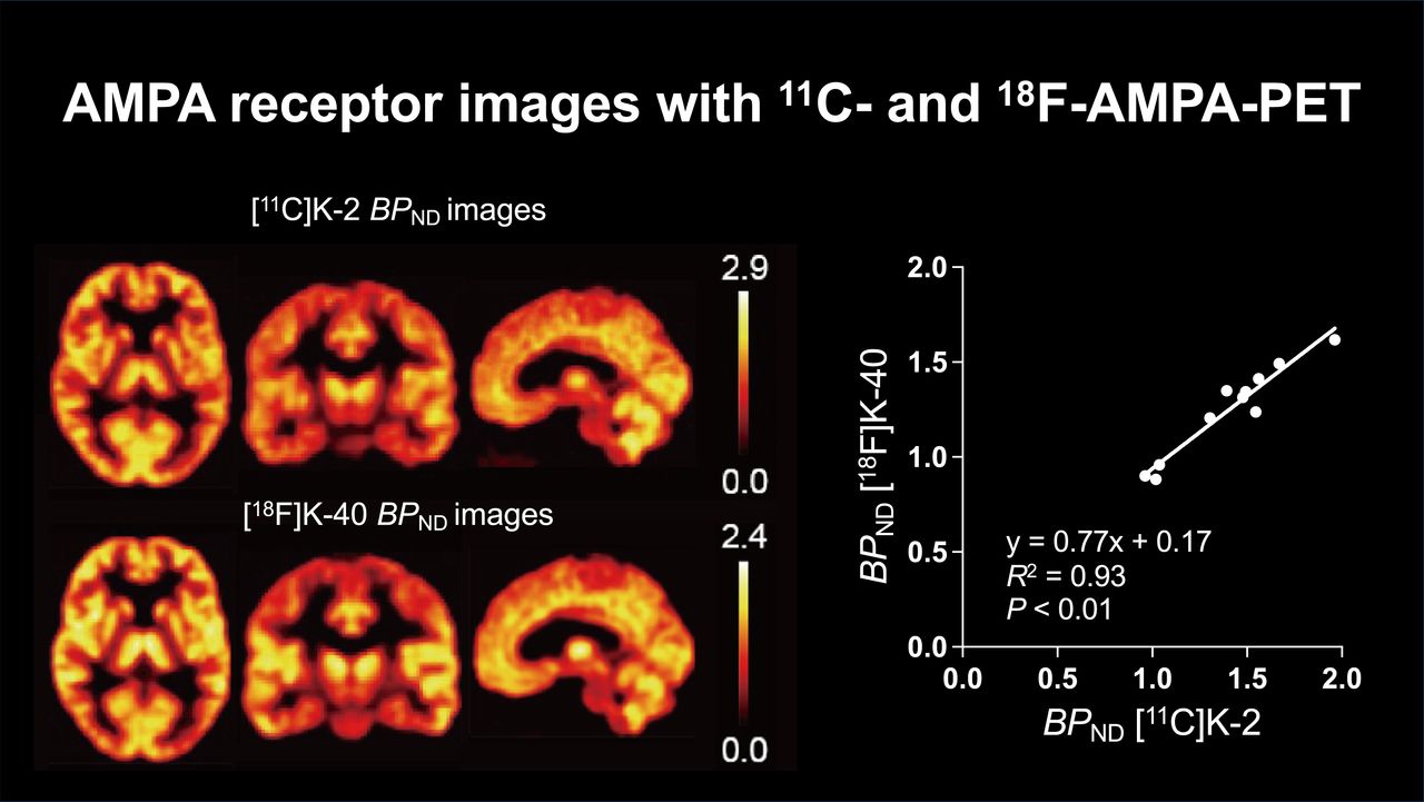
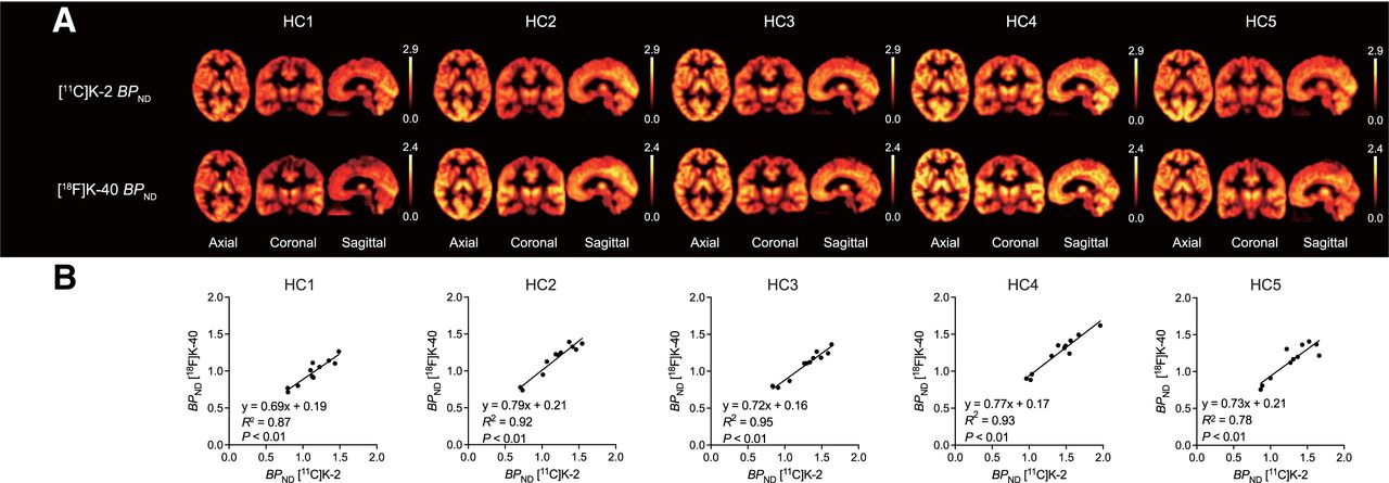
  • FIGURE 5.
    • Download figure
    • Open in new tab
    • Download powerpoint
    FIGURE 5.

    (A) Comparison between [11C]K-2 BPND PET images and [18F]K-40 BPND PET images. (B) Correlation between [11C]K-2 and [18F]K-40 BPND values in brain regions in healthy subjects. HC = healthy control.
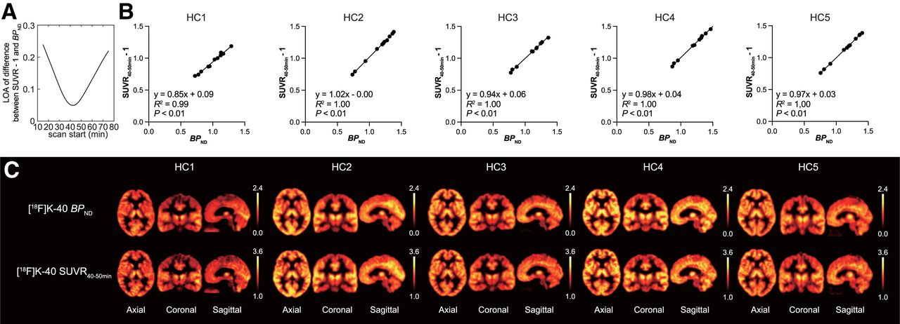
  • FIGURE 6.
    • Download figure
    • Open in new tab
    • Download powerpoint
    FIGURE 6.

    (A) Change in limit of agreement on difference in BPND and SUVR with varying scan start time wherein frame width is fixed at 10 min. (B) Correlation between BPND value and SUVR40–50min−1 in brain regions in healthy subjects. (C) Comparison between [18F]K-40 BPND PET images and [18F]K-40 SUVR PET images at 40–50 min.

Tables

  • Figures
  • Additional Files
    • View popup
    TABLE 1.

    Specification of [18F]K-40

    TestSpecification
    1. Quantity
     1.1 Volume of one batch18–22 mL
     1.2 RadioactivityNLT 1.85 GBq
     1.3 K-40 concentrationNMT 10 μg/mL
     1.4 Specific radioactivityNLT 37 GBq/μmol on EOS
     1.5 Half-lifeNLT 105 min; NMT 115 min
    2. Property
     2.1 AppearanceColorless to light yellow solution
     2.2 ParticleFree of visible particulate matter
    3. Bacterial endotoxinNMT 150 EU/whole solution
    4. SterilityNo growth
    5. pHNLT 5.0; NMT 8.0
    6. Qualitative analysis
     6.1 γ-spectrum*511-keV peak detected
     6.2 Radiochemical identityRadiometric RT ratio of [18F]K-40 to K-40 standard: 1.0 ± 0.1
    7. Purity
     7.1 Other radioisotope*No peak except 511 keV and 1,022 keV
     7.2 Radiochemical purityNMT 95%
    8. Residual solvent
     8.1 MethanolNLT 3,000 ppm
     8.2 AcetonitrileNLT 410 ppm
     8.3 DMSONLT 5,000 ppm
    9. Assay of ethanolNLT 6%; NMT 10%
    • ↵* All tests are required for every production (6.1 and 7.1 are required once/year).

    • NLT = not less than; NMT = not more than; EOS = end of synthesis; RT = retention time; DMSO = dimethylsulfoxide.

    • View popup
    TABLE 2.

    Specification of [11C]K-2

    TestSpecification
    1. Quantity
     1.1 Volume of one batch13–17 mL
     1.2 RadioactivityNLT 1.85 GBq
     1.3 K-2 concentrationNMT 10 μg/mL
     1.4 Specific radioactivityNLT 37 GBq/μmol on EOS
     1.5 Half-lifeNLT 19.0 min; NMT 21.0 min
    2. Property
     2.1 AppearanceColorless to light yellow solution
     2.2 ParticleFree of visible particulate matter
    3. Bacterial endotoxinNMT 150 EU/whole solution
    4. SterilityNo growth
    5. pHNLT 5.0; NMT 8.0
    6. Qualitative analysis
     6.1 γ-spectrum*511-keV peak detected
     6.2 Radiochemical identityRadiometric RT ratio of [11C]K-2 to K-2 standard: 1.0 ± 0.1
    7. Purity
     7.1 Other radioisotope*No peak except 511 keV and 1,022 keV
     7.2 Radiochemical purityNMT 95%
    8. Residual solvent
     8.1 AcetonitrileNLT 410 ppm
     8.2 DMFNLT 880 ppm
    9. Assay of ethanolNLT 3%, NMT 6%
    • ↵* All tests are required for every production (6.1 and 7.1 are required once/year).

    • NLT = not less than; NMT = not more than; EOS = end of synthesis; RT = retention time; DMF = dimethylformamide.

Additional Files

  • Figures
  • Tables
  • Supplemental Data

    Files in this Data Supplement:

    • Supplemental Data
PreviousNext
Back to top

In this issue

Journal of Nuclear Medicine: 66 (6)
Journal of Nuclear Medicine
Vol. 66, Issue 6
June 1, 2025
  • Table of Contents
  • Table of Contents (PDF)
  • About the Cover
  • Index by author
  • Complete Issue (PDF)
Print
Download PDF
Article Alerts
Sign In to Email Alerts with your Email Address
Email Article

Thank you for your interest in spreading the word on Journal of Nuclear Medicine.

NOTE: We only request your email address so that the person you are recommending the page to knows that you wanted them to see it, and that it is not junk mail. We do not capture any email address.

Enter multiple addresses on separate lines or separate them with commas.
First-in-Human Study of 18F-Labeled PET Tracer for Glutamate AMPA Receptor [18F]K-40: A Derivative of [11C]K-2
(Your Name) has sent you a message from Journal of Nuclear Medicine
(Your Name) thought you would like to see the Journal of Nuclear Medicine web site.
Citation Tools
First-in-Human Study of 18F-Labeled PET Tracer for Glutamate AMPA Receptor [18F]K-40: A Derivative of [11C]K-2
Sadamitsu Ichijo, Tetsu Arisawa, Mai Hatano, Waki Nakajima, Tomoyuki Miyazaki, Tsuyoshi Eiro, Yuuki Takada, Ryunosuke Iai, Akane Sano, Masaki Sonoda, Yutaro Takayama, Yuichi Kimura, Takuya Takahashi
Journal of Nuclear Medicine May 2025, jnumed.124.269405; DOI: 10.2967/jnumed.124.269405

Citation Manager Formats

  • BibTeX
  • Bookends
  • EasyBib
  • EndNote (tagged)
  • EndNote 8 (xml)
  • Medlars
  • Mendeley
  • Papers
  • RefWorks Tagged
  • Ref Manager
  • RIS
  • Zotero
Share
First-in-Human Study of 18F-Labeled PET Tracer for Glutamate AMPA Receptor [18F]K-40: A Derivative of [11C]K-2
Sadamitsu Ichijo, Tetsu Arisawa, Mai Hatano, Waki Nakajima, Tomoyuki Miyazaki, Tsuyoshi Eiro, Yuuki Takada, Ryunosuke Iai, Akane Sano, Masaki Sonoda, Yutaro Takayama, Yuichi Kimura, Takuya Takahashi
Journal of Nuclear Medicine May 2025, jnumed.124.269405; DOI: 10.2967/jnumed.124.269405
Twitter logo Facebook logo LinkedIn logo Mendeley logo
  • Tweet Widget
  • Facebook Like
  • Google Plus One
Bookmark this article

Jump to section

  • Article
    • Visual Abstract
    • Abstract
    • MATERIALS AND METHODS
    • RESULTS
    • DISCUSSION
    • CONCLUSION
    • DISCLOSURE
    • Footnotes
    • REFERENCES
  • Figures & Data
  • Supplemental
  • Info & Metrics
  • PDF

Related Articles

  • PubMed
  • Google Scholar

Cited By...

  • No citing articles found.
  • Google Scholar

More in this TOC Section

  • Feasibility of Ultra-Low-Activity 18F-FDG PET/CT Imaging Using a Long–Axial-Field-of-View PET/CT System
  • Is Long–Axial-Field-of-View PET/CT Cost-Effective? An International Health–Economic Analysis
  • Multicycle Dosimetric Behavior and Dose–Effect Relationships in [177Lu]Lu-DOTATATE Peptide Receptor Radionuclide Therapy
Show more Clinical Investigation

Similar Articles

Keywords

  • PET
  • AMPA receptor
  • neuroimaging
  • first-in-human study
SNMMI

© 2025 SNMMI

Powered by HighWire