
Main menu

  • Home
  • Content
    • Current
    • Ahead of print
    • Past Issues
    • JNM Supplement
    • SNMMI Annual Meeting Abstracts
    • Continuing Education
    • JNM Podcasts
  • Subscriptions
    • Subscribers
    • Institutional and Non-member
    • Rates
    • Journal Claims
    • Corporate & Special Sales
  • Authors
    • Submit to JNM
    • Information for Authors
    • Assignment of Copyright
    • AQARA requirements
  • Info
    • Reviewers
    • Permissions
    • Advertisers
  • About
    • About Us
    • Editorial Board
    • Contact Information
  • More
    • Alerts
    • Feedback
    • Help
    • SNMMI Journals
  • SNMMI
    • JNM
    • JNMT
    • SNMMI Journals
    • SNMMI

User menu

  • Subscribe
  • My alerts
  • Log in
  • My Cart

Search

  • Advanced search
Journal of Nuclear Medicine
  • SNMMI
    • JNM
    • JNMT
    • SNMMI Journals
    • SNMMI
  • Subscribe
  • My alerts
  • Log in
  • My Cart
Journal of Nuclear Medicine

Advanced Search

  • Home
  • Content
    • Current
    • Ahead of print
    • Past Issues
    • JNM Supplement
    • SNMMI Annual Meeting Abstracts
    • Continuing Education
    • JNM Podcasts
  • Subscriptions
    • Subscribers
    • Institutional and Non-member
    • Rates
    • Journal Claims
    • Corporate & Special Sales
  • Authors
    • Submit to JNM
    • Information for Authors
    • Assignment of Copyright
    • AQARA requirements
  • Info
    • Reviewers
    • Permissions
    • Advertisers
  • About
    • About Us
    • Editorial Board
    • Contact Information
  • More
    • Alerts
    • Feedback
    • Help
    • SNMMI Journals
  • View or Listen to JNM Podcast
  • Visit JNM on Facebook
  • Join JNM on LinkedIn
  • Follow JNM on Twitter
  • Subscribe to our RSS feeds
Research ArticleTranslational Investigation
Open Access

Quantitative Total-Body Imaging of Blood Flow with High-Temporal-Resolution Early Dynamic 18F-FDG PET Kinetic Modeling

Kevin J. Chung, Abhijit J. Chaudhari, Lorenzo Nardo, Terry Jones, Moon S. Chen, Ramsey D. Badawi, Simon R. Cherry and Guobao Wang
Journal of Nuclear Medicine May 2025, jnumed.124.268706; DOI: https://doi.org/10.2967/jnumed.124.268706
Kevin J. Chung
1Department of Radiology, University of California Davis Health, Sacramento, California;
  • Find this author on Google Scholar
  • Find this author on PubMed
  • Search for this author on this site
Abhijit J. Chaudhari
1Department of Radiology, University of California Davis Health, Sacramento, California;
  • Find this author on Google Scholar
  • Find this author on PubMed
  • Search for this author on this site
Lorenzo Nardo
1Department of Radiology, University of California Davis Health, Sacramento, California;
  • Find this author on Google Scholar
  • Find this author on PubMed
  • Search for this author on this site
Terry Jones
1Department of Radiology, University of California Davis Health, Sacramento, California;
  • Find this author on Google Scholar
  • Find this author on PubMed
  • Search for this author on this site
Moon S. Chen Jr.
2Department of Internal Medicine, University of California Davis Health, Sacramento, California; and
  • Find this author on Google Scholar
  • Find this author on PubMed
  • Search for this author on this site
Ramsey D. Badawi
1Department of Radiology, University of California Davis Health, Sacramento, California;
3Department of Biomedical Engineering, University of California at Davis, Davis, California
  • Find this author on Google Scholar
  • Find this author on PubMed
  • Search for this author on this site
Simon R. Cherry
1Department of Radiology, University of California Davis Health, Sacramento, California;
3Department of Biomedical Engineering, University of California at Davis, Davis, California
  • Find this author on Google Scholar
  • Find this author on PubMed
  • Search for this author on this site
Guobao Wang
1Department of Radiology, University of California Davis Health, Sacramento, California;
  • Find this author on Google Scholar
  • Find this author on PubMed
  • Search for this author on this site
  • Article
  • Figures & Data
  • Supplemental
  • Info & Metrics
  • PDF
Loading

Article Figures & Data

Figures

  • Tables
  • Additional Files
  • Figure
    • Download figure
    • Open in new tab
    • Download powerpoint
  • FIGURE 1.
    • Download figure
    • Open in new tab
    • Download powerpoint
    FIGURE 1.

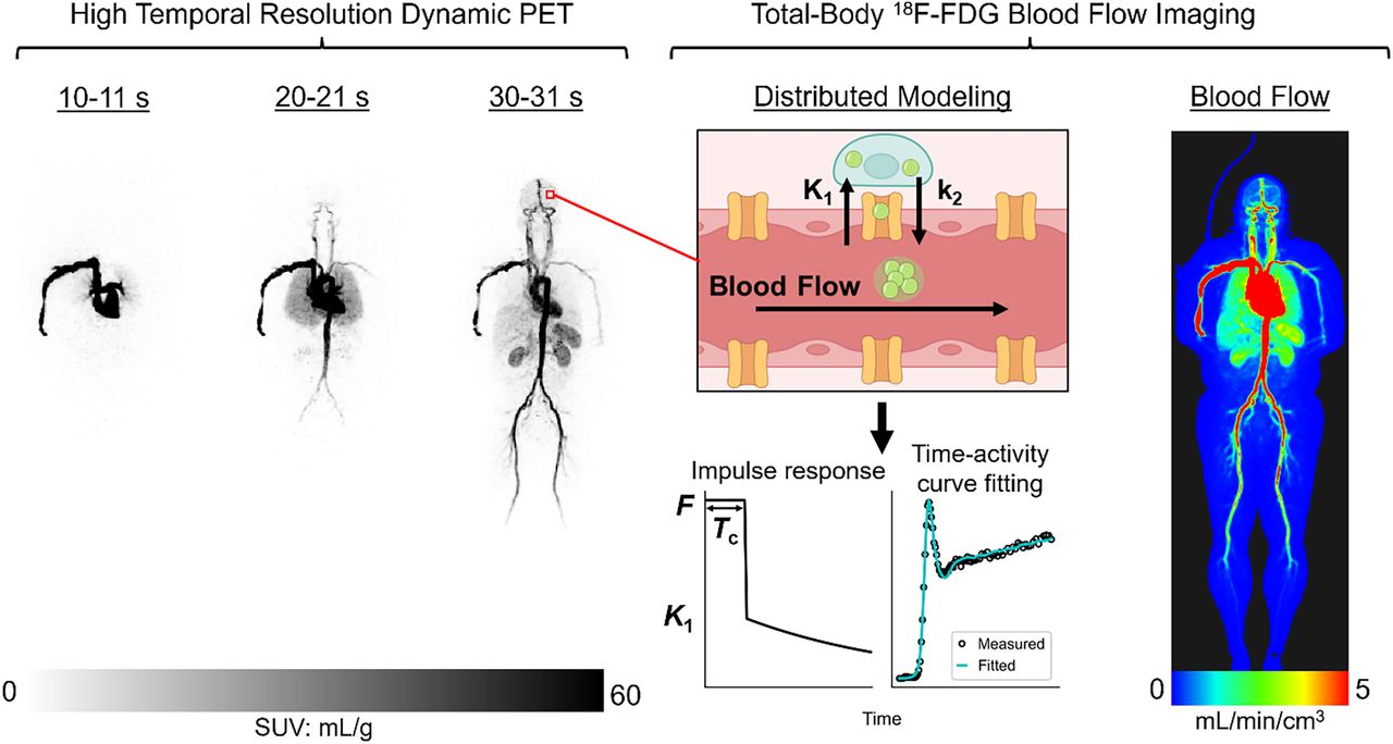
    Modeling intravascular delivery of PET tracers. (A) PET voxel partly comprises arteries, arterioles, capillaries, venules, and veins. PET tracers are initially delivered to and circulate through these vascular volumes via blood flow (F). Tracer transport from blood into (K1) and out of tissue (k2) occurs almost exclusively at capillary level. S1TC model (B) assumes that tracer instantaneously mixes in vascular volume, and effectively mean vascular transit time (Tc) is zero. AATH model (C) accounts for plug flow with single transit time (Tc) for tracer to traverse total vascular volume via blood flow.

  • FIGURE 2.
    • Download figure
    • Open in new tab
    • Download powerpoint
    FIGURE 2.

    Time–activity curve fits in cortical gray matter using S1TC model (A) and AATH model (B) at HTR (60 × 1 s/frame, 30 × 2 s/frame). Dashed red and green lines represent intravascular and extravascular components of fitted curve, respectively. Black arrows indicate areas where S1TC fitting was poor.

  • FIGURE 3.
    • Download figure
    • Open in new tab
    • Download powerpoint
    FIGURE 3.

    Difference in AIC between AATH and S1TC models using original HTR data (60 × 1 s/frame, 30 × 2 s/frame) (A) and at different simulated frame intervals (B). Negative AIC indicates preference toward AATH model. GM = gray matter.

  • FIGURE 4.
    • Download figure
    • Open in new tab
    • Download powerpoint
    FIGURE 4.

    Correlation (left) and Bland–Altman (right) plots comparing 11C-butanol blood flow against 18F-FDG blood flow with AATH model (A) and S1TC model (B) K1 in same participants. GM = gray matter; MD = mean difference.

  • FIGURE 5.
    • Download figure
    • Open in new tab
    • Download powerpoint
    FIGURE 5.

    Total-body parametric imaging of blood flow with early dynamic 18F-FDG method compared with 11C-butanol flow-tracer PET reference in same participant. White arrows indicate sagittal and transverse sinuses in brain.

  • FIGURE 6.
    • Download figure
    • Open in new tab
    • Download powerpoint
    FIGURE 6.

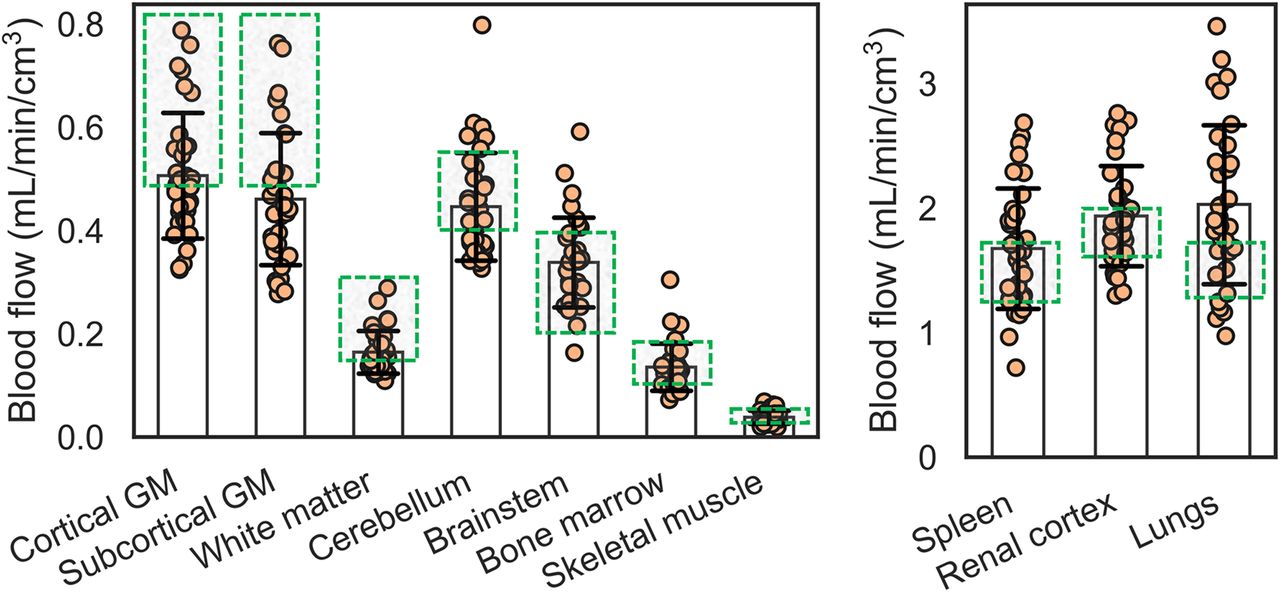
    Regional blood flow in 34 healthy participants estimated with early dynamic 18F-FDG method. Plots are separated by range of blood-flow values. Average estimates mostly fell within range of average blood-flow values reported in literature (Supplemental Table 1), indicated by green boxes. GM = gray matter.

Tables

  • Figures
  • Additional Files
    • View popup
    TABLE 1

    18F-FDG Early Kinetics Across Healthy Participants with AATH Model (n = 34)

    Tissue regionBlood flow (mL/min/cm3)K1 (mL/min/cm3)Extraction fractionvb (mL/cm3)Tc (s)
    Cortical GM0.507 ± 0.1220.136 ± 0.0180.278 ± 0.0460.036 ± 0.0064.4 ± 0.9
    White matter0.165 ± 0.0410.066 ± 0.0090.416 ± 0.0610.018 ± 0.0036.9 ± 1.4
    Subcortical GM0.461 ± 0.1280.143 ± 0.0190.327 ± 0.0690.033 ± 0.0054.6 ± 1.5
    Brain stem0.339 ± 0.0870.125 ± 0.0150.386 ± 0.0820.030 ± 0.0055.6 ± 1.8
    Cerebellum0.447 ± 0.1040.145 ± 0.0160.336 ± 0.0520.037 ± 0.0055.1 ± 1.0
    Spleen1.676 ± 0.4841.204 ± 0.4040.728 ± 0.1510.166 ± 0.0646.5 ± 3.3
    Renal cortex1.938 ± 0.4020.657 ± 0.0910.348 ± 0.0650.318 ± 0.03910.1 ± 1.7
    Skeletal muscle0.039 ± 0.0130.034 ± 0.0120.890 ± 0.0480.017 ± 0.00429.1 ± 8.3
    Bone marrow0.136 ± 0.0460.130 ± 0.0460.954 ± 0.0510.053 ± 0.01824.6 ± 9.0
    Lungs2.031 ± 0.6390.072 (0.059–0.134)*0.041 (0.033–0.067)*0.143 ± 0.0314.4 ± 0.8
    • ↵* Values are expressed as median with interquartile range in parentheses as a result of 2 measurements shifting distribution (Supplemental Fig. 7).

    • AATH = adiabatic approximation to tissue homogeneity; GM = gray matter.

    • Data are expressed as mean ± SD unless otherwise noted.

Additional Files

  • Figures
  • Tables
  • Supplemental Data

    Files in this Data Supplement:

    • Supplemental Data
  • Supplemental Data

    Files in this Data Supplement:

    • Supplemental Data
PreviousNext
Back to top

In this issue

Journal of Nuclear Medicine: 66 (5)
Journal of Nuclear Medicine
Vol. 66, Issue 5
May 1, 2025
  • Table of Contents
  • Table of Contents (PDF)
  • About the Cover
  • Index by author
  • Complete Issue (PDF)
Print
Download PDF
Article Alerts
Sign In to Email Alerts with your Email Address
Email Article

Thank you for your interest in spreading the word on Journal of Nuclear Medicine.

NOTE: We only request your email address so that the person you are recommending the page to knows that you wanted them to see it, and that it is not junk mail. We do not capture any email address.

Enter multiple addresses on separate lines or separate them with commas.
Quantitative Total-Body Imaging of Blood Flow with High-Temporal-Resolution Early Dynamic 18F-FDG PET Kinetic Modeling
(Your Name) has sent you a message from Journal of Nuclear Medicine
(Your Name) thought you would like to see the Journal of Nuclear Medicine web site.
Citation Tools
Quantitative Total-Body Imaging of Blood Flow with High-Temporal-Resolution Early Dynamic 18F-FDG PET Kinetic Modeling
Kevin J. Chung, Abhijit J. Chaudhari, Lorenzo Nardo, Terry Jones, Moon S. Chen, Ramsey D. Badawi, Simon R. Cherry, Guobao Wang
Journal of Nuclear Medicine May 2025, jnumed.124.268706; DOI: 10.2967/jnumed.124.268706

Citation Manager Formats

  • BibTeX
  • Bookends
  • EasyBib
  • EndNote (tagged)
  • EndNote 8 (xml)
  • Medlars
  • Mendeley
  • Papers
  • RefWorks Tagged
  • Ref Manager
  • RIS
  • Zotero
Share
Quantitative Total-Body Imaging of Blood Flow with High-Temporal-Resolution Early Dynamic 18F-FDG PET Kinetic Modeling
Kevin J. Chung, Abhijit J. Chaudhari, Lorenzo Nardo, Terry Jones, Moon S. Chen, Ramsey D. Badawi, Simon R. Cherry, Guobao Wang
Journal of Nuclear Medicine May 2025, jnumed.124.268706; DOI: 10.2967/jnumed.124.268706
Twitter logo Facebook logo LinkedIn logo Mendeley logo
  • Tweet Widget
  • Facebook Like
  • Google Plus One
Bookmark this article

Jump to section

  • Article
    • Visual Abstract
    • Abstract
    • MATERIALS AND METHODS
    • RESULTS
    • DISCUSSION
    • CONCLUSION
    • DISCLOSURE
    • ACKNOWLEDGEMENTS
    • Footnotes
    • REFERENCES
  • Figures & Data
  • Supplemental
  • Info & Metrics
  • PDF

Related Articles

  • PubMed
  • Google Scholar

Cited By...

  • No citing articles found.
  • Google Scholar

Similar Articles

Keywords

  • total-body PET
  • blood-flow/perfusion imaging
  • high-temporal resolution dynamic imaging
  • tracer kinetic modeling
  • distributed kinetic modeling
SNMMI

© 2025 SNMMI

Powered by HighWire