


Article Figures & Data
Figures
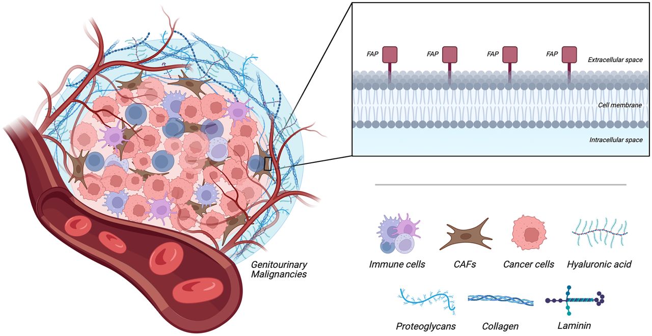
- FIGURE 1.
Graphic illustration of tumor microenvironment with cancer-associated fibroblasts (CAFs) and their overexpression of FAP. (Created with BioRender.com.)
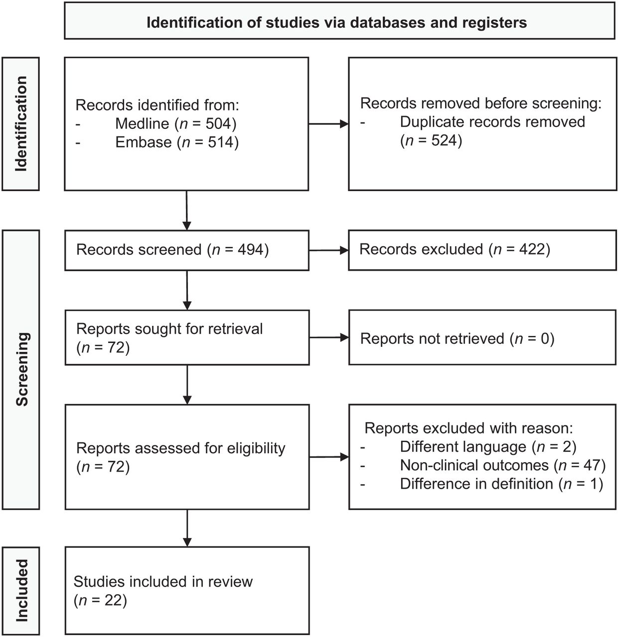
- FIGURE 2.
PRISMA flow diagram showing outcome of searches and selection of full studies included in review.


Tables
Malignancy Study design Patients (n) Type of histology Molecular imaging findings Disease stage Setting Radiotracer Disease evaluation PCa Kratochwil et al. (19) CS 16 Neuroendocrine carcinoma and adenocarcinoma NA NA 68Ga-FAPI-04 No comparative MI modality was performed; FAPI PET/CT was valuable in several highly prevalent cancers, with excellent tumor visualization in primary and metastatic lesions Cai et al. (23) CR 1 Mixed large-cell neuroendocrine carcinoma-acinar adenocarcinoma Metastatic Staging 68Ga-FAPI-46 With more metastatic lesions detected, FAPI PET/CT was superior to PSMA PET/CT; 18F-FDG PET/CT was superior to both FAPI and PSMA PET/CT, as additional metastatic liver lesions were visualized Pang et al. (26) CR 1 Adenocarcinoma Metastatic Staging 68Ga-FAPI-04 With equal metastatic lesions detected, FAPI PET/CT was comparable to PSMA and 18F-FDG PET/CT Tatar et al. (28) CR 1 Signet ringlike cell carcinoma Metastatic Staging 68Ga-FAPI-04 No comparative MI modality was performed; FAPI PET/CT was valuable, with excellent tumor visualization in primary and metastatic lesions Tatar et al. (29) CR 1 Adenocarcinoma Localized Restaging 68Ga-FAPI-04 With no metastatic lesions detected, FAPI PET/CT was comparable to PSMA and 18F-FDG PET/CT; primary lesion was more clearly visualized on 18F-FDG PET/CT Xu et al. (31) CR 1 Adenocarcinoma Unknown* Staging 68Ga-FAPI-04 With equal (metastatic) lesions detected, FAPI PET/CT was comparable to 18F-FDG PET/CT Yang et al. (32) CR 1 Solitary fibrous tumor Localized Staging 68Ga-FAPI-04 With no metastatic lesions detected, FAPI PET/CT was comparable to 18F-FDG PET/CT; primary lesion was more clearly visualized on 18F-FDG PET/CT Aryana et al. (34) CR 1 Adenocarcinoma Metastatic Restaging 68Ga-FAPI-46 With fewer metastatic lesions detected, FAPI PET/CT was inferior to PSMA PET/CT; metastatic lesions were more clearly visualized Kesch et al. (37) CR 1 Adenocarcinoma Metastatic Restaging 68Ga-FAPI-04 No comparative MI modality was performed; FAPI PET/CT was valuable, with excellent tumor visualization in metastatic lesions Kessel et al. (40) CS 6 Neuroendocrine carcinoma and adenocarcinoma Metastatic Restaging 68Ga-FAPI-46 With both more and less metastatic lesions detected, FAPI PET/CT was neither superior nor inferior to 18F-FDG PET/CT Isik et al. (41) CR 2 Adenocarcinoma Metastatic Restaging 68Ga-FAPI-46 With more metastatic lesions detected, FAPI PET/CT was superior to 18F-FDG PET/CT; PSMA PET/CT detected more metastatic lesions in one patient yet less in other patient Khreish et al. (42) CR 1 Adenocarcinoma Metastatic Restaging 68Ga-FAPI-04 With more metastatic lesions detected, FAPI PET/CT was superior to 18F-FDG and PSMA PET/CT Urothelial carcinoma of bladder/upper urinary tract Novruzov et al. (25) CS 8 Urothelial carcinoma Metastatic Staging and restaging 68Ga-FAPI-04 and -46 With more metastatic lesions detected, FAPI PET/CT was superior to 18F-FDG PET/CT Unterrainer et al. (30) CS 15 Urothelial carcinoma Metastatic Staging and restaging 68Ga-FAPI-46 No comparative MI modality was performed. However, FAPI PET/CT was valuable, with excellent tumor visualization in primary and metastatic lesions Dendl et al. (33) CS 4 Urothelial carcinoma Localized and metastatic Staging and restaging 68Ga-FAPI-04, -46, and -74 No comparative MI modality was performed; FAPI PET/CT was valuable, with excellent tumor visualization in primary and metastatic lesions Viergever et al. (43) CS 2 Urothelial carcinoma Metastatic Staging 68Ga-FAPI-04 With more metastatic lesions detected, FAPI PET/CT was superior to 18F-FDG PET/CT RCC Kratochwil et al. (19) CS 1 RCC NA NA 68Ga-FAPI-04 No comparative MI modality was performed; FAPI PET/CT was valuable in several highly prevalent cancers, with excellent tumor visualization in primary and metastatic lesions Dong et al. (24) CR 1 Sarcomatoid RCC Metastatic Staging 68Ga-FAPI-04 No comparative MI modality was performed; FAPI PET/CT was valuable, with excellent tumor visualization in primary and metastatic lesions Pang et al. (27) CR 1 Chromophobe RCC Metastatic Staging 68Ga-FAPI-04 With more metastatic lesions detected, FAPI PET/CT was superior to 18F-FDG PET/CT; in contrast to 18F-FDG PET/CT, primary lesion was visualized on FAPI PET/CT Civan et al. (35) CR 1 Papillary RCC Metastatic Restaging 68Ga-FAPI-04 With equal metastatic lesions detected, FAPI PET/CT was comparable to 18F-FDG PET/CT Xie et al. (38) CR 1 Chromophobe RCC Metastatic Restaging 68Ga-FAPI-04 With more metastatic lesions detected, FAPI PET/CT was superior to 18F-FDG PSMA PET/CT Yang et al. (39) CR 1 Clear cell RCC Metastatic Restaging 68Ga-FAPI-04 No comparative MI modality was performed; FAPI PET/CT was valuable, with excellent tumor visualization in primary and metastatic lesions Testicular cancer: Kaplan et al. (36) CR 1 Mixed germ cell tumor Metastatic Restaging 68Ga-FAPI-04 With equal metastatic lesions detected, FAPI PET/CT was superior to 18F-FDG PET/CT; metastatic lesions were more clearly visualized on FAPI PET/CT ↵* Abnormal uptake was observed in intracranial lesion, though metastatic disease was undetermined.
CS = case series; NA = data not available; CR = case report; MI = molecular imaging.
Extracted data of included studies on methodology, patient population, and molecular imaging findings.
Supplemental Data
Files in this Data Supplement:
In this issue


Jump to section
Related Articles
Cited By...
- No citing articles found.