


Article Figures & Data
Figures
- FIGURE 1.
Phantoms used to derive dose coefficients compared in this work. Solid arrows denote comparisons using different phantoms and different software. Dashed arrows represent comparisons using same phantoms but different software. DC = dose coefficient; ORNL = Oak Ridge National Laboratory; wT = tissue-weighting factor.
- FIGURE 2.
Organ-level absorbed dose coefficients for adult male compared via log relative differences (Eq. 13). (Top) ICRP publication 128 compared against MIRDcalc. (Middle) IDAC-Dose compared against MIRDcalc. (Bottom) OLINDA 2.1 compared against MIRDcalc. Red indicates a dose coefficient estimate higher than that of MIRDcalc; blue indicates lower. Black indicates off-scale values (
 > 300).
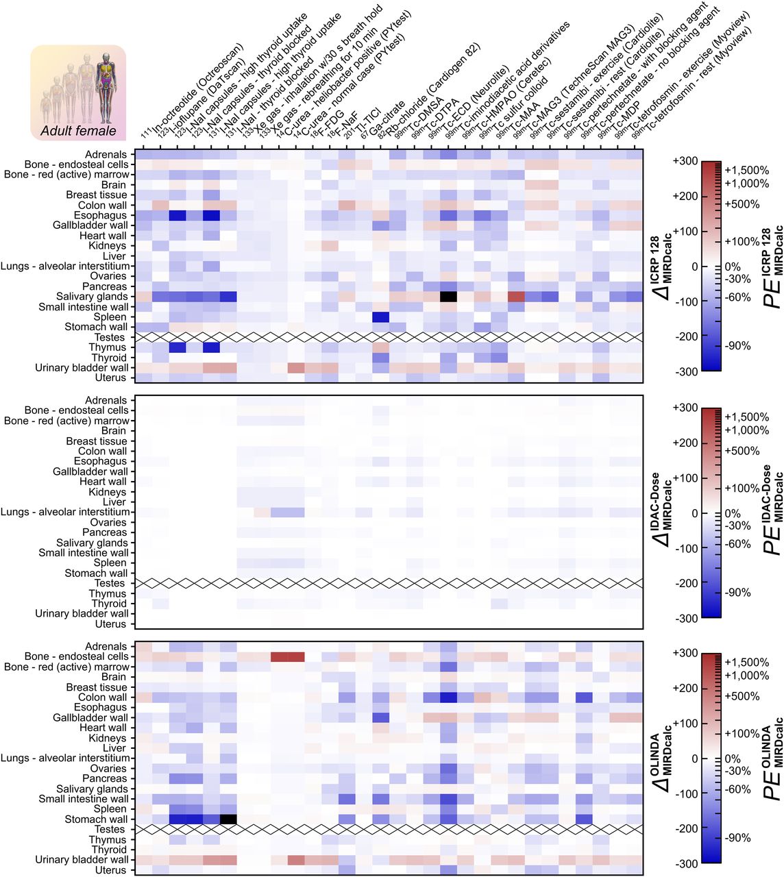
 - FIGURE 3.
Organ-level absorbed dose coefficients for adult female compared via log relative differences (Eq. 13). (Top) ICRP publication 128 compared against MIRDcalc. (Middle) IDAC-Dose compared against MIRDcalc. (Bottom) OLINDA compared against MIRDcalc. Red indicates a dose coefficient estimate higher than that of MIRDcalc; blue indicates lower. Black indicates off-scale values (
> 300). - FIGURE 4.
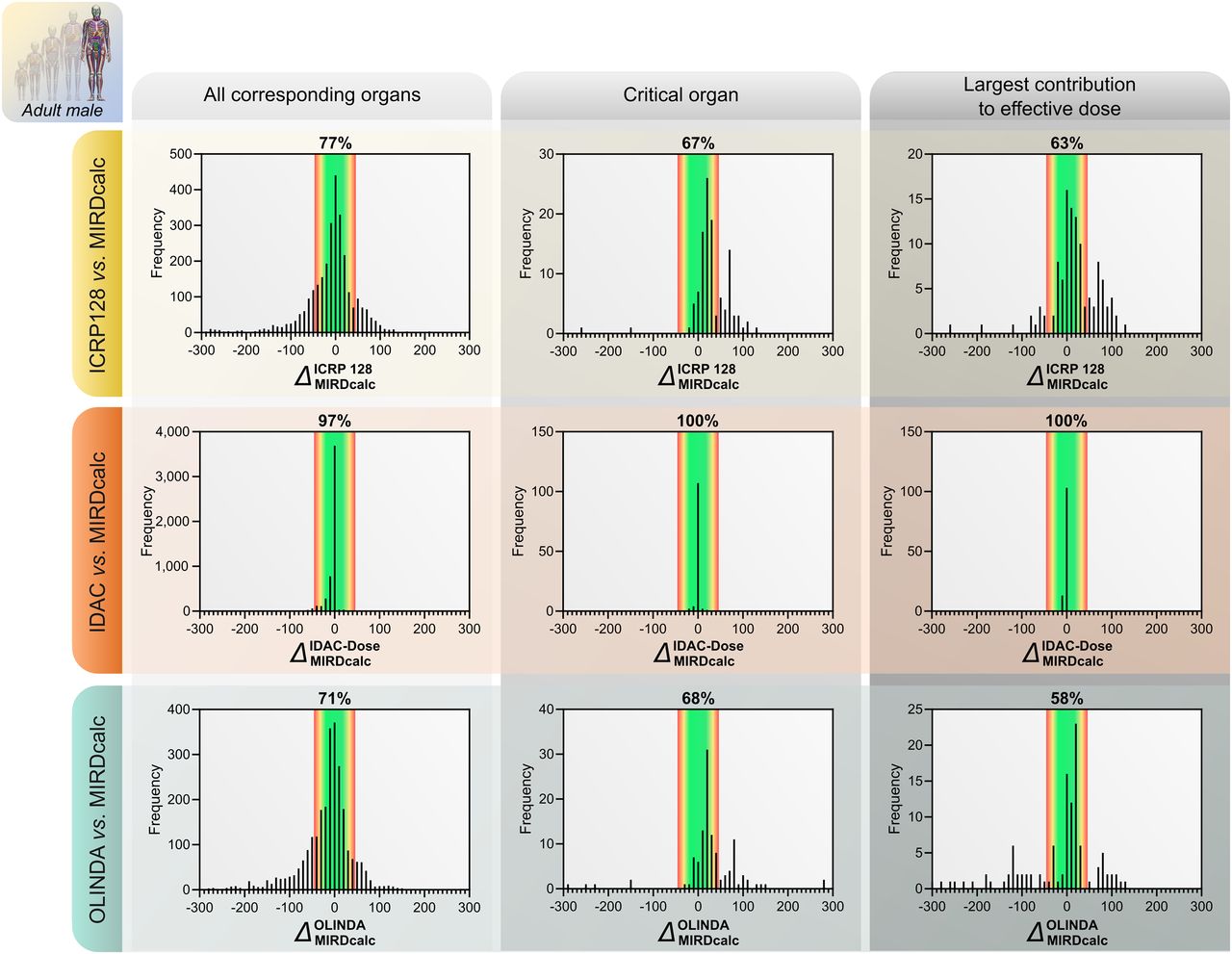
Distribution of log relative differences in organ-level absorbed dose coefficients for adult male phantoms. (Top) ICRP publication 128 compared against MIRDcalc. (Middle) IDAC-Dose compared against MIRDcalc. (Bottom) OLINDA compared against MIRDcalc. Red–green shaded region represents range of reasonable agreement discussed in text; percentage value overlying this region indicates fraction of Δ-values that fall within this range. Histogram bin width is 10.
- FIGURE 5.
Distribution of log relative differences in organ-level absorbed dose coefficients for adult female phantoms. (Top) ICRP publication 128 compared against MIRDcalc. Upper left histogram is negatively skewed because ICRP publication 128 dose estimates are derived from single 70-kg hermaphroditic adult phantom, whereas MIRDcalc, IDAC-Dose, and OLINDA use a 60-kg female phantom. (Middle) IDAC-Dose compared against MIRDcalc. (Bottom) OLINDA compared against MIRDcalc. Red-green shaded region represents range of reasonable agreement discussed in text; percentage value overlying this region indicates fraction of Δ-values that fall within this range. Histogram bin width is 10.


Tables
- TABLE 1.
Adult Effective Dose Coefficients and Relative Differences Compared Among MIRDcalc, IDAC-Dose, and ICRP Publication 128
Radiopharmaceutical Effective dose coefficient (mSv/MBq) MIRDcalc IDAC-Dose OLINDA ICRP 128 111In-octreotide 5.49E−02 5.38E−02 5.89E−02 5.40E−02 −2.0 −2.0% 7.0 7.3% −1.7 −1.6% 123I-ioflupane 2.50E−02 2.36E−02 2.16E−02 2.50E−02 −5.8 −5.6% −15 −14% 0.00 0.00% 123I-NaI capsules (high thyroid uptake) 2.45E−01 2.45E−01 2.77E−01 3.00E−01 0.00 0.00% 12 13% 20 22% 123I-NaI capsules (thyroid blocked) 3.04E−02 3.02E−02 2.04E−02 3.70E−02 −0.66 −0.7% −40 −33% 20 22% 131I-NaI capsules (high thyroid uptake) 2.17E+01 2.16E+01 2.64E+01 2.90E+01 −0.46 −0.50% 20 22% 29 34% 131I-NaI (thyroid blocked) 2.10E−01 2.08E−01 1.55E−01 2.80E−01 −0.96 −1.0% −30 −26% 29 33% 133Xe gas 1.99E−04 1.64E−04 1.86E−04 1.80E−04 −19 −18% −6.8 −6.5% −10 −9.5% 133Xe gas (rebreathing for 10 min) 1.27E−03 1.21E−03 1.11E−03 1.10E−03 −4.8 −4.7% −13 −13% −14 −13% 14C-urea, Heliobacter positive 8.68E−02 7.75E−02 8.93E−02 8.10E−02 −11 −11% 2.8 2.9% −6.9 −6.7% 14C-urea, normal case 2.42E−02 2.16E−02 2.93E−02 3.10E−02 −11 −11% 19 21% 25 28% 18F-FDG 1.67E−02 1.61E−02 1.92E−02 1.90E−02 −3.7 −3.6% 14 15% 13 14% 18F-NaF 1.30E−02 1.28E−02 1.65E−02 1.70E−02 −1.6 −1.5% 24 27% 27 31% 201Tl-TlCl 1.13E−01 1.09E−01 8.47E−02 1.40E−01 −3.6 −3.5% −29 −25% 21 24% 67Ga-citrate 9.43E−02 8.97E−02 8.72E−02 1.00E−01 −5.0 −4.9% −7.8 −7.5% 5.9 6.0% 82Rb-chloride 1.05E−03 1.00E−03 7.77E−04 1.10E−03 −4.9 −4.8% −30 −26% 4.7 4.8% 99mTc-DMSA 7.02E−03 6.92E−03 7.24E−03 8.80E−03 −1.4 −1.4% 3.1 3.1% 23 25% 99mTc-DTPA 3.37E−03 3.26E−03 4.57E−03 4.90E−03 −3.3 −3.3% 30 36% 37 45% 99mTc-ECD 5.56E−03 5.52E−03 5.08E−03 7.70E−03 −0.72 −0.70% −9.0 −8.6% 33 39% 99mTc-iminodiacetic acid derivatives 9.44E−03 9.55E−03 3.84E−03 1.60E−02 1.2 1.2% −90 −59% 53 70% 99mTc-macroaggregated albumin 1.40E−02 1.15E−02 1.37E−02 1.10E−02 −20 −18% −2.2 −2.1% −24 −21% 99mTc-MAG3 4.12E−03 4.09E−03 6.38E−03 7.00E−03 −0.73 −0.70% 44 55% 53 70% 99mTc-sestamibi, exercise 6.19E−03 6.03E−03 3.74E−03 7.90E−03 −2.6 −2.6% −50 −40% 24 28% 99mTc-sestamibi, rest 7.09E−03 6.89E−03 4.64E−03 9.00E−03 −2.9 −2.8% −42 −35% 24 27% 99mTc-HMPAO 8.35E−03 7.99E−03 7.10E−03 9.30E−03 −4.4 −4.3% −16 −15% 11 11% 99mTc sulfur colloid 1.10E−02 1.09E−02 1.11E−02 9.10E−03 −0.91 −0.90% 0.90 0.90% −19 −17% 99mTc-pertechnetate, with blocking agent 4.07E−03 3.78E−03 4.47E−03 4.60E−03 −7.4 −7.1% 9.4 9.8% 12 13% 99mTc-pertechnetate, no blocking agent 9.99E−03 9.87E−03 4.24E−03 1.30E−02 −1.2 −1.2% −86 −58% 26 30% 99mTc-MDP 4.32E−03 4.25E−03 5.10E−03 4.90E−03 −1.6 −1.6% 17 18% 13 13% 99mTc-tetrofosmin, exercise 5.63E−03 5.37E−03 4.51E−03 6.90E−03 −4.7 −4.6% −22 −20% 20 23% 99mTc-tetrofosmin, rest 6.09E−03 5.89E−03 4.56E−03 8.00E−03 −3.3 −3.3% −29 −25% 27 31% Fluorodeoxyglucose (FDG), dimercaptosuccinic acid (DMSA), diethylenetriaminepentaacetic acid (DTPA), ethylenedicysteine diester (ECD), mercaptoacetyltriglycine (MAG3), hexamethylpropyleneamine oxime (HMPAO), methyl diphosphonate (MDP).


Supplemental Data
Files in this Data Supplement:
In this issue


Jump to section
- Article
- Abstract
- COMPUTATION OF DOSIMETRIC QUANTITIES
- DOSIMETRY SOFTWARE AND PHANTOMS
- REFERENCE DOSE COEFFICIENTS AND BIOKINETIC DATA
- MODIFICATIONS OF ICRP PUBLICATION 128 REFERENCE BIOKINETIC DATA FOR USE IN OTHER PHANTOMS
- COMPARISON OF DOSE COEFFICIENTS
- COMPARISON OF MIRDCALC AND IDAC-DOSE
- COMPARISON OF MIRDCALC AND ICRP PUBLICATION 128
- COMPARISON OF MIRDCALC AND OLINDA
- COMPARISON OF RESULTS: WHAT CONSTITUTES REASONABLE AGREEMENT?
- LIMITATIONS AND FUTURE WORK
- CONCLUSION
- DISCLOSURE
- ACKNOWLEGMENTS
- Footnotes
- REFERENCES
- Figures & Data
- Supplemental
- Info & Metrics
Related Articles
Cited By...
- MIRD Pamphlet No. 30: MIRDfit--A Tool for Fitting of Biodistribution Time-Activity Data for Internal Dosimetry
- Single Chelator-Minibody Theranostic Agents for 89Zr PET Imaging and 177Lu Radiopharmaceutical Therapy of PSMA-Expressing Prostate Cancer
- The MIRD Schema for Radiopharmaceutical Dosimetry: A Review
- Addendum to MIRD Pamphlet No. 28