
Main menu

  • Home
  • Content
    • Current
    • Ahead of print
    • Past Issues
    • JNM Supplement
    • SNMMI Annual Meeting Abstracts
    • Continuing Education
    • JNM Podcasts
  • Subscriptions
    • Subscribers
    • Institutional and Non-member
    • Rates
    • Journal Claims
    • Corporate & Special Sales
  • Authors
    • Submit to JNM
    • Information for Authors
    • Assignment of Copyright
    • AQARA requirements
  • Info
    • Reviewers
    • Permissions
    • Advertisers
  • About
    • About Us
    • Editorial Board
    • Contact Information
  • More
    • Alerts
    • Feedback
    • Help
    • SNMMI Journals
  • SNMMI
    • JNM
    • JNMT
    • SNMMI Journals
    • SNMMI

User menu

  • Subscribe
  • My alerts
  • Log in
  • My Cart

Search

  • Advanced search
Journal of Nuclear Medicine
  • SNMMI
    • JNM
    • JNMT
    • SNMMI Journals
    • SNMMI
  • Subscribe
  • My alerts
  • Log in
  • My Cart
Journal of Nuclear Medicine

Advanced Search

  • Home
  • Content
    • Current
    • Ahead of print
    • Past Issues
    • JNM Supplement
    • SNMMI Annual Meeting Abstracts
    • Continuing Education
    • JNM Podcasts
  • Subscriptions
    • Subscribers
    • Institutional and Non-member
    • Rates
    • Journal Claims
    • Corporate & Special Sales
  • Authors
    • Submit to JNM
    • Information for Authors
    • Assignment of Copyright
    • AQARA requirements
  • Info
    • Reviewers
    • Permissions
    • Advertisers
  • About
    • About Us
    • Editorial Board
    • Contact Information
  • More
    • Alerts
    • Feedback
    • Help
    • SNMMI Journals
  • View or Listen to JNM Podcast
  • Visit JNM on Facebook
  • Join JNM on LinkedIn
  • Follow JNM on Twitter
  • Subscribe to our RSS feeds
Research ArticleClinical Investigations

An Investigation of Lesion Detection Accuracy for Artificial Intelligence–Based Denoising of Low-Dose 64Cu-DOTATATE PET Imaging in Patients with Neuroendocrine Neoplasms

Mathias Loft, Claes N. Ladefoged, Camilla B. Johnbeck, Esben A. Carlsen, Peter Oturai, Seppo W. Langer, Ulrich Knigge, Flemming L. Andersen and Andreas Kjaer
Journal of Nuclear Medicine May 2023, 264826; DOI: https://doi.org/10.2967/jnumed.122.264826
Mathias Loft
1Department of Clinical Physiology and Nuclear Medicine & Cluster for Molecular Imaging, Copenhagen University Hospital–Rigshospitalet & Department of Biomedical Sciences, University of Copenhagen, Copenhagen, Denmark;
2ENETS Neuroendocrine Tumor Center of Excellence, Copenhagen University Hospital–Rigshospitalet, Copenhagen, Denmark;
  • Find this author on Google Scholar
  • Find this author on PubMed
  • Search for this author on this site
Claes N. Ladefoged
1Department of Clinical Physiology and Nuclear Medicine & Cluster for Molecular Imaging, Copenhagen University Hospital–Rigshospitalet & Department of Biomedical Sciences, University of Copenhagen, Copenhagen, Denmark;
  • Find this author on Google Scholar
  • Find this author on PubMed
  • Search for this author on this site
Camilla B. Johnbeck
1Department of Clinical Physiology and Nuclear Medicine & Cluster for Molecular Imaging, Copenhagen University Hospital–Rigshospitalet & Department of Biomedical Sciences, University of Copenhagen, Copenhagen, Denmark;
2ENETS Neuroendocrine Tumor Center of Excellence, Copenhagen University Hospital–Rigshospitalet, Copenhagen, Denmark;
  • Find this author on Google Scholar
  • Find this author on PubMed
  • Search for this author on this site
Esben A. Carlsen
1Department of Clinical Physiology and Nuclear Medicine & Cluster for Molecular Imaging, Copenhagen University Hospital–Rigshospitalet & Department of Biomedical Sciences, University of Copenhagen, Copenhagen, Denmark;
2ENETS Neuroendocrine Tumor Center of Excellence, Copenhagen University Hospital–Rigshospitalet, Copenhagen, Denmark;
  • Find this author on Google Scholar
  • Find this author on PubMed
  • Search for this author on this site
Peter Oturai
1Department of Clinical Physiology and Nuclear Medicine & Cluster for Molecular Imaging, Copenhagen University Hospital–Rigshospitalet & Department of Biomedical Sciences, University of Copenhagen, Copenhagen, Denmark;
2ENETS Neuroendocrine Tumor Center of Excellence, Copenhagen University Hospital–Rigshospitalet, Copenhagen, Denmark;
  • Find this author on Google Scholar
  • Find this author on PubMed
  • Search for this author on this site
Seppo W. Langer
2ENETS Neuroendocrine Tumor Center of Excellence, Copenhagen University Hospital–Rigshospitalet, Copenhagen, Denmark;
3Department of Oncology, Copenhagen University Hospital–Rigshospitalet, Copenhagen, Denmark;
4Department of Clinical Medicine, University of Copenhagen, Copenhagen, Denmark; and
  • Find this author on Google Scholar
  • Find this author on PubMed
  • Search for this author on this site
Ulrich Knigge
2ENETS Neuroendocrine Tumor Center of Excellence, Copenhagen University Hospital–Rigshospitalet, Copenhagen, Denmark;
5Departments of Clinical Endocrinology and Surgical Gastroenterology, Copenhagen University Hospital–Rigshospitalet, Copenhagen, Denmark
  • Find this author on Google Scholar
  • Find this author on PubMed
  • Search for this author on this site
Flemming L. Andersen
1Department of Clinical Physiology and Nuclear Medicine & Cluster for Molecular Imaging, Copenhagen University Hospital–Rigshospitalet & Department of Biomedical Sciences, University of Copenhagen, Copenhagen, Denmark;
  • Find this author on Google Scholar
  • Find this author on PubMed
  • Search for this author on this site
Andreas Kjaer
1Department of Clinical Physiology and Nuclear Medicine & Cluster for Molecular Imaging, Copenhagen University Hospital–Rigshospitalet & Department of Biomedical Sciences, University of Copenhagen, Copenhagen, Denmark;
2ENETS Neuroendocrine Tumor Center of Excellence, Copenhagen University Hospital–Rigshospitalet, Copenhagen, Denmark;
  • Find this author on Google Scholar
  • Find this author on PubMed
  • Search for this author on this site
  • Article
  • Figures & Data
  • Supplemental
  • Info & Metrics
  • PDF
Loading

Article Figures & Data

Figures

  • Tables
  • Additional Files
  • Figure
    • Download figure
    • Open in new tab
    • Download powerpoint
  • FIGURE 1.
    • Download figure
    • Open in new tab
    • Download powerpoint
    FIGURE 1.

    Joint histogram of PET intensity values for PET25% (top) and PETAI (bottom) versus reference PET100%. Green line is identity line, and R2 is shown above each image. Analysis was performed on training sets (n = 38).

  • FIGURE 2.
    • Download figure
    • Open in new tab
    • Download powerpoint
    FIGURE 2.

    Image similarity metrics: PSNR (top), MSE (middle), and SSIM (bottom). Error bars mark 95% CI. Analysis was performed on training sets (n = 38).

  • FIGURE 3.
    • Download figure
    • Open in new tab
    • Download powerpoint
    FIGURE 3.

    Distribution of Likert scale–defined image quality scores—3, moderate; 4, good; and 5, excellent (Likert scale–defined image quality scores 4 and 5 are considered diagnostic image quality)—on PET100% and PETAI. No patient had Likert scale–defined image quality score below 3. Analysis was performed on patient subset for clinical image analysis consisting of patients with ≤20 lesions per organ (n = 33).

  • FIGURE 4.
    • Download figure
    • Open in new tab
    • Download powerpoint
    FIGURE 4.

    Examples of full-dose PET100%, low-dose PET25%, and denoised PETAI.
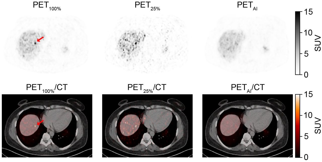
  • FIGURE 5.
    • Download figure
    • Open in new tab
    • Download powerpoint
    FIGURE 5.

    Patient with FN liver lesion. Patient had additional concordant TP liver lesions. Arrows mark lesion location on PET100% and PET100%/CT PET25% shown for reference.

  • FIGURE 6.
    • Download figure
    • Open in new tab
    • Download powerpoint
    FIGURE 6.

    Patient with FP liver lesion on PETAI. Patient had no lesions detected on PET100%. Arrows mark lesion location on PETAI and PETAI/CT PET25% shown for reference.
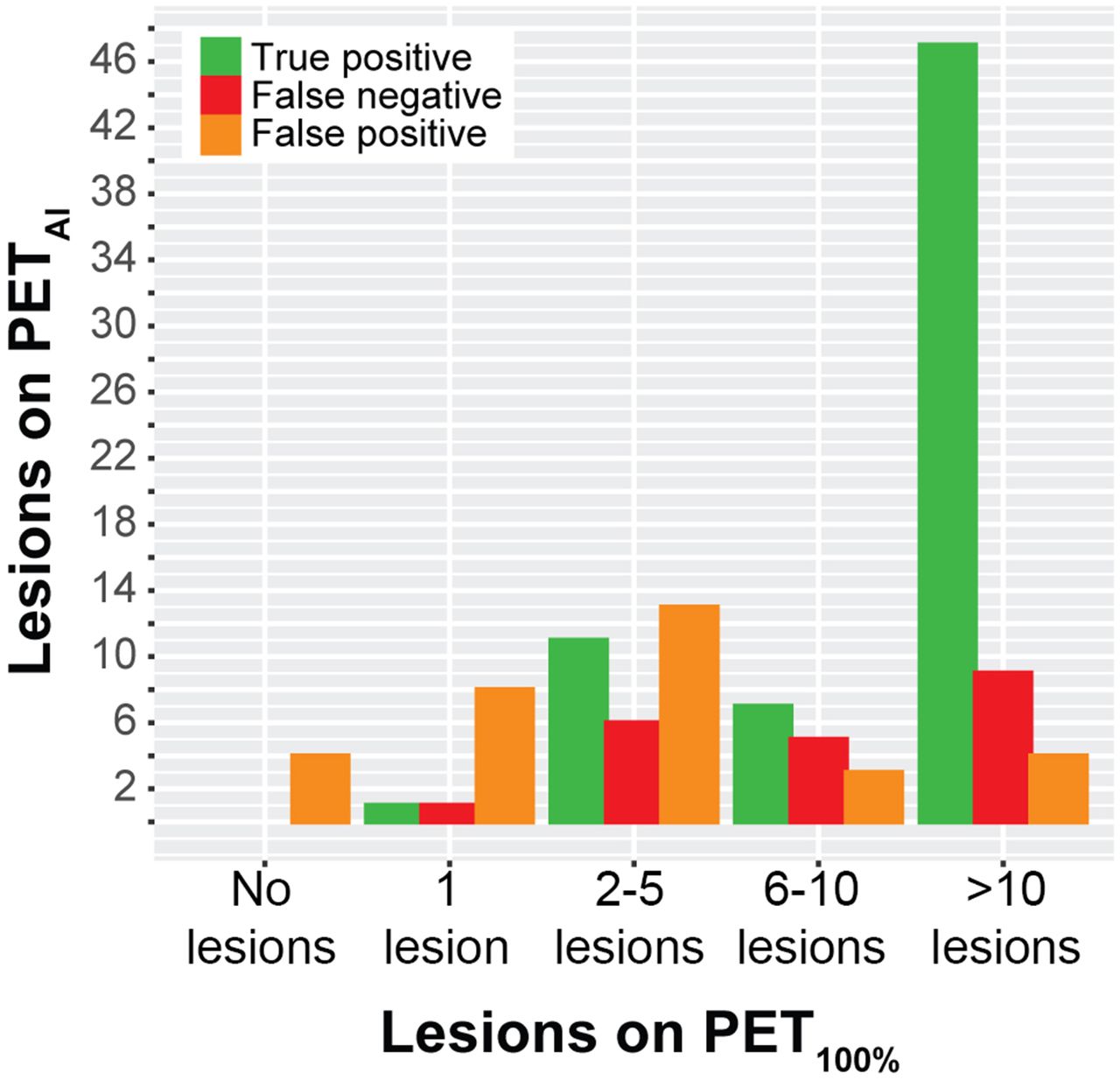
  • FIGURE 7.
    • Download figure
    • Open in new tab
    • Download powerpoint
    FIGURE 7.

    Distribution of TP, FP, and FN on PETAI corresponding to number of lesions detected on PET100%. Analysis was performed on patient subset for clinical image analysis consisting of patients with ≤20 lesions per organ (n = 33).

Tables

  • Figures
  • Additional Files
    • View popup
    TABLE 1.

    Patient Characteristics

    CharacteristicData (n = 38)Subset for clinical image analysis (n = 33)*
    Sex
     Female21 (55)19 (58)
     Male17 (45)14 (42)
    Age (y)
     Median6464
     Range37–8437–84
    Site of primary tumor
     Small intestine21 (55)16 (49)
     Pancreas11 (29)11 (33)
     Lung3 (8)3 (9)
     Other3 (8)3 (9)
    Previous treatment†
     Surgery29 (76)27 (82)
     Somatostatin analogs23 (61)18 (55)
     Peptide receptor radionuclide therapy12 (32)8 (24)
     Chemotherapy10 (26)7 (21)
     Radiofrequency ablation (liver metastases)2 (5)2 (6)
    Ki-67 proliferation index
     <3%9 (24)8 (24)
     3%–20%26 (68)22 (67)
     >20%3 (8)3 (9)
    Dose (MBq)‡
     PET100%191 (169–209)191 (172–209)
     PET25%/PETAI48 (42–52)48 (43–52)
    • ↵* Patients with >20 lesions per organ (n = 5) were excluded for clinical image analysis. Patients used for clinical image analysis (n = 33) thus represent subset of all 38 patients included in training sets.

    • ↵† Some patients received multiple treatments. Therefore, total number of treatments exceeds number of patients.

    • ↵‡ Dose at PET100% is 64Cu-DOTATATE activity dose given to patient for PET/CT. PET25% and PETAI dose is derived from simulated equivalent dose at 25% of PET100% dose.

    • Data are number followed by percentage in parentheses, except for age and dose (median and range).

    • View popup
    TABLE 2.

    Number of Lesions Grouped by Organs and Regions in 33 Patients with NENs

    Organ or regionNo. of lesions PET100%No. lesions PETAITPFPFNSensitivity*FDR*
    Liver363817211947 (30–65)55 (38–71)
    Pancreas67610100 (54–100)14 (0–58)
    Abdominal494736111373 (59–85)23 (12–38)
    Extraabdominal LNs56510100 (48–100)17 (0–64)
    Bone1712102759 (33–82)17 (2–48)
    Other5541180 (28–99)20 (1–72)
    Overall11811578374066 (57–75)32 (24–42)
    • ↵* Data for sensitivity and FDR are percentages followed by 95% CI in parentheses.

    • Abdominal = intestines, intraabdominal carcinosis, and intraabdominal lymph nodes (LNs); other = brain (1), ovary (1), thyroid or parathyroid (1), and skin (2). Analysis is performed on patient subset for clinical image analysis consisting of patients with ≤20 lesions per organ (n = 33).

    • View popup
    TABLE 3.

    Certainty of Detected Lesions in 33 Patients with NENs

    Organ or regionAll lesionsTPP*FNFP
    TotalC1C0TotalC1C0TotalC1C0TotalC1C0
    Liver
     PET100%36315171701.019145N/AN/AN/A
     PETAI3829917170N/AN/AN/A21129
    Pancreas
     PET100%6606601.0000N/AN/AN/A
     PETAI770660N/AN/AN/A110
    Abdominal
     PET100%49454363511.013103N/AN/AN/A
     PETAI4743436360N/AN/AN/A1174
    Extraabdominal LNs
     PET100%5505501.0000N/AN/AN/A
     PETAI651550N/AN/AN/A101
    Bone
     PET100%17161101001.0761N/AN/AN/A
     PETAI121111091N/AN/AN/A220
    Other
     PET100%5504401.0110
     PETAI550440N/AN/AN/A110
    Overall
     PET100%11810810787710.540319N/AN/AN/A
     PETAI1151001578771N/AN/AN/A372314
    • ↵* P values calculated using McNemar test for paired proportions of distribution of C1 and C0 lesion scores in TP lesions on PET100% vs. PETAI.

    • Abdominal = intestines, intraabdominal carcinosis, and intraabdominal lymph nodes (LNs); N/A = not applicable; other = brain (1), ovary (1), thyroid or parathyroid (1), and skin (2); — = ▪▪▪. Analysis is performed on patient subset for clinical image analysis consisting of patients with ≤20 lesions per organ (n = 33).

    • View popup
    TABLE 4.

    Characteristics of 33 Patients with NENs Based on Lesion Type

    ParameterTP-only or no lesions (n = 11)FN (n = 16)P*FP (n = 15)P*
    Injected dose (MBq)188 (181.5–201.5)190.5 (184–198.9)0.94192.0 (184.0–195.6)0.94
    Weight (kg)76.0 (67–81.5)73.0 (64.3)0.8786.7 (74.0–97.5)0.09
    Dose/weight (MBq/kg)2.5 (2.4–2.9)2.6 (2.0–3.1)0.822.3 (2.0–2.6)0.07
    Liver SUVmean5.0 (4.7–6.6)5.0 (4.7–6.1)0.796.1 (5.0–6.9)0.22
    • ↵* Mann–Whitney U test for comparison with reference (TP-only or no lesions group).

    • Data are shown as medians with interquartile range in parentheses. Analysis is performed on patient subset for clinical image analysis consisting of patients with ≤20 lesions per organ (n = 33). n refers to number of patients in each group. Patients may appear in both FN and FP groups if they have both FP and FN lesions. Accordingly, total number of patients exceeds 33.

Additional Files

  • Figures
  • Tables
  • Supplemental Data

    Files in this Data Supplement:

    • Supplemental Data
PreviousNext
Back to top

In this issue

Journal of Nuclear Medicine: 66 (5)
Journal of Nuclear Medicine
Vol. 66, Issue 5
May 1, 2025
  • Table of Contents
  • Table of Contents (PDF)
  • About the Cover
  • Index by author
  • Complete Issue (PDF)
Print
Download PDF
Article Alerts
Sign In to Email Alerts with your Email Address
Email Article

Thank you for your interest in spreading the word on Journal of Nuclear Medicine.

NOTE: We only request your email address so that the person you are recommending the page to knows that you wanted them to see it, and that it is not junk mail. We do not capture any email address.

Enter multiple addresses on separate lines or separate them with commas.
An Investigation of Lesion Detection Accuracy for Artificial Intelligence–Based Denoising of Low-Dose 64Cu-DOTATATE PET Imaging in Patients with Neuroendocrine Neoplasms
(Your Name) has sent you a message from Journal of Nuclear Medicine
(Your Name) thought you would like to see the Journal of Nuclear Medicine web site.
Citation Tools
An Investigation of Lesion Detection Accuracy for Artificial Intelligence–Based Denoising of Low-Dose 64Cu-DOTATATE PET Imaging in Patients with Neuroendocrine Neoplasms
Mathias Loft, Claes N. Ladefoged, Camilla B. Johnbeck, Esben A. Carlsen, Peter Oturai, Seppo W. Langer, Ulrich Knigge, Flemming L. Andersen, Andreas Kjaer
Journal of Nuclear Medicine May 2023, 264826; DOI: 10.2967/jnumed.122.264826

Citation Manager Formats

  • BibTeX
  • Bookends
  • EasyBib
  • EndNote (tagged)
  • EndNote 8 (xml)
  • Medlars
  • Mendeley
  • Papers
  • RefWorks Tagged
  • Ref Manager
  • RIS
  • Zotero
Share
An Investigation of Lesion Detection Accuracy for Artificial Intelligence–Based Denoising of Low-Dose 64Cu-DOTATATE PET Imaging in Patients with Neuroendocrine Neoplasms
Mathias Loft, Claes N. Ladefoged, Camilla B. Johnbeck, Esben A. Carlsen, Peter Oturai, Seppo W. Langer, Ulrich Knigge, Flemming L. Andersen, Andreas Kjaer
Journal of Nuclear Medicine May 2023, 264826; DOI: 10.2967/jnumed.122.264826
Twitter logo Facebook logo LinkedIn logo Mendeley logo
  • Tweet Widget
  • Facebook Like
  • Google Plus One
Bookmark this article

Jump to section

  • Article
    • Visual Abstract
    • Abstract
    • MATERIALS AND METHODS
    • RESULTS
    • DISCUSSION
    • CONCLUSION
    • DISCLOSURE
    • Footnotes
    • REFERENCES
  • Figures & Data
  • Supplemental
  • Info & Metrics
  • PDF

Related Articles

  • PubMed
  • Google Scholar

Cited By...

  • No citing articles found.
  • Google Scholar

More in this TOC Section

  • Feasibility of Ultra-Low-Activity 18F-FDG PET/CT Imaging Using a Long–Axial-Field-of-View PET/CT System
  • Cardiac Presynaptic Sympathetic Nervous Function Evaluated by Cardiac PET in Patients with Chronotropic Incompetence Without Heart Failure
  • Validation and Evaluation of a Vendor-Provided Head Motion Correction Algorithm on the uMI Panorama PET/CT System
Show more Clinical Investigations

Similar Articles

Keywords

  • 64Cu-DOTATATE
  • somatostatin receptor imaging
  • PET/CT
  • neuroendocrine neoplasms
  • artificial intelligence
SNMMI

© 2025 SNMMI

Powered by HighWire