
Main menu

  • Home
  • Content
    • Current
    • Ahead of print
    • Past Issues
    • JNM Supplement
    • SNMMI Annual Meeting Abstracts
    • Continuing Education
    • JNM Podcasts
  • Subscriptions
    • Subscribers
    • Institutional and Non-member
    • Rates
    • Journal Claims
    • Corporate & Special Sales
  • Authors
    • Submit to JNM
    • Information for Authors
    • Assignment of Copyright
    • AQARA requirements
  • Info
    • Reviewers
    • Permissions
    • Advertisers
  • About
    • About Us
    • Editorial Board
    • Contact Information
  • More
    • Alerts
    • Feedback
    • Help
    • SNMMI Journals
  • SNMMI
    • JNM
    • JNMT
    • SNMMI Journals
    • SNMMI

User menu

  • Subscribe
  • My alerts
  • Log in
  • My Cart

Search

  • Advanced search
Journal of Nuclear Medicine
  • SNMMI
    • JNM
    • JNMT
    • SNMMI Journals
    • SNMMI
  • Subscribe
  • My alerts
  • Log in
  • My Cart
Journal of Nuclear Medicine

Advanced Search

  • Home
  • Content
    • Current
    • Ahead of print
    • Past Issues
    • JNM Supplement
    • SNMMI Annual Meeting Abstracts
    • Continuing Education
    • JNM Podcasts
  • Subscriptions
    • Subscribers
    • Institutional and Non-member
    • Rates
    • Journal Claims
    • Corporate & Special Sales
  • Authors
    • Submit to JNM
    • Information for Authors
    • Assignment of Copyright
    • AQARA requirements
  • Info
    • Reviewers
    • Permissions
    • Advertisers
  • About
    • About Us
    • Editorial Board
    • Contact Information
  • More
    • Alerts
    • Feedback
    • Help
    • SNMMI Journals
  • View or Listen to JNM Podcast
  • Visit JNM on Facebook
  • Join JNM on LinkedIn
  • Follow JNM on Twitter
  • Subscribe to our RSS feeds
Research ArticleBRIEF COMMUNICATION
Open Access

Bioanalytic Hybrid System Merging 3-Dimensional Cell Culture and Chromatographic Precision for Unprecedented Preclinical Insights in Molecular Imaging

Verena Pichler, Verena Schwingenschlögl-Maisetschläger, Irem Duman, Xavier Monforte, Stefanie Ponti, Lukas Zimmermann, Elma Joldic, Monika Dumanic, Chrysoula Vraka, Marcus Hacker, Christian Kraule and Andreas Herbert Teuschl-Woller
Journal of Nuclear Medicine May 2025, 66 (5) 813-816; DOI: https://doi.org/10.2967/jnumed.124.269133
Verena Pichler
1Department of Pharmaceutical Chemistry, University of Vienna, Vienna, Austria;
  • Find this author on Google Scholar
  • Find this author on PubMed
  • Search for this author on this site
Verena Schwingenschlögl-Maisetschläger
1Department of Pharmaceutical Chemistry, University of Vienna, Vienna, Austria;
2Department of Biomedical-Imaging and Image-Guided Therapy, Medical University of Vienna, Vienna, Austria;
3PhaNuSpo Doctoral School, University of Vienna, Vienna, Austria;
  • Find this author on Google Scholar
  • Find this author on PubMed
  • Search for this author on this site
Irem Duman
1Department of Pharmaceutical Chemistry, University of Vienna, Vienna, Austria;
3PhaNuSpo Doctoral School, University of Vienna, Vienna, Austria;
  • Find this author on Google Scholar
  • Find this author on PubMed
  • Search for this author on this site
Xavier Monforte
4Department of Life Science Engineering, University of Applied Sciences Technikum Wien, Vienna, Austria;
  • Find this author on Google Scholar
  • Find this author on PubMed
  • Search for this author on this site
Stefanie Ponti
2Department of Biomedical-Imaging and Image-Guided Therapy, Medical University of Vienna, Vienna, Austria;
  • Find this author on Google Scholar
  • Find this author on PubMed
  • Search for this author on this site
Lukas Zimmermann
5Division of Medical Radiation Physics, Department of Radiation Oncology, Medical University of Vienna, Vienna, Austria; and
  • Find this author on Google Scholar
  • Find this author on PubMed
  • Search for this author on this site
Elma Joldic
1Department of Pharmaceutical Chemistry, University of Vienna, Vienna, Austria;
  • Find this author on Google Scholar
  • Find this author on PubMed
  • Search for this author on this site
Monika Dumanic
2Department of Biomedical-Imaging and Image-Guided Therapy, Medical University of Vienna, Vienna, Austria;
  • Find this author on Google Scholar
  • Find this author on PubMed
  • Search for this author on this site
Chrysoula Vraka
2Department of Biomedical-Imaging and Image-Guided Therapy, Medical University of Vienna, Vienna, Austria;
  • Find this author on Google Scholar
  • Find this author on PubMed
  • Search for this author on this site
Marcus Hacker
2Department of Biomedical-Imaging and Image-Guided Therapy, Medical University of Vienna, Vienna, Austria;
  • Find this author on Google Scholar
  • Find this author on PubMed
  • Search for this author on this site
Christian Kraule
6DOC medikus GmbH, Krems, Austria
  • Find this author on Google Scholar
  • Find this author on PubMed
  • Search for this author on this site
Andreas Herbert Teuschl-Woller
4Department of Life Science Engineering, University of Applied Sciences Technikum Wien, Vienna, Austria;
  • Find this author on Google Scholar
  • Find this author on PubMed
  • Search for this author on this site
  • Article
  • Figures & Data
  • Supplemental
  • Info & Metrics
  • PDF
Loading

Article Figures & Data

Figures

  • Additional Files
  • Figure
    • Download figure
    • Open in new tab
    • Download powerpoint
  • FIGURE 1.
    • Download figure
    • Open in new tab
    • Download powerpoint
    FIGURE 1.

    Technical setup of silk sponge as stationary phase and column body. (A) Technical drawing of column body including flow direction (cranial to caudal). (B) Stepwise improvement by changing from noncompartmentalized to compartmentalized system. From top to bottom: final composition of column including silk, frits, and caps; PET/CT image of preliminary setup without compartments; CT image of final setup including compartments separated by frits. (C) Logistic setup of column preparation, radiotracer production, transfer to radioactive area, and measurement within 24-h time frame (created in bioRender). (D) Setup for automated administration of radiotracer to column and schematic representation of overall system. (E) Pressure limitation test of system for empty and filled columns. No additional pressure was introduced in system using silk scaffolds. HPLC = high-performance liquid chromatography; QC = quality control.
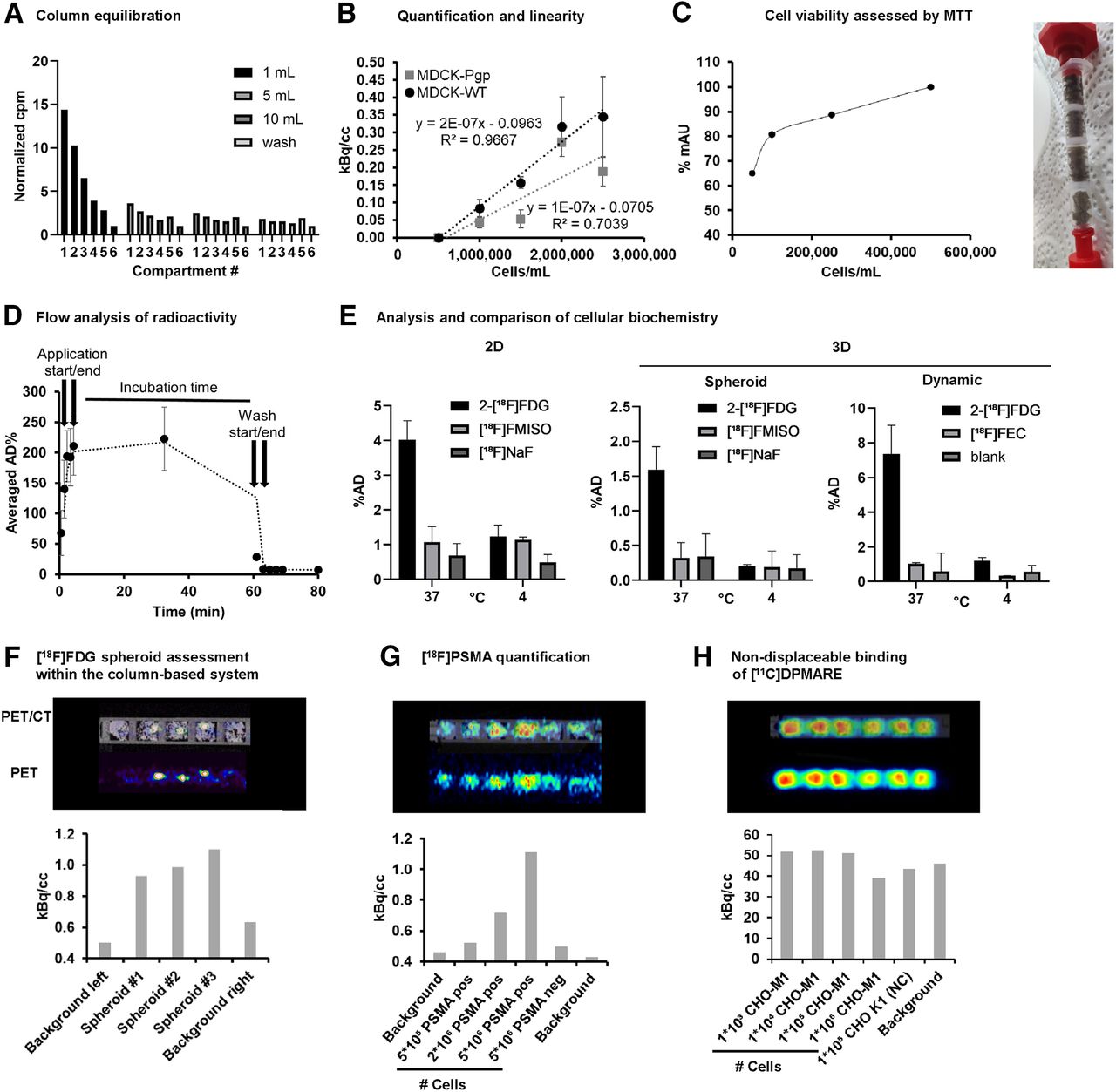
  • FIGURE 2.
    • Download figure
    • Open in new tab
    • Download powerpoint
    FIGURE 2.

    Radiotracer interaction studies in dynamic 3D cell culture system. (A) Analysis of void volume and administration volume of system to ensure equal medium distribution for incubation. (B) Quantification of cellular accumulation of radiotracer [18F]FDG as marker for cell viability and proliferation in MDCK-WT and MDCK-Pgp cells (n = 3). Cell line–dependent accumulation was observed, with higher reproducibility for MDCK-WT than for MDCK-Pgp cells. (C) Cell viability assessment of seeded cells in dependency on cell count to estimate ideal cell seeding capacity to avoid overgrowth in HCT116 cells after 5 d (n > 3), and MTT (3-[4,5-dimethylthiazol-2-yl]-2,5 diphenyl tetrazolium bromide) assay performed to analyze cell viability within column. (D) Dynamic scan to analyze fluidic behavior of administered radiotracer. (E) Comparison of behavior of HT29 cells in monolayer, multicellular tumor spheroid, and our dynamic system exposed to [18F]FDG (active transport), [18F]fluoromisonidazole (passive distribution), [18F]fluoroethylcholine (active transport), and [18F]NaF (passive distribution) at 37°C and 4°C. (F) Small-animal PET/CT scan of 600-μm-sized HT1080 spheroids incubated with [18F]FDG (n = 4, 1 representative dataset is shown to visualize direct readout), and analysis of cell viability by [18F]FDG. (G) Quantification of PET tracer with high specific binding and low nondisplaceable binding. Radiotracer interaction of [18F]FPSMA was investigated on PC3-PSMA–positive cells (increasing cell number in 5 × 10⁴ to 5 × 10⁶ cells per sponge) in comparison to PC3-PSMA–negative cells and silk without cells as background (n = 3, 1 representative dataset is shown). (H) Quantification of investigational PET tracer with high specific binding and high nondisplaceable binding. [11C]DPMARE (investigational radiotracer toward muscarinic acetylcholine receptors) was investigated in CHO-M1, expressing human M1 subtype of muscarinic acetylcholine receptors, and CHO-K1 cells as negative control (n = 2, 1 representative dataset is shown). AD = applied dose; FEC = fluoroethylcholine; FMISO = fluoromisonidazole.

Additional Files

  • Figures
  • Supplemental Data

    Files in this Data Supplement:

    • Supplemental Data
PreviousNext
Back to top

In this issue

Journal of Nuclear Medicine: 66 (5)
Journal of Nuclear Medicine
Vol. 66, Issue 5
May 1, 2025
  • Table of Contents
  • Table of Contents (PDF)
  • About the Cover
  • Index by author
  • Complete Issue (PDF)
Print
Download PDF
Article Alerts
Sign In to Email Alerts with your Email Address
Email Article

Thank you for your interest in spreading the word on Journal of Nuclear Medicine.

NOTE: We only request your email address so that the person you are recommending the page to knows that you wanted them to see it, and that it is not junk mail. We do not capture any email address.

Enter multiple addresses on separate lines or separate them with commas.
Bioanalytic Hybrid System Merging 3-Dimensional Cell Culture and Chromatographic Precision for Unprecedented Preclinical Insights in Molecular Imaging
(Your Name) has sent you a message from Journal of Nuclear Medicine
(Your Name) thought you would like to see the Journal of Nuclear Medicine web site.
Citation Tools
Bioanalytic Hybrid System Merging 3-Dimensional Cell Culture and Chromatographic Precision for Unprecedented Preclinical Insights in Molecular Imaging
Verena Pichler, Verena Schwingenschlögl-Maisetschläger, Irem Duman, Xavier Monforte, Stefanie Ponti, Lukas Zimmermann, Elma Joldic, Monika Dumanic, Chrysoula Vraka, Marcus Hacker, Christian Kraule, Andreas Herbert Teuschl-Woller
Journal of Nuclear Medicine May 2025, 66 (5) 813-816; DOI: 10.2967/jnumed.124.269133

Citation Manager Formats

  • BibTeX
  • Bookends
  • EasyBib
  • EndNote (tagged)
  • EndNote 8 (xml)
  • Medlars
  • Mendeley
  • Papers
  • RefWorks Tagged
  • Ref Manager
  • RIS
  • Zotero
Share
Bioanalytic Hybrid System Merging 3-Dimensional Cell Culture and Chromatographic Precision for Unprecedented Preclinical Insights in Molecular Imaging
Verena Pichler, Verena Schwingenschlögl-Maisetschläger, Irem Duman, Xavier Monforte, Stefanie Ponti, Lukas Zimmermann, Elma Joldic, Monika Dumanic, Chrysoula Vraka, Marcus Hacker, Christian Kraule, Andreas Herbert Teuschl-Woller
Journal of Nuclear Medicine May 2025, 66 (5) 813-816; DOI: 10.2967/jnumed.124.269133
Twitter logo Facebook logo LinkedIn logo Mendeley logo
  • Tweet Widget
  • Facebook Like
  • Google Plus One
Bookmark this article

Jump to section

  • Article
    • Visual Abstract
    • Abstract
    • MATERIALS AND METHODS
    • RESULTS
    • DISCUSSION
    • CONCLUSION
    • DISCLOSURE
    • ACKNOWLEDGMENTS
    • Footnotes
    • REFERENCES
  • Figures & Data
  • Supplemental
  • Info & Metrics
  • PDF

Related Articles

  • PubMed
  • Google Scholar

Cited By...

  • No citing articles found.
  • Google Scholar

More in this TOC Section

  • Measuring Total Metabolic Tumor Volume from 18F-FDG PET: A Reality Check
  • Radiances of Cerenkov-Emitting Radionuclides on the In Vivo Imaging System
Show more BRIEF COMMUNICATION

Similar Articles

Keywords

  • tissue engineering
  • radiotracer development
  • artificial extracellular matrix
SNMMI

© 2025 SNMMI

Powered by HighWire