


Abstract
241942
Introduction: Respiratory motion (RM) presents a significant challenge in PET/CT imaging, often leading to artifacts that can adversely affect clinical diagnosis. Respiratory gating is a widely-used strategy to mitigate RM artifacts, yet it can still result in image blurring due to unpredictable residual intra-gate motion, particularly in patients with pronounced RM. Additionally, gating techniques tend to increase image noise, further degrading image quality. In response to these challenges, we previously developed a novel respiratory gating method known as Distance Gating (DG) [1]. Unlike conventional gating methods, which typically rely on a desired count-level for input, DG specifically targets a maximal tolerable intra-gate motion (MTIM). Building upon the DG method, we introduce the Virtual Breath-Hold (VBH) technique. VBH synergistically combines DG with the Total Variation Regularized Expectation Maximization (TVREM) approach, enabling simultaneous motion management and noise reduction in PET images. The effectiveness of the VBH approach was preliminarily tested using the state-of-the-art long axial field-of-view (148 cm) uMI Panorama GS system.
Methods: The proposed VBH method consists of four key steps: AI-based RM detection, RM signal calibration and MITM data selection, TVREM reconstruction and alignment between PET and CT images. Specifically, (1) A pre-trained AI model segments a region of interest (ROI) covering the thoracic and abdominopelvic areas. The RM signal, specifically the centroid-of-distribution (COD), is derived from PET raw data within this ROI. (2) The DG method calibrates the COD signal, and a predetermined MITM value is used to filter qualified imaging events. (3) The HYPER Iterative [2] TVREM algorithm reconstructs images from selected events, minimizing noise from discarded data. For attenuation correction (AC), a neural network synthesizes deep learning-based attenuation maps (DL-μ) from PET image without AC. (4) Finally, the reconstructed PET image is aligned with the CT scan by registering the DL-μ with the CT attenuation map, thereby simplifying multi-modality registration to a single modality task. Pilot data were acquired on the uMI Panorama GS. Three subjects were administered with ~0.1 mCi/kg of 18F-FDG injection and underwent a 3-min scan while instructed to perform deep breathing for 2 mins. The clinical study was approved by the Independent Ethics Committee of the First People's Hospital of Kunshan (2023-03-055-K01).
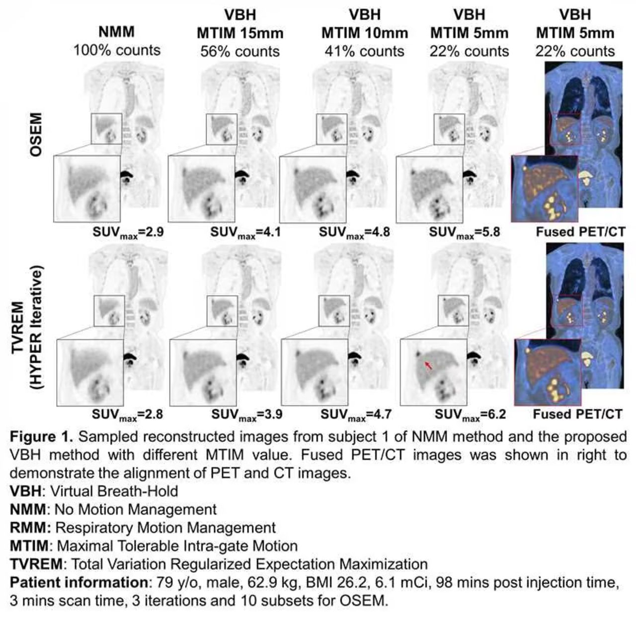
Results: Figure 1 displays reconstructed images of subject 1. Images without motion management (NMM) show noticeable blurring from respiration, a challenge effectively addressed by the VBH method through reducing MITM value. As a result, image artifacts are removed, leading to improved clarity in organ and lesion boundaries, along with an increase in SUV. However, a trade-off is observed: as fewer counts are used, image noise rises. This noise is subsequently eliminated following TVREM reconstruction, leading to superior image quality. Additionally, the SUV results from TVREM are on par with those from OSEM. The fusion image further confirms the PET and CT alignment using VBH. Figure 2, showcasing the images from three subjects, echoes these findings. An analysis of 22 high-uptake regions, segmented by a pre-trained AI model, reveals the VBH method increased the average SUVmax from 4.5 to 6.9 (a 53.3% increase) compared to NMM. Small SUV discrepancy (~5%) between TVREM and OSEM is observed.
Conclusions: We present a Virtual Breath-Hold (VBH) method designed for effective respiratory motion management in PET/CT imaging, specifically tailored for the uMI Panorama GS system. Our preliminary findings underscore the efficacy of this approach in simultaneously eliminating motion artifacts and ensuring high-quality, quantitatively accurate images with minimal noise.


In this issue


Jump to section
Related Articles
Cited By...
- No citing articles found.