


Abstract
242424
Introduction: With the release of our open-source python-based reconstruction library PyTomography, we aim to create a central platform for community-engaged development, validation, and deployment of novel reconstruction algorithms. Here, we include AI-based frameworks in PyTomography. Specifically, the deep image prior reconstruction algorithm ("DIPRecon"), proposed originally by Gong et al., was implemented, to make AI-based recon (i) accessible to a large audience, and (ii) robust enough for use of custom neural network architectures within the algorithm. The algorithm was designed within PyTomography such that it is imaging modality independent; this work explores its efficacy in both SPECT and PET use cases.
Methods: The DIPRecon reconstruction algorithm was developed in PyTomography to be compatible with PyTorch neural network architectures. The DIPRecon algorithm employed a UNet neural network with (i) 4,8,16,32 64 channels (PET) and 2,4,8,16,32 channels (SPECT) in each of the 5 layers of the encoder/decoder path, (ii) skip connections to link the encoder/decoder paths, and (iii) bi-linear interpolation up-sampling in the decoder. The selected hyperparameters of the algorithm were 2 subiterations for iterative updates, 10 subiterations for network training, and a value of ρ=50 (SPECT) and ρ=50000 (PET). Additional parameters for each use case can be found in the publicly available GitHub code.
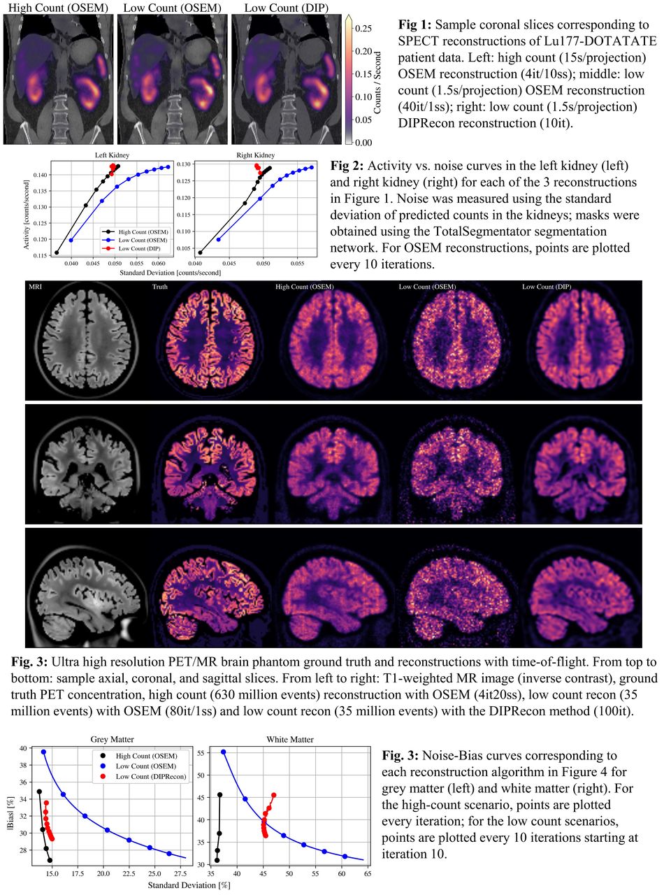
The SPECT use case considers publicly available Lu177-DOTATATE data from the Deep Blue data repository. Data were split into a "high-count" scenario (consisting of all counts or 15 s/projection) and a "low-count" scenario (1.5 s/projection) where the low count data were obtained by subsampling counts from the full dataset. All reconstructions employed attenuation, PSF, and scatter correction. The high-count data was reconstructed using OSEM (up to 10it/10ss), and the low-count data was reconstructed using (i) OSEM (up to 100it/1ss) and (ii) DIPRecon (up to 10it) where the prior image was obtained via training the network on low-count reconstruction with OSEM (40it/1ss).
The PET use case considered simulated GATE data acquired via a scanner with geometry representative of a Siemens mMR. An ultra-high-resolution PET/MR brain phantom was used as the digital phantom; in this case, an additional calibration scan using a thin cylindrical shell was also simulated to obtain normalization coefficients. List-mode reconstructions were performed in 3 ways: (i) using the full dataset with OSEM (4it/20ss), (ii) using 6% of the dataset with OSEM (80it/1ss), and (iii) using 6% of the dataset with DIPRecon (up to 100it), where the prior image was obtained via training the network on the reconstruction from case (ii).
Results: In the SPECT low-count use case, the DIPRecon method yields images with smoother activity distribution in the kidneys than OSEM (Figure 1). Activity-noise curves (dependent on iteration number) of each kidney reveal similar characteristics between low-count DIPRecon images and high-count OSEM images (Figure 2). In the PET use case, the low-count DIPRecon images have reduced noise compared to the low-count OSEM images and appear to closely resemble the high-count OSEM (Figure 3). Analysis of bias-noise curves (Figure 4) reveal that while the DIPRecon can reduce noise close to the levels of the high-count OSEM images in grey matter, the bias in both white and grey matter is still larger relative to the low-count OSEM images.
Conclusions: The DIPRecon tomographic reconstruction algorithm was implemented in PyTomography and demonstrated on both SPECT and PET use cases. In both cases, the DIPRecon method was able to reduce noise in low-count images to similar levels of the corresponding high-count images. All code is publicly available; users are encouraged to further experiment with network architectures and algorithm hyperparameters to improve on cases demonstrated here.

In this issue


Jump to section
Related Articles
Cited By...
- No citing articles found.