


Abstract
241396
Introduction: In PET imaging, conventional analytic and iterative algorithms, as well as deep-learning based methods, discretize both the projection space and the source image space. The discrete nature of coincidence detector bins intrinsically quantifies PET image resolution by one-half the crystal size. However, practical issues of backprojecting of projections into a discretized image space and vice versa introduce inconsistencies into proposed reconstruction processes, consequently degrading spatial resolution. Taking advantage of the expressive power of deep neural networks, this study proposes faithful representation of a continuous-domain image using a neural radiance field (NeRF). Additionally, a relative certainty-weighted sampling strategy is developed to project the continuous-domain image into the discrete projection data. By optimizing the goodness-of-fit of data, the NeRF is learned to reconstruct the continuous-domain function of source PET image, potentially leading to improved spatial resolution compared to other reconstruction methods.
Methods: Our algorithm represents a static radiotracer distribution using a fully-connected deep neural network. The network takes a single continuous three-dimensional (3D) coordinate as input. We traverse a set of measured lines of response (LORs) within each direct or oblique imaging plane, generating a sampled set of 3D points. These points serve as input to the neural network, producing an output set of activities and relative certainties. Interpreting relative certainty as the probability of a pair of photons emitted at an infinitesimal location and detected by specific detector bins, we employ relative-certainty weighted forward projection to accumulate activities along the LORs into a two-dimensional projection image (sinogram). Since these processes are differentiable, we utilize gradient descent to optimize the NeRF by minimizing the error between measured projections and the corresponding sinograms from our representation. To enhance projection efficiency and avoid sampling free space that does not contribute to the projection image, we employ a hierarchical sampling strategy. In addition to stratified sampling along each LOR, this approach allocates samples proportionally to their effects on the final projection. Moreover, to enhance the representation of high-frequency variation in activity and the complex geometry of a PET system, we transform input 3D coordinates to higher frequency input using positional encoding.
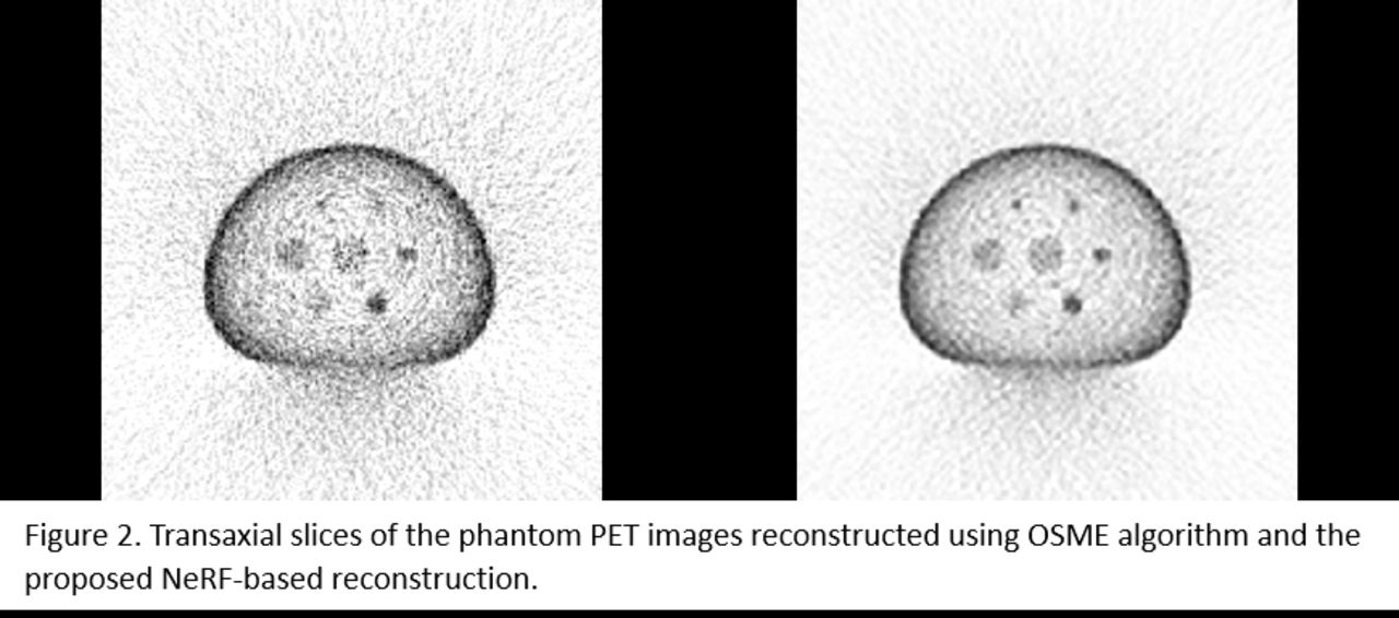
Results: To evaluate the proposed NeRF-based reconstruction method, we performed a simulation study using the Siemens Biograph 16 PET acquisition and a National Electrical Manufacturers Association (NEMA) International Electrotechnical Commission (IEC) body phantom, implemented by Geant4 Application Tomography Emission (GATE) software. The GATE simulation output was converted into a sinogram with dimensions 624x312x407, utilizing a span of 7, a maximum ring difference of 24, and 77 direct slices. The Software for Tomographic Image Reconstruction was employed to reconstruct the sinogram with and without NeRF synthesization into PET images using the ordered subsets expectation maximization (OSEM) algorithm. The reconstruction did not incorporate corrections for attenuation, scatter, and random events. The resulting PET images have dimensions of 413x413x77 with a voxel volume of 2.05 mm x 2.05 mm x 2.05 mm. The proposed method results in less noise and better hot-to-background contrast recovery than OSEM reconstruction.
Conclusions: Our work addresses the PET image reconstruction problem in a novel manner by directly optimizing parameters of a continuous 3D object representation. In a phantom simulation study, we demonstrate that the proposed NeRF generates PET images with higher resolution compared to conventional reconstruction methods. Future work will involve evaluating the proposed method on clinical patient data and integrating corrections for various physical effects.


In this issue


Jump to section
Related Articles
Cited By...
- No citing articles found.