


Abstract
241355
Introduction: Total-Body Positron Emission Tomography/Computed Tomography (TB PET/CT) enables the collection of imaging signals synchronously from the entire body with high sensitivity. In situations of lower back pain, increased blood flow has been described using muscle sonography. However, ultrasound has limitations regarding its penetration depth. Moreover, only one site of pain can be examined with ultra-sound simultaneously making comparisons over time to normal tissue limited. Yet it has been unclear if sites of lower back pain show inflammatory changes. Therefore, the aim of this work was to demonstrate the feasibility of dynamic TB PET/CT imaging using a dual tracer approach, with [11C]Butanol as a marker of blood flow and [18F]FDG as a marker of glucose metabolism, for the assessment of chronic low back pain.
Methods: Patients with chronic low back pain, who had at least one palpable myofascial nodule or taut band were recruited in this ongoing prospective single-center, observational, cross-sectional study. Participants were scanned on a TB PET/CT starting immediately after the intravenous injection of 262-275 MBq of 1[11C]Butanol, for 20 minutes dynamically. A separate dynamic acquisition using 99-111 MBq [18F]FDG for a total of 70 minutes was obtained in an additional scan within a median interval of 12 [0-14] days. Voxel wise kinetic analysis was performed using one- and two-tissue compartment models for [11C]Butanol and [18F]FDG for parametric imaging. For both radiotracers, plasma-to-tissue transport rate (K1) and standardized uptake value (SUV) images were reconstructed. SUV images (single frame at 2-5 min for [11C]Butanol and at 60-70 min for 18FDG) were reviewed for any abnormality. For SUV and K1images, volumes of interest (VOI) were placed on myofascial tissues in the low back at known sites of pain and in control non-painful tissues. Peak values (mean within one cubic centimeter (cc) sphere) were quantified.
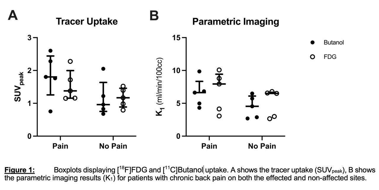
Results: In this initial evaluation, five patients have been included (♀ = 2, ♂ = 3; median: 63 y/o). There was no adverse events during scans to report. The biodistribution of each radiotracer was consistent with the literature. SUVpeak of [11C]Butanol and [18F]FDG were slightly higher at sites of pain compared to control sites without pain ([11C]Butanol median [range]: 1.8 [0.75-2.6] vs. 0.96 [0.68-2.1] and [18F]FDG: 1.4 [1.2-2.3] vs. 1.2 [0.79-1.5]). Similar results were seen for K1 metrics ([11C]Butanol: 6.6 [4.3-9.9] vs. 4.5 [2.7-6.5]; and [18F]FDG: 7.9 [3.1-10.1] vs. 6.5 [2.7-6.8] ml/min/100cc).
Conclusions: This ongoing study demonstrated the feasibility of performing dynamic TB PET/CT with [11C]Butanol and [18F]FDG in participants with chronic low back pain. Initial results demonstrate that the regional blood flow as assessed from both [11C]Butanol and [18F]FDG scans, and the steady-state glucose metabolism from the [18F]FDG scan are elevated in painful tissues versus non-painful tissues. These initial findings indicate that parametric imaging with both [11C]Butanol and [18F]FDG can help identifying blood flow and metabolism changes in patients with chronic low back pain.


In this issue


Jump to section
Related Articles
Cited By...
- No citing articles found.