
Main menu

  • Home
  • Content
    • Current
    • Ahead of print
    • Past Issues
    • JNM Supplement
    • SNMMI Annual Meeting Abstracts
    • Continuing Education
    • JNM Podcasts
  • Subscriptions
    • Subscribers
    • Institutional and Non-member
    • Rates
    • Journal Claims
    • Corporate & Special Sales
  • Authors
    • Submit to JNM
    • Information for Authors
    • Assignment of Copyright
    • AQARA requirements
  • Info
    • Reviewers
    • Permissions
    • Advertisers
  • About
    • About Us
    • Editorial Board
    • Contact Information
  • More
    • Alerts
    • Feedback
    • Help
    • SNMMI Journals
  • SNMMI
    • JNM
    • JNMT
    • SNMMI Journals
    • SNMMI

User menu

  • Subscribe
  • My alerts
  • Log in
  • My Cart

Search

  • Advanced search
Journal of Nuclear Medicine
  • SNMMI
    • JNM
    • JNMT
    • SNMMI Journals
    • SNMMI
  • Subscribe
  • My alerts
  • Log in
  • My Cart
Journal of Nuclear Medicine

Advanced Search

  • Home
  • Content
    • Current
    • Ahead of print
    • Past Issues
    • JNM Supplement
    • SNMMI Annual Meeting Abstracts
    • Continuing Education
    • JNM Podcasts
  • Subscriptions
    • Subscribers
    • Institutional and Non-member
    • Rates
    • Journal Claims
    • Corporate & Special Sales
  • Authors
    • Submit to JNM
    • Information for Authors
    • Assignment of Copyright
    • AQARA requirements
  • Info
    • Reviewers
    • Permissions
    • Advertisers
  • About
    • About Us
    • Editorial Board
    • Contact Information
  • More
    • Alerts
    • Feedback
    • Help
    • SNMMI Journals
  • View or Listen to JNM Podcast
  • Visit JNM on Facebook
  • Join JNM on LinkedIn
  • Follow JNM on Twitter
  • Subscribe to our RSS feeds
Research ArticleBasic Science Investigation

Deep Semisupervised Transfer Learning for Fully Automated Whole-Body Tumor Quantification and Prognosis of Cancer on PET/CT

Kevin H. Leung, Steven P. Rowe, Moe S. Sadaghiani, Jeffrey P. Leal, Esther Mena, Peter L. Choyke, Yong Du and Martin G. Pomper
Journal of Nuclear Medicine April 2024, 65 (4) 643-650; DOI: https://doi.org/10.2967/jnumed.123.267048
Kevin H. Leung
1Russell H. Morgan Department of Radiology and Radiological Science, Johns Hopkins University School of Medicine, Baltimore, Maryland;
  • Find this author on Google Scholar
  • Find this author on PubMed
  • Search for this author on this site
Steven P. Rowe
2Department of Radiology, University of North Carolina at Chapel Hill School of Medicine, Chapel Hill, North Carolina; and
  • Find this author on Google Scholar
  • Find this author on PubMed
  • Search for this author on this site
Moe S. Sadaghiani
1Russell H. Morgan Department of Radiology and Radiological Science, Johns Hopkins University School of Medicine, Baltimore, Maryland;
  • Find this author on Google Scholar
  • Find this author on PubMed
  • Search for this author on this site
Jeffrey P. Leal
1Russell H. Morgan Department of Radiology and Radiological Science, Johns Hopkins University School of Medicine, Baltimore, Maryland;
  • Find this author on Google Scholar
  • Find this author on PubMed
  • Search for this author on this site
Esther Mena
3Center for Cancer Research, National Cancer Institute, Bethesda, Maryland
  • Find this author on Google Scholar
  • Find this author on PubMed
  • Search for this author on this site
Peter L. Choyke
3Center for Cancer Research, National Cancer Institute, Bethesda, Maryland
  • Find this author on Google Scholar
  • Find this author on PubMed
  • Search for this author on this site
Yong Du
1Russell H. Morgan Department of Radiology and Radiological Science, Johns Hopkins University School of Medicine, Baltimore, Maryland;
  • Find this author on Google Scholar
  • Find this author on PubMed
  • Search for this author on this site
Martin G. Pomper
1Russell H. Morgan Department of Radiology and Radiological Science, Johns Hopkins University School of Medicine, Baltimore, Maryland;
  • Find this author on Google Scholar
  • Find this author on PubMed
  • Search for this author on this site
  • Article
  • Figures & Data
  • Supplemental
  • Info & Metrics
  • PDF
Loading

Article Figures & Data

Figures

  • Tables
  • Additional Files
  • Figure
    • Download figure
    • Open in new tab
    • Download powerpoint
  • FIGURE 1.
    • Download figure
    • Open in new tab
    • Download powerpoint
    FIGURE 1.

    Incomplete manual tumor segmentations compared with predicted segmentations on maximum-intensity projections of PSMA PET scans of 6 patients with prostate cancer.

  • FIGURE 2.
    • Download figure
    • Open in new tab
    • Download powerpoint
    FIGURE 2.

    Predicted segmentations on maximum-intensity projections of 18F-FDG PET scans of lung cancer, melanoma, lymphoma, head and neck cancer (H & N), and breast cancer. Pre- and posttherapy scans of breast cancer are shown (bottom row), with first 2 patients from left to right having pCR and the others being nonresponders.

  • FIGURE 3.
    • Download figure
    • Open in new tab
    • Download powerpoint
    FIGURE 3.

    (A and B) Lesionwise (A) and voxelwise (B) analysis of tumor detection and segmentation. (C and D) Prostate cancer detection rates by DeepSSTL approach throughout different stages of training progression (C) and compared with baseline models (D). DSC = Dice similarity coefficient; FDR = false-discovery rate; NPV = negative predictive value; PPV = positive predictive value; TNR = true-negative rate; TPR = true-positive rate.

  • FIGURE 4.
    • Download figure
    • Open in new tab
    • Download powerpoint
    FIGURE 4.

    (A and B) Receiver-operating-characteristic curves for radiomics classifier (A) and risk model (B) for prostate cancer. (C and D) Box plots of predicted risk scores vs. overall PSMA-RADS scores (C) and post-PSMA PET therapies (D).
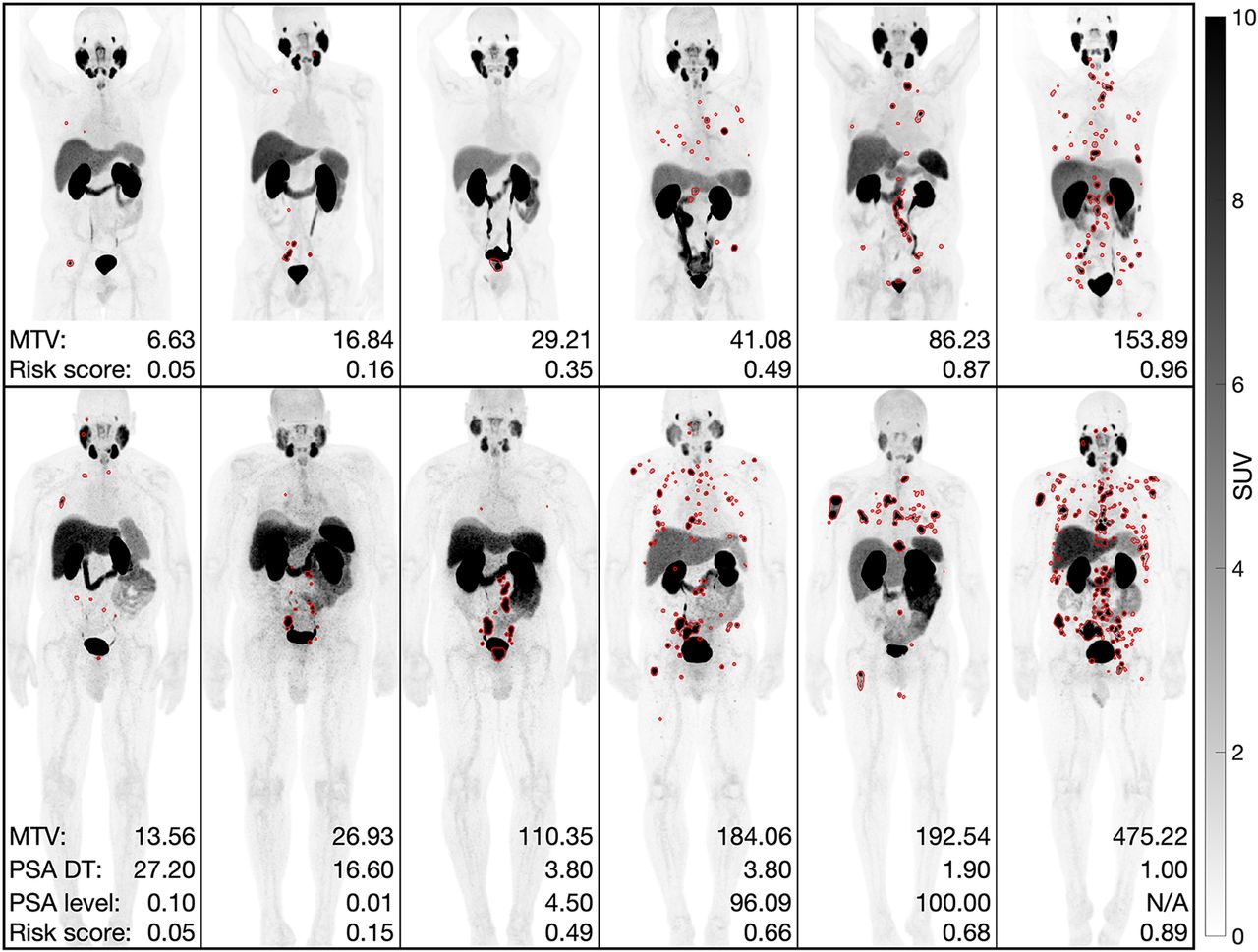
  • FIGURE 5.
    • Download figure
    • Open in new tab
    • Download powerpoint
    FIGURE 5.

    Predicted segmentations on maximum-intensity projections of PSMA PET scans of prostate cancer from datasets 1 (top row) and 2 (bottom row). MTV, PSA doubling times (DT), and follow-up PSA levels were measured in cubic centimeters, months, and ng/mL, respectively.
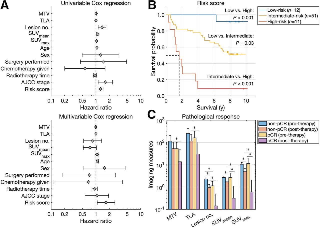
  • FIGURE 6.
    • Download figure
    • Open in new tab
    • Download powerpoint
    FIGURE 6.

    (A) Forest plots of univariable and multivariable Cox regression analysis. (B) Kaplan–Meier survival curves for head and neck cancer. (C) Imaging measures quantified from pre- and posttherapy scans of breast cancer.

Tables

  • Figures
  • Additional Files
    • View popup
    Table 1

    Patient Characteristics

    CharacteristicDataCharacteristicData
    Dataset 1*Dataset 3†
     Age (y) (mean ± SD)65.67 ± 7.97 Age (y) (mean ± SD)60.11 ± 16.51
     Sex Sex
      Men270  Men290
      Women0  Women211
     Overall PSMA-RADS scoreDataset 4‡
      NA12 Age (y) (mean ± SD)62.47 ± 7.78
      13 Sex
      224  Men62
      363  Women12
      448 AJCC stage
      5120  I14
    Dataset 2*  II5
     Age (y) (mean ± SD)66.46 ± 7.35  III13
     Sex  IV42
      Men138 Surgery
      Women0  No70
     Gleason score  Yes4
      NA2 Chemotherapy
      ≤611  No61
      743  Yes13
      829 Radiotherapy time (d)37 (31–47)
      944Dataset 5§
      109 Age (y) (mean ± SD)48.69 ± 10.33
     Initial PSA level (ng/mL)6.38 (0.02–5,000.00) Sex
     Follow-up PSA level (ng/mL)2.24 (0.00–7,270.00)  Men0
     PSA doubling time (mo)5.20 (0.23–81.70)  Women36
     Post-PSMA PET therapy Pathologic response
      NA37  pCR10
      None7  Non-pCR26
      Local18
      Systemic androgen-targeted56
      Systemic and cytotoxic20
    • ↵* Prostate cancer.

    • ↵† Lung cancer, melanoma, and lymphoma.

    • ↵‡ Head and neck cancer.

    • ↵§ Breast cancer.

    • NA = not applicable.

    • Qualitative data are number; continuous data are median and range, except for age.

    • View popup
    TABLE 2.

    Cox Regression Analysis and Respective C-Index Values

    Univariable Cox regressionMultivariable Cox regression
    ParameterHazard ratio95% CIPHazard ratio95% CIPC index
    MTV1.011.01–1.02<0.0010.990.97–1.020.570.68
    TLA1.001.00–1.00<0.0011.001.00–1.000.270.69
    Lesion no.1.381.13–1.690.0020.840.53–1.340.470.69
    SUVmean1.111.04–1.180.0020.740.53–1.030.070.65
    SUVmax1.041.02–1.06<0.0011.060.96–1.170.270.65
    Age1.061.01–1.100.021.081.02–1.140.010.61
    Sex1.270.49–3.280.621.560.52–4.670.430.47
    Surgery1.480.45–4.830.520.710.18–2.760.620.50
    Chemotherapy0.570.20–1.610.290.670.12–3.950.660.47
    Radiotherapy time0.950.88–1.030.230.940.82–1.080.380.46
    AJCC stage1.621.12–2.340.011.050.57–1.920.880.61
    Risk score1.271.11–1.45<0.0011.691.07–2.660.020.71
    • View popup
    TABLE 3.

    Predicting pCR

    Model and parameterAccuracyAUCArea under precision-recall curveTrue-positive ratePositive predictive valueTrue-negative rateNegative predictive value
    MTV0.420.550.281.000.320.191.00
    TLA0.670.580.300.500.420.730.79
    Lesion no.0.470.670.390.800.320.350.82
    SUVmean0.720.440.280.300.500.880.77
    SUVmax0.640.440.270.400.360.730.76
    Decision tree 10.720.720.510.500.500.810.81
    Decision tree 20.840.760.670.431.001.000.82

Additional Files

  • Figures
  • Tables
  • Supplemental Data

    Files in this Data Supplement:

    • Supplemental Data
PreviousNext
Back to top

In this issue

Journal of Nuclear Medicine: 65 (4)
Journal of Nuclear Medicine
Vol. 65, Issue 4
April 1, 2024
  • Table of Contents
  • Table of Contents (PDF)
  • About the Cover
  • Index by author
  • Complete Issue (PDF)
Print
Download PDF
Article Alerts
Sign In to Email Alerts with your Email Address
Email Article

Thank you for your interest in spreading the word on Journal of Nuclear Medicine.

NOTE: We only request your email address so that the person you are recommending the page to knows that you wanted them to see it, and that it is not junk mail. We do not capture any email address.

Enter multiple addresses on separate lines or separate them with commas.
Deep Semisupervised Transfer Learning for Fully Automated Whole-Body Tumor Quantification and Prognosis of Cancer on PET/CT
(Your Name) has sent you a message from Journal of Nuclear Medicine
(Your Name) thought you would like to see the Journal of Nuclear Medicine web site.
Citation Tools
Deep Semisupervised Transfer Learning for Fully Automated Whole-Body Tumor Quantification and Prognosis of Cancer on PET/CT
Kevin H. Leung, Steven P. Rowe, Moe S. Sadaghiani, Jeffrey P. Leal, Esther Mena, Peter L. Choyke, Yong Du, Martin G. Pomper
Journal of Nuclear Medicine Apr 2024, 65 (4) 643-650; DOI: 10.2967/jnumed.123.267048

Citation Manager Formats

  • BibTeX
  • Bookends
  • EasyBib
  • EndNote (tagged)
  • EndNote 8 (xml)
  • Medlars
  • Mendeley
  • Papers
  • RefWorks Tagged
  • Ref Manager
  • RIS
  • Zotero
Share
Deep Semisupervised Transfer Learning for Fully Automated Whole-Body Tumor Quantification and Prognosis of Cancer on PET/CT
Kevin H. Leung, Steven P. Rowe, Moe S. Sadaghiani, Jeffrey P. Leal, Esther Mena, Peter L. Choyke, Yong Du, Martin G. Pomper
Journal of Nuclear Medicine Apr 2024, 65 (4) 643-650; DOI: 10.2967/jnumed.123.267048
Twitter logo Facebook logo LinkedIn logo Mendeley logo
  • Tweet Widget
  • Facebook Like
  • Google Plus One
Bookmark this article

Jump to section

  • Article
    • Visual Abstract
    • Abstract
    • MATERIALS AND METHODS
    • RESULTS
    • DISCUSSION
    • CONCLUSION
    • DISCLOSURE
    • ACKNOWLEDGMENT
    • Footnotes
    • REFERENCES
  • Figures & Data
  • Supplemental
  • Info & Metrics
  • PDF

Related Articles

  • PubMed
  • Google Scholar

Cited By...

  • Validation of an Artificial Intelligence-Based Prediction Model Using 5 External PET/CT Datasets of Diffuse Large B-Cell Lymphoma
  • Google Scholar

More in this TOC Section

  • Establishing In Vitro Dosimetric Models and Dose–Effect Relationships for 177Lu-DOTATATE in Neuroendocrine Tumors
  • Designed Ankyrin Repeat Protein–Mediated Peptide Nucleic Acid–Based Pretargeting: A Proof-of-Principle Study
  • [11C]ZTP-1: An Effective Short-Lived Radioligand for PET of Rat and Monkey Brain Phosphodiesterase Type 4 Subtype B
Show more Basic Science Investigation

Similar Articles

Keywords

  • deep learning
  • semisupervised transfer learning
  • PET/CT
  • tumor segmentation
  • cancer prognosis
SNMMI

© 2025 SNMMI

Powered by HighWire