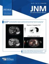
Prostate-specific membrane antigen (PSMA) is a transmembrane glycoprotein highly overexpressed in many cancers and benign conditions, and [68Ga]Ga-PSMA-11 PET/CT represents a successful diagnostic imaging method in nuclear medicine for prostate cancer.
This is a case of a 64-y-old woman enrolled in the Monocentric Observational Experience with PSMA study in Bologna, Italy, a prospective study examining in vivo and in vitro PSMA expression in patients with nonprostatic neoplasms, with histopathology as the reference standard. The study protocol was approved by the local ethics committee (code 538/2021/Oss/AOUBo), and subjects provided written informed consent. The patient’s contrast-enhanced MRI revealed a lesion in segment 5 of the liver, with late enhancement in wash-in and wash-out phases, consistent with hepatocellular carcinoma. [68Ga]Ga-PSMA-11 PET/CT was performed 17 d later (injected activity, 146 MBq; uptake time, 85 min), showing increased PSMA uptake at segment 5 (SUVmax, 11.3; SUVmax liver background, 7.9) and matching the MRI lesion. A wedge resection of segment 5 of the liver was executed 8 days later.
A yellowish well-defined 2-cm nodule made of an inflammatory component (mainly comprising histiocytic xanthogranulomatous cells and multinucleated giant cell macrophages containing biliary pigment, with stromal fibrotic and fibroblastic proliferation) was observed within noncirrhotic liver parenchyma. Eosinophilic nodular deposits surrounded by plasma cells, foamy macrophages or neutrophils, and activated myofibroblasts were also observed. Immunohistochemistry showed no cytokeratin-positive epithelial cells within the lesion, whereas the histiocytic component was CD68PGM1-positive/s100-negative with an IgG4-to-IgG ratio of less than 40%. The final diagnosis was a hepatic inflammatory pseudotumor (fibrohistiocytic type) (1).
Both PSMA immunohistochemistry and immunofluorescence were performed. The results are shown in Figure 1.
(A) Maximum-intensity projection image showing increased [68Ga]Ga-PSMA-11 uptake (arrowhead) in liver (SUVmax, 11.3). (B–E) Gadolinium ethoxybenzyl diethylenetriamine pentaacetic acid MRI showing nodule in segment 5 (arrow) hyperintense in T2-weighted image (B), highly hyperintense in diffusion-weighted image (C), highly hyperintense in arterial phase (D), and with targetoid sign in transitional phase (E). (F) Axial-fused [68Ga]Ga-PSMA-11 PET/CT showing increased PSMA uptake (SUVmax, 11.3) at segment 5. (G) PSMA immunohistochemistry image (×20) with sparce positive endothelial cells in intralesional microvascular component (circles) and negative inflammatory cells. (H) Negative PSMA immunofluorescence image (×10) in normal liver parenchyma (asterisk) with adjacent inflammatory infiltrate and sparce but strongly positive endothelial cells in intralesional microvascular component (circles).
Interestingly, only endothelial cells within the pseudotumor expressed PSMA, whereas inflammatory cells and normal liver endothelial cells were negative for PSMA. Hypothetically, endothelial cells within the lesion are stimulated by the tumor microenvironment to express PSMA, even if the mechanism beyond is still not known.
Hepatic inflammatory pseudotumor is a rare disease with less than 300 cases described in the literature. Differential diagnosis between a pseudotumor and hepatocellular carcinoma with CT and MRI is challenging. One multicentric study showed that 50% of patients with a final diagnosis of pseudotumor received a previous hepatocellular carcinoma misdiagnosis at CT or MRI (2). However, the prognosis and the treatment of pseudotumors are different and often resolvable with more conservative approaches, avoiding unnecessary invasive procedures.
The hereby described clinical case highlights that characterization of liver lesions is challenging with PSMA ligands because of the presence of several PSMA-avid diseases (e.g., hepatocellular carcinoma, prostate cancer metastases, and hemangioma) along with the physiologic PSMA background uptake in the liver parenchyma. Moreover, unlike prostate cancer, in which PSMA is overexpressed directly by cancer cells, PSMA in an inflammatory pseudotumor is expressed by endothelial cells. Therefore, PSMA images necessitate careful interpretation when evaluating the liver, as different mechanisms of PSMA overexpression can mimic neoplastic lesions.
DISCLOSURE
No potential conflict of interest relevant to this article was reported.
Footnotes
Published online Jun. 13, 2024.
- © 2024 by the Society of Nuclear Medicine and Molecular Imaging.
- Received for publication February 5, 2024.
- Accepted for publication May 13, 2024.


