


Article Figures & Data
Figures
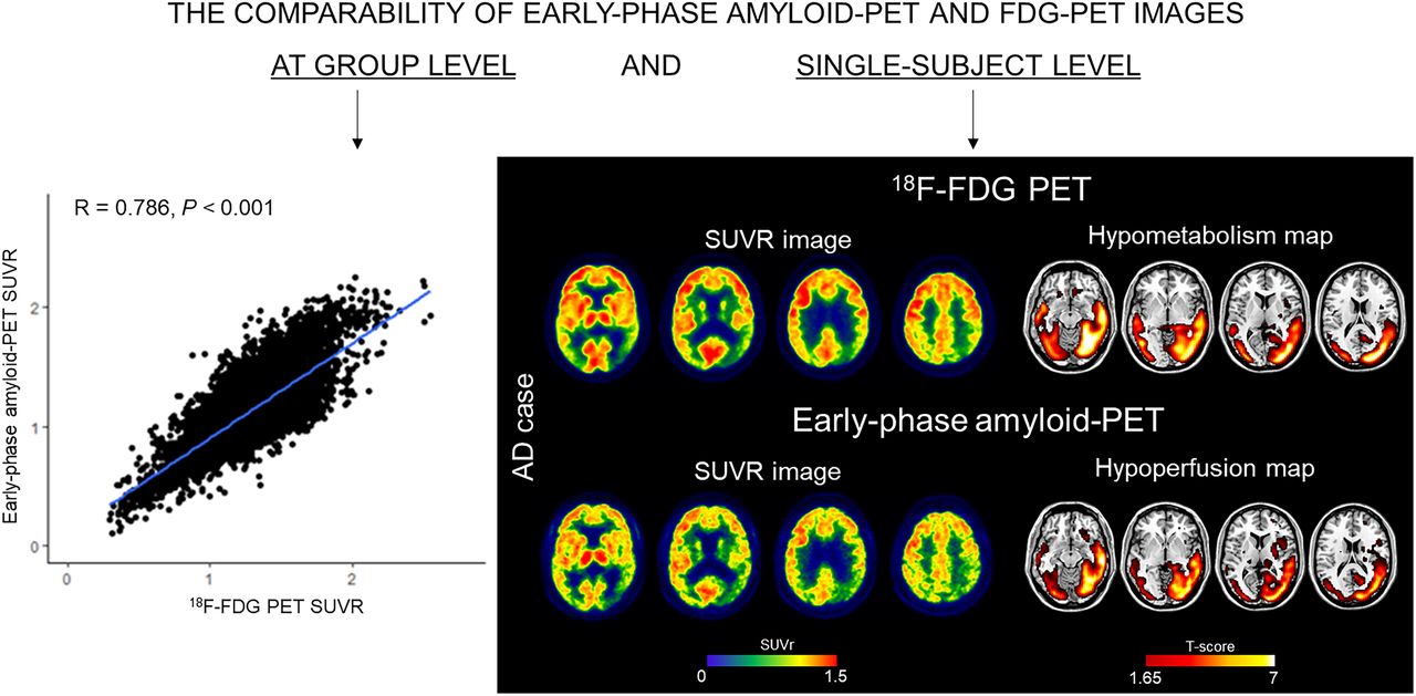
- FIGURE 1.
Correlation between eFBP/eFMM and 18F-FDG PET SUVR. Scatterplots showing association between eFBP/eFMM SUVR (y-axis) in AAL regions and their respective 18F-FDG SUVR (x-axis). Results are presented for whole sample and separately for subgroups divided according to Aβ status. Lines resulting from linear regression are shown in blue. R and P values are given in the upper left corner. FBP = florbetapir; FMM = flutemetamol; eFBP = early FBP; eFMM = early FMM; Aβ− = amyloid negative; Aβ+ = amyloid positive.
- FIGURE 2.
Hypometabolic and hypoperfusion patterns at the single-subject level. (A) Patterns of 18F-FDG PET hypometabolism and eFBP/eFMM hypoperfusion in single individuals. Hypometabolism maps, hypoperfusion maps, and their overlap were imposed on standard Montreal Neurological Institute template. These maps were obtained from binarization of single-subject 18F-FDG PET SPM T-maps and eFBP/eFMM SPM T-maps (P < 0.05 uncorrected, k > 100). The Dice similarity index is reported to the right of the brain template of each subject. (B) Clinical groups ordered according to degree of similarity between brain hypometabolism and hypoperfusion, as measured by Dice similarity index average. Lower-to-higher values of Dice indicate increasing degree of overlap. DEM = dementia; eFBP = early florbetapir; eFMM = early flutemetamol; Aβ+ = amyloid positive; Aβ− = amyloid negative; AD = Alzheimer disease; MCI = mild cognitive impairment.
- FIGURE 3.
Discriminative performance of eFBP/eFMM and 18F-FDG PET SUVR. ROC curves showing diagnostic performance of 18F-FDG PET and eFBP/eFMM SUVR in AD composite meta-ROI for distinguishing AD patients from HC. AUCs for eFBP/eFMM and 18F-FDG PET are shown in blue and green, respectively. Results of De Long test comparing 2 AUCs (eFBP/eFMM vs. 18F-FDG PET) are given in bottom box. A+ = Aβ-positive; N+ = neurodegeneration-positive; AUC = area under the curve; FBP = florbetapir; FMM = flutemetamol; AD = Alzheimer disease; HC = healthy controls.


Tables
Characteristic Whole sample FBP group FMM group P* n 166 94 72 Age 73.18 ± 6.35 74.27 ± 5.548 71.76 ± 7.068 0.012 Sex 0.425 Female 98 58 40 Male 68 36 32 MMSE 25.92 ± 4.00 26.12 ± 3.857 25.66 ± 4.202 0.471 Aβ status 0.980 Negative 70 39 31 Positive 93 52 41 Clinical groups according to Aβ status Aβ+ AD dementia 18 13 5 Aβ− dementia 3 2 1 Aβ+ MCI 52 31 22 Aβ− MCI 21 9 11 Aβ+ CU 30 11 19 Aβ− CU (HC) 42 28 14 ↵* From t test comparing data from eFBP and eFMM subgroups.
MMSE = Mini-Mental State Examination; FBP = florbetapir; FMM = flutemetamol; n = number; Aβ− = amyloid negative; Aβ+ = amyloid positive; AD = Alzheimer disease; MCI = mild cognitive impairment; CU = cognitively unimpaired; HC = healthy controls.
Qualitative data are number; continuous data are mean ± SD.
- TABLE 2.
Contingency Table Reporting Frequency of Different Hypometabolism and Hypoperfusion Patterns in Whole Sample
Hypoperfusion pattern Hypometabolism pattern AD-like FTD-like DLB-like Limbic-like Unclassified Normal Total AD-like 30 1 0 4 2 2 39 FTD-like 0 9 0 1 0 0 10 DLB-like 0 0 3 0 0 0 3 Limbic-like 0 0 0 14 0 0 14 Unclassified 0 0 0 1 24 1 26 Normal 2 0 0 0 1 29 32 Total 32 11 2 19 28 32 124 AD = Alzheimer disease; FTD = frontotemporal dementia; DLB = dementia with Lewy bodies.
- TABLE 3.
Distribution of Hypometabolism Patterns and Their Voxel-by-Voxel Concordance with Hypoperfusion Maps in Clinical Groups
18F-FDG hypometabolism pattern Dementia MCI CU Whole group Sample (n = 21) Dice* % visual match Sample (n = 73) Dice* % visual match Sample (n = 30) Dice* % visual match Sample (n = 124) Dice % visual match AD-like 10 (all Aβ+) 0.632 ± 0.159 90 25 (all Aβ+) 0.459 ± 0.178 68 4 (all Aβ+) 0.611 ± 0.135 100 39 (all Aβ+) 0.516 ± 0.185 77 FTD-like 5 (3 Aβ+, 2 Aβ−) 0.483 ± 0.201 80 5 (3 Aβ+, 2 Aβ−) 0.531 ± 0.128 100 0 10 (6 Aβ+, 4 Aβ−) 0.507 ± 0.161 90 DLB-like 0 3 (2 Aβ+, 1 Aβ−) 0.467 ± 0.236 100 0 3 (2 Aβ+, 1 Aβ−) 0.467 ± 0.236 100 Limbic-like 0 13 (9 Aβ+, 4 Aβ−) 0.504 ± 0.078 100 1 (all Aβ+) 0.521 100 14 (10 Aβ+, 4 Aβ−) 0.504 ± 0.075 100 Unclassified 6 (5 Aβ+, 1 Aβ−) 0.621 ± 0.071 83 13 (8 Aβ+, 5 Aβ−) 0.498 ± 0.205 100 7 (all Aβ+) 0.381 ± 0.293 86 26 (20 Aβ+, 6 Aβ−) 0.499 ± 0.217 92 Normal 0 14 (6 Aβ+, 8 Aβ−) 86† 18 (all Aβ+) 94† 32 (24 Aβ+, 8 Aβ−) 90†
Supplemental Data
Files in this Data Supplement:


