


Article Figures & Data
Figures
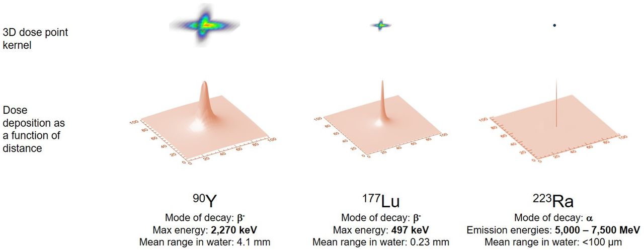
- FIGURE 1.
Graphical representation of dose deposition ranges delivered by different radionuclides having varying modes of decay. Top row shows relative geometric dose deposition delivered by point source of activity. Bottom row shows same data represented with 2-dimensional curve, illustrated as point spread function. Figure illustrates variable dose deposition properties and is not to scale.
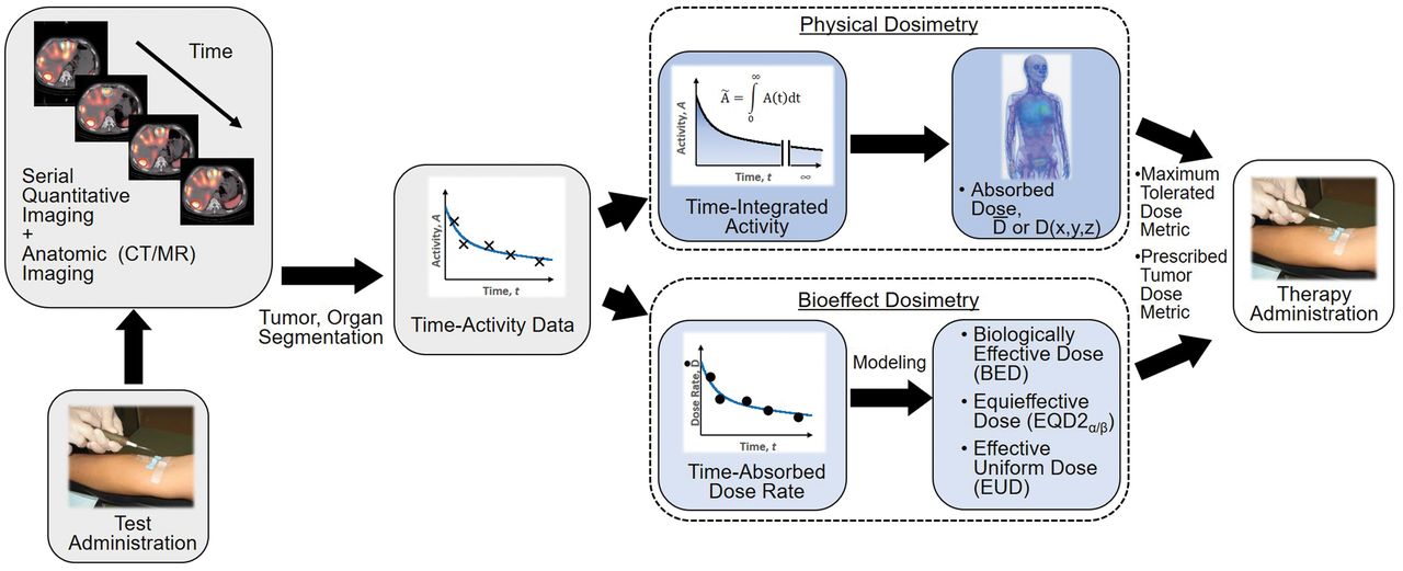
- FIGURE 2.
General workflow for RPT dosimetry. Process begins with test administration (may be either pretherapy administration or first cycle of multidose therapy regimen). Serial quantitation measurements can then support calculation of absorbed doses, either in terms of tumor and organ mean doses (D) or dose distributions (D[x,y,z]). Dose estimation per unit of administered activity can then be used to tailor treatment. The term dose metric may refer to absorbed dose (for physical dosimetry) or biologically effective dose (BED), equieffective dose (EQD2α/β), or effective uniform dose (EUD) (for bioeffect dosimetry).
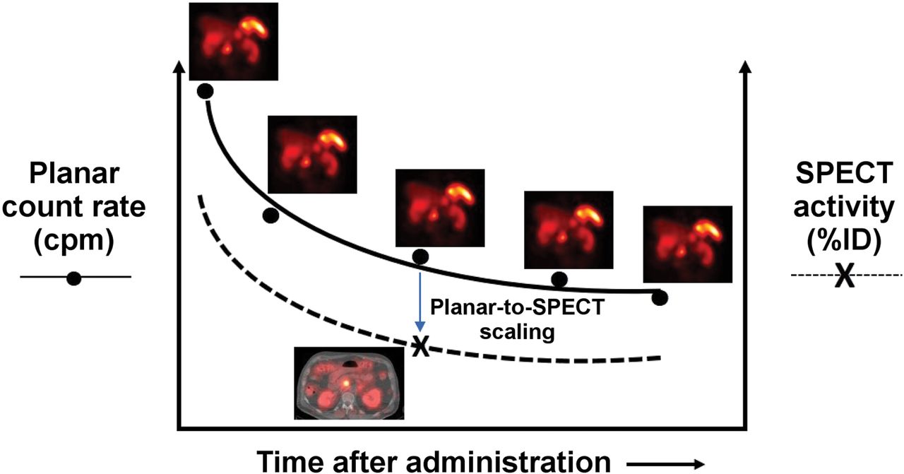
- FIGURE 3.
Hybrid SPECT/planar imaging approach to imaging-based measurement of time–activity data (55).


Tables
Isotope Primary emission Half-life LET Maximum range in tissue (therapeutic radiation) 211At α 7.21 h High 80 μm 212Pb α 10.6 h High 100 μm 213Bi α 45.6 min High 100 μm 223Ra α 11.4 d High 70 μm 225Ac α 10.0 d High 85 μm 227Th α 18.7 d High 70 μm 67Cu β 61.8 h Low 2.1 mm 90Y β 64.1 h Low 11 mm 131I β 8.02 d Low 3.3 mm 153Sm β 46.5 h Low 3.3 mm 177Lu β 6.65 d Low 1.8 mm
Supplemental Data
Files in this Data Supplement:
In this issue


Jump to section
Related Articles
Cited By...
- Computational Nuclear Oncology Toward Precision Radiopharmaceutical Therapies: Current Tools, Techniques, and Uncharted Territories
- Dosimetry of [177Lu]Lu-DOTATATE in Patients with Advanced Midgut Neuroendocrine Tumors: Results from a Substudy of the Phase III NETTER-1 Trial
- The MIRD Schema for Radiopharmaceutical Dosimetry: A Review
- Dosimetry in Radiopharmaceutical Therapy
- Reply: Dosimetry in Radiopharmaceutical Therapy