
Main menu

  • Home
  • Content
    • Current
    • Ahead of print
    • Past Issues
    • JNM Supplement
    • SNMMI Annual Meeting Abstracts
    • Continuing Education
    • JNM Podcasts
  • Subscriptions
    • Subscribers
    • Institutional and Non-member
    • Rates
    • Journal Claims
    • Corporate & Special Sales
  • Authors
    • Submit to JNM
    • Information for Authors
    • Assignment of Copyright
    • AQARA requirements
  • Info
    • Reviewers
    • Permissions
    • Advertisers
  • About
    • About Us
    • Editorial Board
    • Contact Information
  • More
    • Alerts
    • Feedback
    • Help
    • SNMMI Journals
  • SNMMI
    • JNM
    • JNMT
    • SNMMI Journals
    • SNMMI

User menu

  • Subscribe
  • My alerts
  • Log in
  • My Cart

Search

  • Advanced search
Journal of Nuclear Medicine
  • SNMMI
    • JNM
    • JNMT
    • SNMMI Journals
    • SNMMI
  • Subscribe
  • My alerts
  • Log in
  • My Cart
Journal of Nuclear Medicine

Advanced Search

  • Home
  • Content
    • Current
    • Ahead of print
    • Past Issues
    • JNM Supplement
    • SNMMI Annual Meeting Abstracts
    • Continuing Education
    • JNM Podcasts
  • Subscriptions
    • Subscribers
    • Institutional and Non-member
    • Rates
    • Journal Claims
    • Corporate & Special Sales
  • Authors
    • Submit to JNM
    • Information for Authors
    • Assignment of Copyright
    • AQARA requirements
  • Info
    • Reviewers
    • Permissions
    • Advertisers
  • About
    • About Us
    • Editorial Board
    • Contact Information
  • More
    • Alerts
    • Feedback
    • Help
    • SNMMI Journals
  • View or Listen to JNM Podcast
  • Visit JNM on Facebook
  • Join JNM on LinkedIn
  • Follow JNM on Twitter
  • Subscribe to our RSS feeds
Meeting ReportOral - PhysicianPharm

The impact of noise level mismatch between training and testing images for deep learning-based PET denoising

Qiong Liu, Hui Liu, Mirian Niloufar, Sijin Ren and Chi Liu
Journal of Nuclear Medicine May 2021, 62 (supplement 1) 109;
Qiong Liu
1Yale University New Haven CT United States
  • Find this author on Google Scholar
  • Find this author on PubMed
  • Search for this author on this site
Hui Liu
1Yale University New Haven CT United States
  • Find this author on Google Scholar
  • Find this author on PubMed
  • Search for this author on this site
Mirian Niloufar
1Yale University New Haven CT United States
  • Find this author on Google Scholar
  • Find this author on PubMed
  • Search for this author on this site
Sijin Ren
1Yale University New Haven CT United States
  • Find this author on Google Scholar
  • Find this author on PubMed
  • Search for this author on this site
Chi Liu
1Yale University New Haven CT United States
  • Find this author on Google Scholar
  • Find this author on PubMed
  • Search for this author on this site
  • Article
  • Figures & Data
  • Info & Metrics
Loading

Abstract

109

Objectives: Deep-learning methods have been applied in PET image denoising. However, when the noise level in the training and testing images are different, a deep-learning model could introduce spatial blurring and affect denoising performance. In this study, the impact of the unmatched noise level in training and testing images for denoising low dose PET images was investigated and potential solutions are proposed.

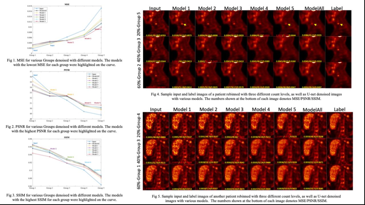
Methods: 168 patients 18F-FDG images acquired on a Siemens mCT PET/CT were included in this study. Low-count PET images were generated by rebinning the list-mode data at 20% (2 samples), 40% (2 samples), and 60% (1 sample) by uniform down-sampling. Among all patients, 100 studies were randomly selected as the training datasets, and the rest as testing data. 500 training images (100 patients x 5 count samples) were sorted into five noise level groups, which were surrogated by the image-based measurement of normalized standard deviation (NSTD) within a 3-cm diameter 3D spherical ROI within the liver. 340 testing images (68 patients x 5 count samples) were sorted into 5 groups with the same NSTD range in the training groups. The NSTD ranges in each group are 0-0.12, 0.12-0.14, 0.14-0.17, 0.17-0.20, 0.20-0.50. Models trained on each group were named with the corresponding group number, e.g. Model 1 was trained on Group 1 images. Model/Group 1 corresponded to the lowest noise group while Model/Group 5 corresponded to the highest noise group. We also applied a control group covering the full range of noise level with NSTD of 0-0.5, and the model trained on this group was named as ModelAll. 3D U-Net was used in this study. The total numbers of images in each group are the same with 100 noisy images as input and the corresponding full-dose images as the label. Mean squared error (MSE), peak signal to noise ratio (PSNR), and structural similarity (SSIM) as compared to full-dose images were used for evaluation.

Results: For each group, different U-Net models resulted in denoised images with different tradeoffs between noise reduction and resolution. Trained on less noisy images and tested on noisier images, the model showed limited denoising performance but better preserved resolution. Trained on noisier images and tested on less noisy images, the model achieved greater noise reduction but introduced substantial blurring. Models 2, 3, and 4 led to the highest SSIM for Groups 2, 3, and 4 testing images, respectively, while such a trend is not clear for MSE and PSNR. Among all the evaluation parameters, ModelAll did not outperform other individual models on all the testing groups.

Conclusions: Unmatched training and testing datasets resulted in different trade-offs between denoising and preserving resolution. Our results suggested that U-Net models trained on images with noise level ranges that match with those of testing images are generally desirable, though different imaging tasks and evaluation metrics could require different optimized models. A one-size-fits-all model trained with all noise levels is not recommended. A personalized strategy is needed for deep learning-based PET denoising.

Figure
  • Download figure
  • Open in new tab
  • Download powerpoint
Previous
Back to top

In this issue

Journal of Nuclear Medicine
Vol. 62, Issue supplement 1
May 1, 2021
  • Table of Contents
  • Index by author
Article Alerts
Sign In to Email Alerts with your Email Address
Email Article

Thank you for your interest in spreading the word on Journal of Nuclear Medicine.

NOTE: We only request your email address so that the person you are recommending the page to knows that you wanted them to see it, and that it is not junk mail. We do not capture any email address.

Enter multiple addresses on separate lines or separate them with commas.
The impact of noise level mismatch between training and testing images for deep learning-based PET denoising
(Your Name) has sent you a message from Journal of Nuclear Medicine
(Your Name) thought you would like to see the Journal of Nuclear Medicine web site.
Citation Tools
The impact of noise level mismatch between training and testing images for deep learning-based PET denoising
Qiong Liu, Hui Liu, Mirian Niloufar, Sijin Ren, Chi Liu
Journal of Nuclear Medicine May 2021, 62 (supplement 1) 109;

Citation Manager Formats

  • BibTeX
  • Bookends
  • EasyBib
  • EndNote (tagged)
  • EndNote 8 (xml)
  • Medlars
  • Mendeley
  • Papers
  • RefWorks Tagged
  • Ref Manager
  • RIS
  • Zotero
Share
The impact of noise level mismatch between training and testing images for deep learning-based PET denoising
Qiong Liu, Hui Liu, Mirian Niloufar, Sijin Ren, Chi Liu
Journal of Nuclear Medicine May 2021, 62 (supplement 1) 109;
Twitter logo Facebook logo LinkedIn logo Mendeley logo
  • Tweet Widget
  • Facebook Like
  • Google Plus One
Bookmark this article

Jump to section

  • Article
  • Figures & Data
  • Info & Metrics

Related Articles

  • No related articles found.
  • Google Scholar

Cited By...

  • No citing articles found.
  • Google Scholar

More in this TOC Section

Oral - PhysicianPharm

  • Dynamic 68Ga-DOTA PET/CT and Compartmental Modelling to Non-invasively Estimate the Glomerular Filtration Rate
  • Myocardial glucose suppression interferes with the detection of inflammatory cells with FDG-PET in a canine model of myocardial infarction
  • Global cardiac atherosclerotic burden assessed by fast automated artificial intelligence-based heart segmentation in 18F-sodium fluoride PET/CT scans: head-to-head comparison with manual segmentation
Show more Oral - PhysicianPharm

Advances in Data Sciences and Image Analysis

  • Linking radiomic PET features with metabolic tissue parameters using a hybrid mathematical model of tumor growth
  • Latent feature representation with the variational auto-encoder for the 18 F-flortaucipir, AV-1451, tau PET imaging biomarker in Alzheimer's disease
  • Machine learning improves interpretation of coronary artery disease using Rb-82 PET quantification of myocardial blood flow
Show more Advances in Data Sciences and Image Analysis

Similar Articles

SNMMI

© 2025 SNMMI

Powered by HighWire