
Main menu

  • Home
  • Content
    • Current
    • Ahead of print
    • Past Issues
    • JNM Supplement
    • SNMMI Annual Meeting Abstracts
    • Continuing Education
    • JNM Podcasts
  • Subscriptions
    • Subscribers
    • Institutional and Non-member
    • Rates
    • Journal Claims
    • Corporate & Special Sales
  • Authors
    • Submit to JNM
    • Information for Authors
    • Assignment of Copyright
    • AQARA requirements
  • Info
    • Reviewers
    • Permissions
    • Advertisers
  • About
    • About Us
    • Editorial Board
    • Contact Information
  • More
    • Alerts
    • Feedback
    • Help
    • SNMMI Journals
  • SNMMI
    • JNM
    • JNMT
    • SNMMI Journals
    • SNMMI

User menu

  • Subscribe
  • My alerts
  • Log in
  • Log out
  • My Cart

Search

  • Advanced search
Journal of Nuclear Medicine
  • SNMMI
    • JNM
    • JNMT
    • SNMMI Journals
    • SNMMI
  • Subscribe
  • My alerts
  • Log in
  • Log out
  • My Cart
Journal of Nuclear Medicine

Advanced Search

  • Home
  • Content
    • Current
    • Ahead of print
    • Past Issues
    • JNM Supplement
    • SNMMI Annual Meeting Abstracts
    • Continuing Education
    • JNM Podcasts
  • Subscriptions
    • Subscribers
    • Institutional and Non-member
    • Rates
    • Journal Claims
    • Corporate & Special Sales
  • Authors
    • Submit to JNM
    • Information for Authors
    • Assignment of Copyright
    • AQARA requirements
  • Info
    • Reviewers
    • Permissions
    • Advertisers
  • About
    • About Us
    • Editorial Board
    • Contact Information
  • More
    • Alerts
    • Feedback
    • Help
    • SNMMI Journals
  • View or Listen to JNM Podcast
  • Visit JNM on Facebook
  • Join JNM on LinkedIn
  • Follow JNM on Twitter
  • Subscribe to our RSS feeds
Meeting ReportOral - PhysicianPharm

Latent feature representation with the variational auto-encoder for the 18 F-flortaucipir, AV-1451, tau PET imaging biomarker in Alzheimer's disease

Jimin Hong, Axel Rominger, Kuangyu Shi and Hongyoon Choi
Journal of Nuclear Medicine May 2021, 62 (supplement 1) 111;
Jimin Hong
2University of Bern Bern Switzerland
1Inselspital Bern Switzerland
  • Find this author on Google Scholar
  • Find this author on PubMed
  • Search for this author on this site
Axel Rominger
1Inselspital Bern Switzerland
  • Find this author on Google Scholar
  • Find this author on PubMed
  • Search for this author on this site
Kuangyu Shi
2University of Bern Bern Switzerland
1Inselspital Bern Switzerland
  • Find this author on Google Scholar
  • Find this author on PubMed
  • Search for this author on this site
Hongyoon Choi
3Seoul National University Hospital Seoul Korea, Republic of
  • Find this author on Google Scholar
  • Find this author on PubMed
  • Search for this author on this site
  • Article
  • Figures & Data
  • Info & Metrics
Loading

Abstract

111

Objectives: The hyperphosphorylation and abnormal aggregation of tau protein is one of the main hallmarks for Alzheimer’s disease (AD). The post mortem studies of AD revealed that tau spreads in a certain pattern along with the disease progression, namely Braak stages. Tau quantification has not yet reached consensus to be introduced into the biomarker scheme, as recent findings provided conflicting results as opposed to Braak staging. Here, we propose a data-driven approach, without any priori assumption such as Braak model, to reproduce the tau progression in AD and support biomarker scheme of tau PET using variational autoencoder (VAE) and hierarchical agglomerative clustering as a building block. We believe that latent or hidden patterns inherently exist in tau PET images. By clustering such essence of features in latent space, we derived and examined the spatial pattern which is assumed to correspond to a certain stage of the AD progression.

Methods: 1080 pairs of T1 MRI image and AV-1451 PET were recruited in total (78 AD, 483 MCI, 519 CN). PET image were spatially normalized to the Montreal Neurological Institute (MNI) space using statistical parametric mapping (SPM8, www.fil.ion.ucl.ac.uk/spm). Our method consists of two building blocks, VAE and hierarchical clustering. In this work, each encoder and generator was built with five convolutional layers and a latent feature dimension of 100. Before being fed into the network, the original data were divided by mean uptake of the cerebellum and down-sampled to half in each dimension. The hierarchical agglomerative clustering was performed in latent space. The significance of the group difference between clusters was evaluated using the Chi-Squared analysis for the categorical clinical phenotype variables. One-way analysis of variance (ANOVA) was performed for continuous clinical phenotype variables, followed by Tukey’s post hoc pairwise test for multiple comparisons.

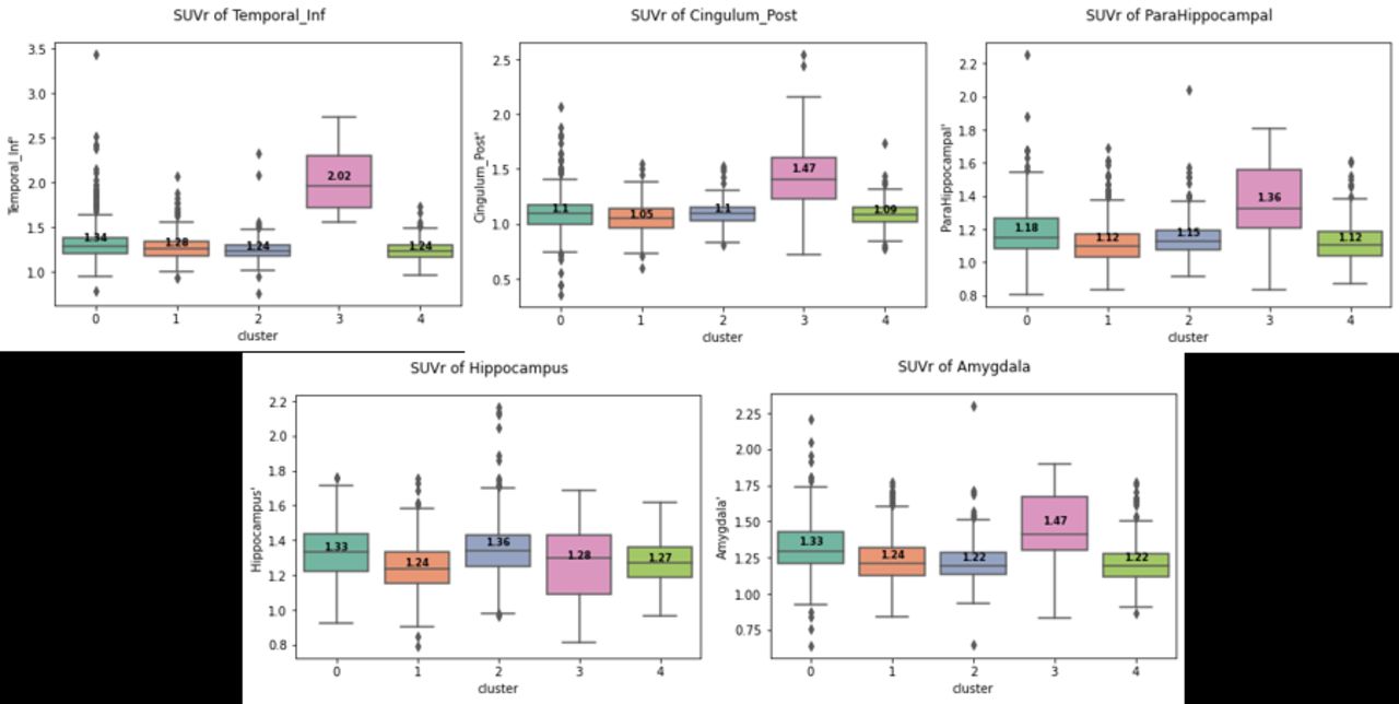
Results: t-SNE plot as well as contingency table illustrated the clustering result and the diagnosis of 1080 data. The tau distribution of each cluster was arranged in orders, with help of clinical information such as diagnosis, age, MMSE, and APOE4 and the series of cluster 4, cluster 0, and cluster 3 resembled the Braak stages. Standardized uptake value ratio (SUVr) for each ROI was calculated with cerebellum grey matter as a reference region. Across the majority of regions, cluster 3 presented the highest average SUVr. Amongst AD-signature regions, including amygdala, hippocampus, heschl, fusiform, inferior, middle and superior temporal gyrus, insula, and anterior, middle, and posterior cingulate, the most sensitive region was amygdala.

Conclusions: The data-driven approach, without any priori information on tau progress, corresponded well with the Braak staging. Our findings suggest that amygdala is the most sensitive region across the clusters, indicating the most ideal for the early detection of AD. Figure 1 t-SNE plot with clustering reult (left) and diagnosis (middle), and heatmap of contingency table of clustering result and diagnosis (right). conv (MCI-converter) and nonconv (MCI-nonconverter) Figure 2 Average image of cluster 4,0 and 3 Figure 3 SUVr of ROIs, including temporal_inf, cingulum_post, parahippocampal, hippocampus, and amygdala, between clusters

Figure
  • Download figure
  • Open in new tab
  • Download powerpoint
Figure
  • Download figure
  • Open in new tab
  • Download powerpoint
Figure
  • Download figure
  • Open in new tab
  • Download powerpoint
Previous
Back to top

In this issue

Journal of Nuclear Medicine
Vol. 62, Issue supplement 1
May 1, 2021
  • Table of Contents
  • Index by author
Article Alerts
Sign In to Email Alerts with your Email Address
Email Article

Thank you for your interest in spreading the word on Journal of Nuclear Medicine.

NOTE: We only request your email address so that the person you are recommending the page to knows that you wanted them to see it, and that it is not junk mail. We do not capture any email address.

Enter multiple addresses on separate lines or separate them with commas.
Latent feature representation with the variational auto-encoder for the 18 F-flortaucipir, AV-1451, tau PET imaging biomarker in Alzheimer's disease
(Your Name) has sent you a message from Journal of Nuclear Medicine
(Your Name) thought you would like to see the Journal of Nuclear Medicine web site.
Citation Tools
Latent feature representation with the variational auto-encoder for the 18 F-flortaucipir, AV-1451, tau PET imaging biomarker in Alzheimer's disease
Jimin Hong, Axel Rominger, Kuangyu Shi, Hongyoon Choi
Journal of Nuclear Medicine May 2021, 62 (supplement 1) 111;

Citation Manager Formats

  • BibTeX
  • Bookends
  • EasyBib
  • EndNote (tagged)
  • EndNote 8 (xml)
  • Medlars
  • Mendeley
  • Papers
  • RefWorks Tagged
  • Ref Manager
  • RIS
  • Zotero
Share
Latent feature representation with the variational auto-encoder for the 18 F-flortaucipir, AV-1451, tau PET imaging biomarker in Alzheimer's disease
Jimin Hong, Axel Rominger, Kuangyu Shi, Hongyoon Choi
Journal of Nuclear Medicine May 2021, 62 (supplement 1) 111;
Twitter logo Facebook logo LinkedIn logo Mendeley logo
  • Tweet Widget
  • Facebook Like
  • Google Plus One
Bookmark this article

Jump to section

  • Article
  • Figures & Data
  • Info & Metrics

Related Articles

  • No related articles found.
  • Google Scholar

Cited By...

  • No citing articles found.
  • Google Scholar

More in this TOC Section

Oral - PhysicianPharm

  • Pretargeted SPECT/CT imaging of CD11b expression allows for detecting instable aorta aneurysm that full of inflammation
  • The Area of Fibroblast Activation Exceeds the Hypoperfused Infarct Region in Patients with Acute Myocardial Infarction
  • Discordant low amyloid-β PET and high neocortical tau PET retention
Show more Oral - PhysicianPharm

Advances in Data Sciences and Image Analysis

  • Linking radiomic PET features with metabolic tissue parameters using a hybrid mathematical model of tumor growth
  • Machine learning improves interpretation of coronary artery disease using Rb-82 PET quantification of myocardial blood flow
Show more Advances in Data Sciences and Image Analysis

Similar Articles

SNMMI

© 2025 SNMMI

Powered by HighWire