
Main menu

  • Home
  • Content
    • Current
    • Ahead of print
    • Past Issues
    • JNM Supplement
    • SNMMI Annual Meeting Abstracts
    • Continuing Education
    • JNM Podcasts
  • Subscriptions
    • Subscribers
    • Institutional and Non-member
    • Rates
    • Journal Claims
    • Corporate & Special Sales
  • Authors
    • Submit to JNM
    • Information for Authors
    • Assignment of Copyright
    • AQARA requirements
  • Info
    • Reviewers
    • Permissions
    • Advertisers
  • About
    • About Us
    • Editorial Board
    • Contact Information
  • More
    • Alerts
    • Feedback
    • Help
    • SNMMI Journals
  • SNMMI
    • JNM
    • JNMT
    • SNMMI Journals
    • SNMMI

User menu

  • Subscribe
  • My alerts
  • Log in
  • Log out
  • My Cart

Search

  • Advanced search
Journal of Nuclear Medicine
  • SNMMI
    • JNM
    • JNMT
    • SNMMI Journals
    • SNMMI
  • Subscribe
  • My alerts
  • Log in
  • Log out
  • My Cart
Journal of Nuclear Medicine

Advanced Search

  • Home
  • Content
    • Current
    • Ahead of print
    • Past Issues
    • JNM Supplement
    • SNMMI Annual Meeting Abstracts
    • Continuing Education
    • JNM Podcasts
  • Subscriptions
    • Subscribers
    • Institutional and Non-member
    • Rates
    • Journal Claims
    • Corporate & Special Sales
  • Authors
    • Submit to JNM
    • Information for Authors
    • Assignment of Copyright
    • AQARA requirements
  • Info
    • Reviewers
    • Permissions
    • Advertisers
  • About
    • About Us
    • Editorial Board
    • Contact Information
  • More
    • Alerts
    • Feedback
    • Help
    • SNMMI Journals
  • View or Listen to JNM Podcast
  • Visit JNM on Facebook
  • Join JNM on LinkedIn
  • Follow JNM on Twitter
  • Subscribe to our RSS feeds
Research ArticleBasic Science Investigation

18F-6-Fluoro-l-Dopa PET/CT Imaging of Congenital Hyperinsulinism

Lisa J. States, J. Christopher Davis, Steven M. Hamel, Susan A. Becker and Hongming Zhuang
Journal of Nuclear Medicine July 2021, 62 (Supplement 2) 51S-56S; DOI: https://doi.org/10.2967/jnumed.120.246033
Lisa J. States
1Radiology Department, Children’s Hospital of Philadelphia, Philadelphia, Pennsylvania; and
2Perelman School of Medicine, University of Pennsylvania, Philadelphia, Pennsylvania, and
  • Find this author on Google Scholar
  • Find this author on PubMed
  • Search for this author on this site
J. Christopher Davis
1Radiology Department, Children’s Hospital of Philadelphia, Philadelphia, Pennsylvania; and
2Perelman School of Medicine, University of Pennsylvania, Philadelphia, Pennsylvania, and
  • Find this author on Google Scholar
  • Find this author on PubMed
  • Search for this author on this site
Steven M. Hamel
1Radiology Department, Children’s Hospital of Philadelphia, Philadelphia, Pennsylvania; and
  • Find this author on Google Scholar
  • Find this author on PubMed
  • Search for this author on this site
Susan A. Becker
1Radiology Department, Children’s Hospital of Philadelphia, Philadelphia, Pennsylvania; and
  • Find this author on Google Scholar
  • Find this author on PubMed
  • Search for this author on this site
Hongming Zhuang
1Radiology Department, Children’s Hospital of Philadelphia, Philadelphia, Pennsylvania; and
2Perelman School of Medicine, University of Pennsylvania, Philadelphia, Pennsylvania, and
  • Find this author on Google Scholar
  • Find this author on PubMed
  • Search for this author on this site
  • Article
  • Figures & Data
  • Supplemental
  • Info & Metrics
  • PDF
Loading

Article Figures & Data

Figures

  • Tables
  • Additional Files
  • FIGURE 1.
    • Download figure
    • Open in new tab
    • Download powerpoint
    FIGURE 1.

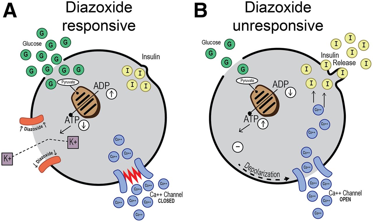
    (A) Diazoxide-responsive β-cell with normal KATP channel (orange) shows diazoxide keeping channel open and causing hyperpolarization of membrane and inhibition of insulin release. (B) Diazoxide-unresponsive β-cell with failure of KATP channel assembly and tracking to plasma membrane because of ABCC8 or KCNJ11 mutation allows depolarization of membrane with opening of calcium channel causing influx of calcium and unregulated release of insulin. (Courtesy of Serene McLaughlin.)
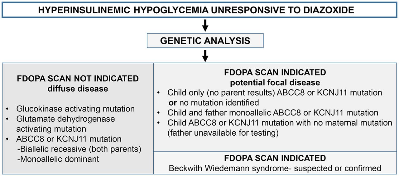
  • FIGURE 2.
    • Download figure
    • Open in new tab
    • Download powerpoint
    FIGURE 2.

    Indications for 18F-FDOPA PET/CT using genetic analysis.
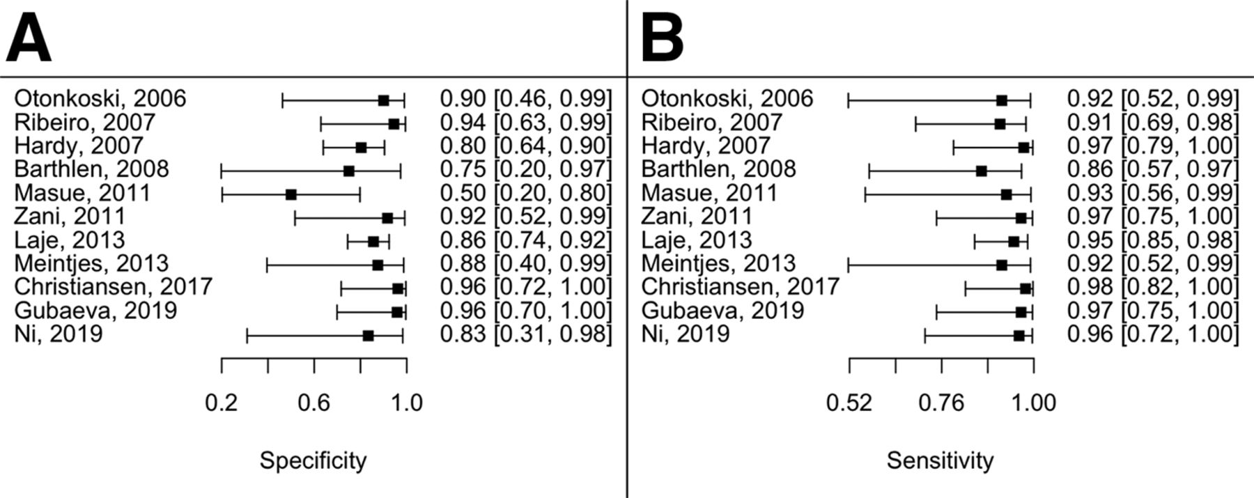
  • FIGURE 3.
    • Download figure
    • Open in new tab
    • Download powerpoint
    FIGURE 3.

    Specificity (A) and sensitivity (B) of 18F-FDOPA PET/CT for detection of focal lesions.
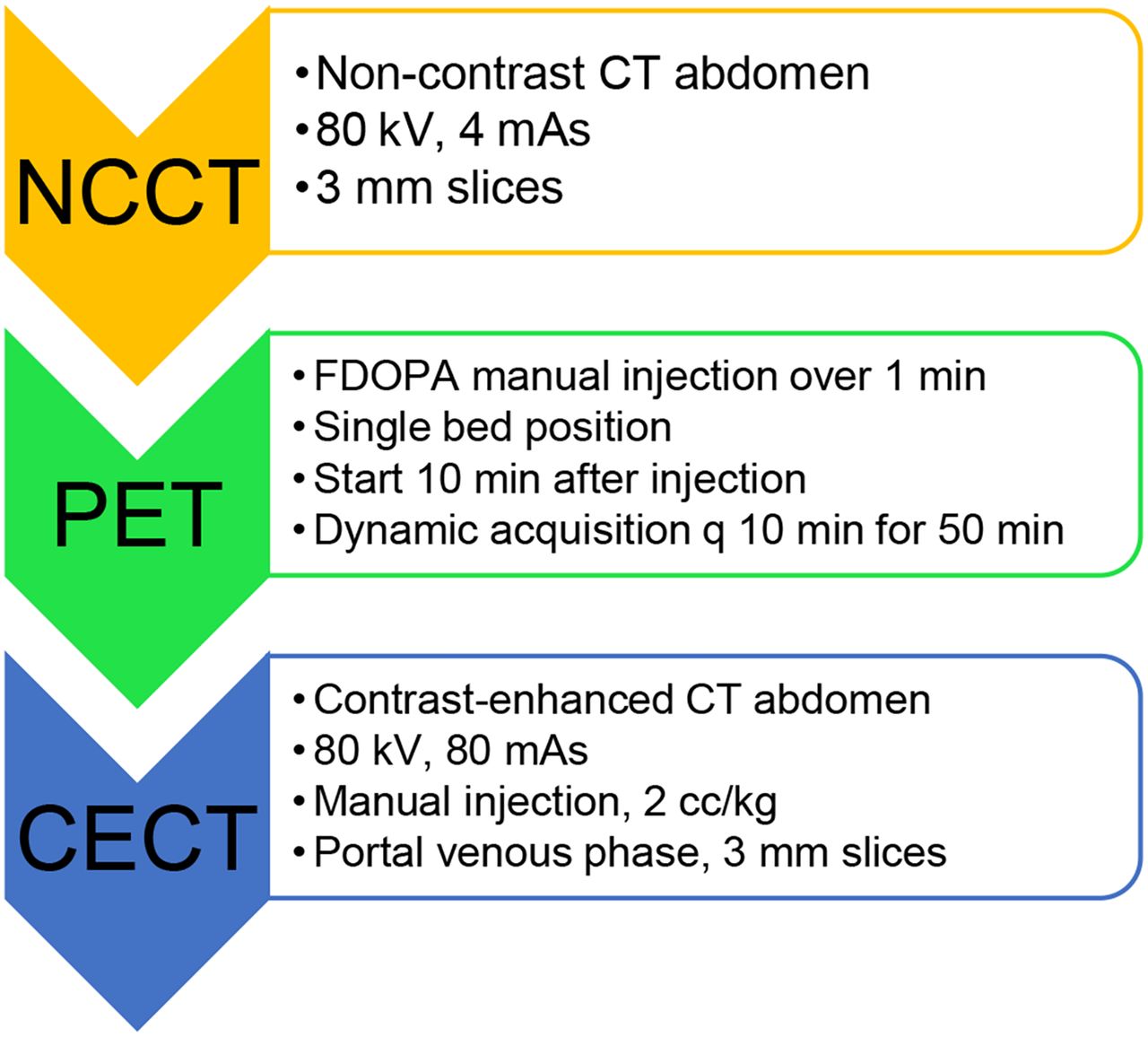
  • FIGURE 4.
    • Download figure
    • Open in new tab
    • Download powerpoint
    FIGURE 4.

    Sample imaging protocol. CECT = contrast-enhanced CT; NCCT = noncontrast CT.

  • FIGURE 5.
    • Download figure
    • Open in new tab
    • Download powerpoint
    FIGURE 5.

    Focal disease. 3-mo-old female with diazoxide-unresponsive HI with ABCC8 mutation. 18F-FDOPA 3-dimensional MIP image at 50 min shows 2 focal lesions, 1 within head (arrow) and other at pancreatic body/tail junction (arrowhead). Lesions were excised with 10% pancreatectomy. Finding 2 lesions is rare occurrence.
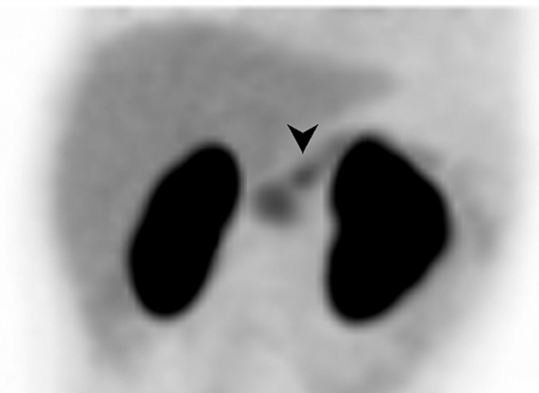
  • FIGURE 6.
    • Download figure
    • Open in new tab
    • Download powerpoint
    FIGURE 6.

    Focal lesion in pancreatic body. 3-mo-old female with ABCC8 mutation and paternal mutation. 18F-FDOPA 3-dimensional MIP image shows mild increased activity in pancreatic head and small lesion (arrowhead) in pancreatic body. Focal lesion was found in pancreatic body, abutting vasculature and requiring 50% pancreatectomy.
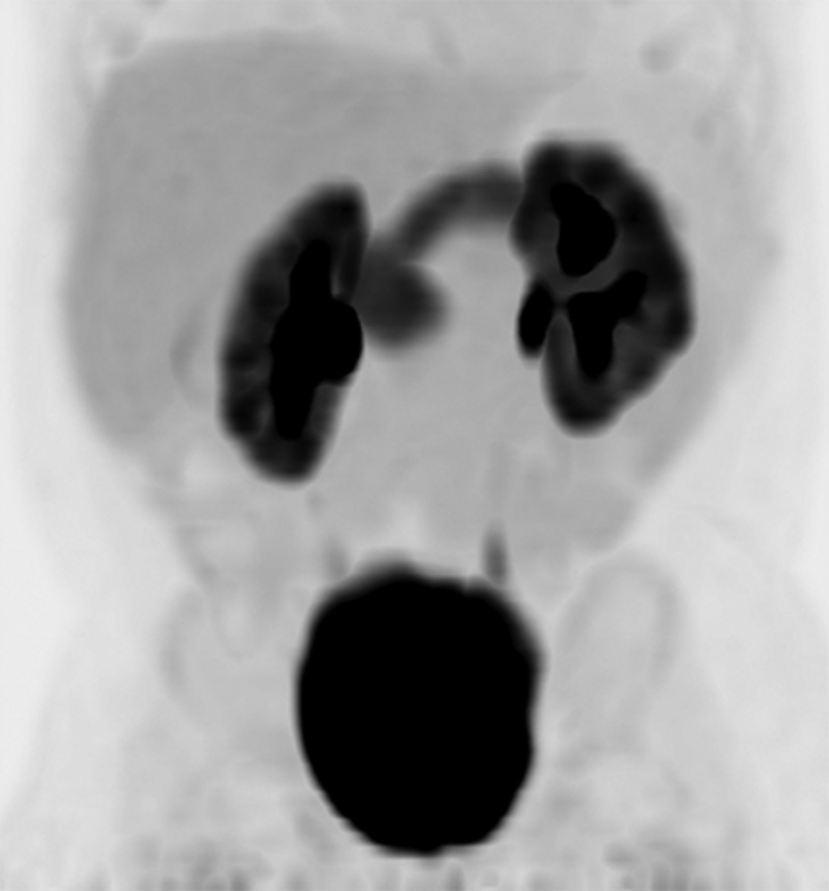
  • FIGURE 7.
    • Download figure
    • Open in new tab
    • Download powerpoint
    FIGURE 7.

    BWS. 1-mo-old male with suspected BWS and presenting with HI. 18F-FDOPA 3-dimensional MIP image at 10 min shows uptake within markedly enlarged pancreas, typical of BWS. Increased uptake within enlarged kidneys is also related to patient’s syndrome. Patient underwent 95% pancreatectomy.

Tables

  • Figures
  • Additional Files
    • View popup
    TABLE 1

    Publications with Surgical Histologic Confirmation of PET Results

    No. of patients
    Undergoing surgeryWith focal PET/histology resultsWith diffuse PET/histology resultsNo. of atypical casesAccuracy of localization (%)
    Study
    Otonkoski et al. (25) (2006; Finland)95/54/4100
    Ribeiro et al. (32) (2007; France)2415/158/93 atypical92
    Hardy et al. (19) (2007; United States)5018/2426/261 large focal; 2 localized islet nuclear enlargement100
    Barthlen et al. (33) (2008; Germany)119/91/21 atypical100
    Masue et al. (26) (2011; Japan)126/93/33 large focal33
    Zani et al. (49) (2011; United Kingdom)1914/145/51 large focal79
    Laje et al. (28) (2013; United States)10545/5350/52100
    Meintjes et al. (15) (2013; United Kingdom)85/53/3100
    Christiansen et al. (35) (2018; Denmark)3422/2212/121 atypical; 1 normal; 1 ectopic91
    Gubaeva et al. (50) (2019; Russia)2514/1411/111 giant100
    Ni et al. (51) (2019; China)1412/122/2Atypical100
    Total311165/182 (91%)125/129 (97%)

Additional Files

  • Figures
  • Tables
  • Supplemental Data

    Files in this Data Supplement:

    • Supplemental Data
PreviousNext
Back to top

In this issue

Journal of Nuclear Medicine: 62 (Supplement 2)
Journal of Nuclear Medicine
Vol. 62, Issue Supplement 2
July 1, 2021
  • Table of Contents
  • Table of Contents (PDF)
  • About the Cover
  • Index by author
Print
Download PDF
Article Alerts
Sign In to Email Alerts with your Email Address
Email Article

Thank you for your interest in spreading the word on Journal of Nuclear Medicine.

NOTE: We only request your email address so that the person you are recommending the page to knows that you wanted them to see it, and that it is not junk mail. We do not capture any email address.

Enter multiple addresses on separate lines or separate them with commas.
18F-6-Fluoro-l-Dopa PET/CT Imaging of Congenital Hyperinsulinism
(Your Name) has sent you a message from Journal of Nuclear Medicine
(Your Name) thought you would like to see the Journal of Nuclear Medicine web site.
Citation Tools
18F-6-Fluoro-l-Dopa PET/CT Imaging of Congenital Hyperinsulinism
Lisa J. States, J. Christopher Davis, Steven M. Hamel, Susan A. Becker, Hongming Zhuang
Journal of Nuclear Medicine Jul 2021, 62 (Supplement 2) 51S-56S; DOI: 10.2967/jnumed.120.246033

Citation Manager Formats

  • BibTeX
  • Bookends
  • EasyBib
  • EndNote (tagged)
  • EndNote 8 (xml)
  • Medlars
  • Mendeley
  • Papers
  • RefWorks Tagged
  • Ref Manager
  • RIS
  • Zotero
Share
18F-6-Fluoro-l-Dopa PET/CT Imaging of Congenital Hyperinsulinism
Lisa J. States, J. Christopher Davis, Steven M. Hamel, Susan A. Becker, Hongming Zhuang
Journal of Nuclear Medicine Jul 2021, 62 (Supplement 2) 51S-56S; DOI: 10.2967/jnumed.120.246033
Twitter logo Facebook logo LinkedIn logo Mendeley logo
  • Tweet Widget
  • Facebook Like
  • Google Plus One
Bookmark this article

Jump to section

  • Article
    • Abstract
    • PATIENT SELECTION
    • 18F-FDOPA MECHANISM AND HISTOPATHOLOGY
    • REVIEW OF LITERATURE
    • PROTOCOL AND PREPARATION
    • IMAGE INTERPRETATION
    • SAFETY
    • FUTURE CONSIDERATIONS
    • CONCLUSION
    • DISCLOSURE
    • ACKNOWLEDGMENTS
    • REFERENCES
  • Figures & Data
  • Supplemental
  • Info & Metrics
  • PDF

Related Articles

  • PubMed
  • Google Scholar

Cited By...

  • 68Ga-NODAGA-Exendin-4 PET Scanning for Focal Congenital Hyperinsulinism: Need for Replication
  • Google Scholar

More in this TOC Section

  • [11C]ZTP-1: An Effective Short-Lived Radioligand for PET of Rat and Monkey Brain Phosphodiesterase Type 4 Subtype B
  • Reduced Renal Uptake of Various Radiopharmaceuticals with Sodium Paraaminohippurate Coadministration in a Rat Model
  • In Vivo Visualization and Quantification of Brain Heat Shock Protein 90 with [11C]HSP990 in Healthy Aging and Neurodegeneration
Show more Basic Science Investigation

Similar Articles

Keywords

  • 18F-FDOPA
  • hyperinsulinism
  • congenital hyperinsulinism
SNMMI

© 2025 SNMMI

Powered by HighWire