


Article Figures & Data
Figures
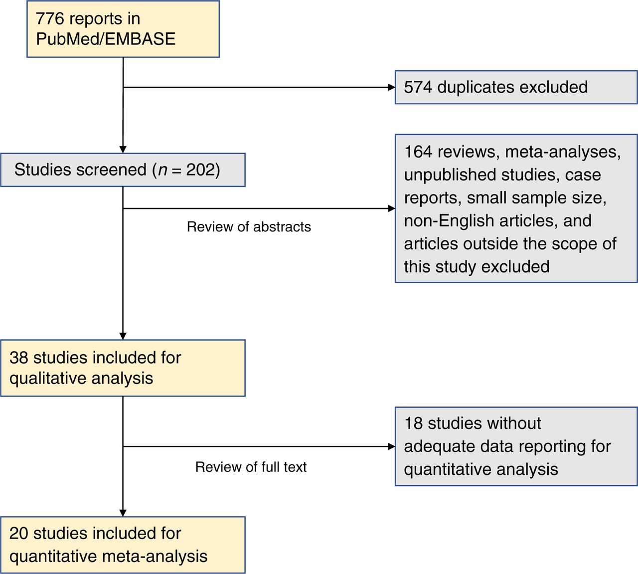
- FIGURE 1.
PRISMA (Preferred Reporting Items for Systematic Review and Metaanalysis) flow diagram depicting process for selecting papers included in this metaanalysis.
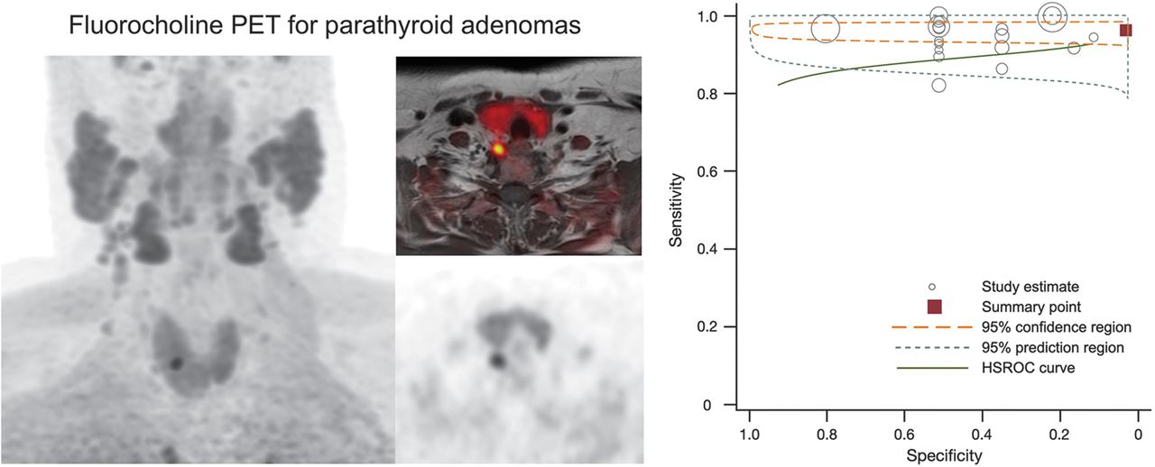
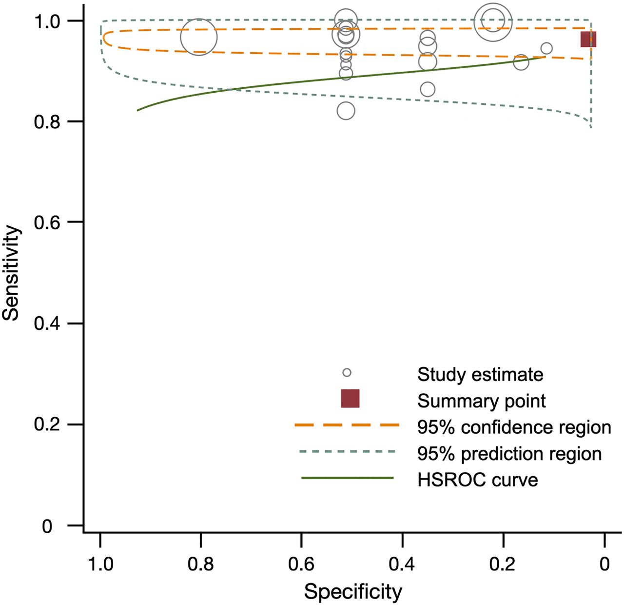
- FIGURE 2.
Summary of sensitivity, specificity, and hierarchic summary receiver-operating-characteristic (HSROC) plot of sensitivity and specificity for 18F-FCH vs. pathology overall. Effect sizes for sensitivity and specificity were 0.97 (95% CI, 0.96–0.98) and 0.23 (95% CI, 0.11–0.35), respectively. Size of circles represents size of individual studies.
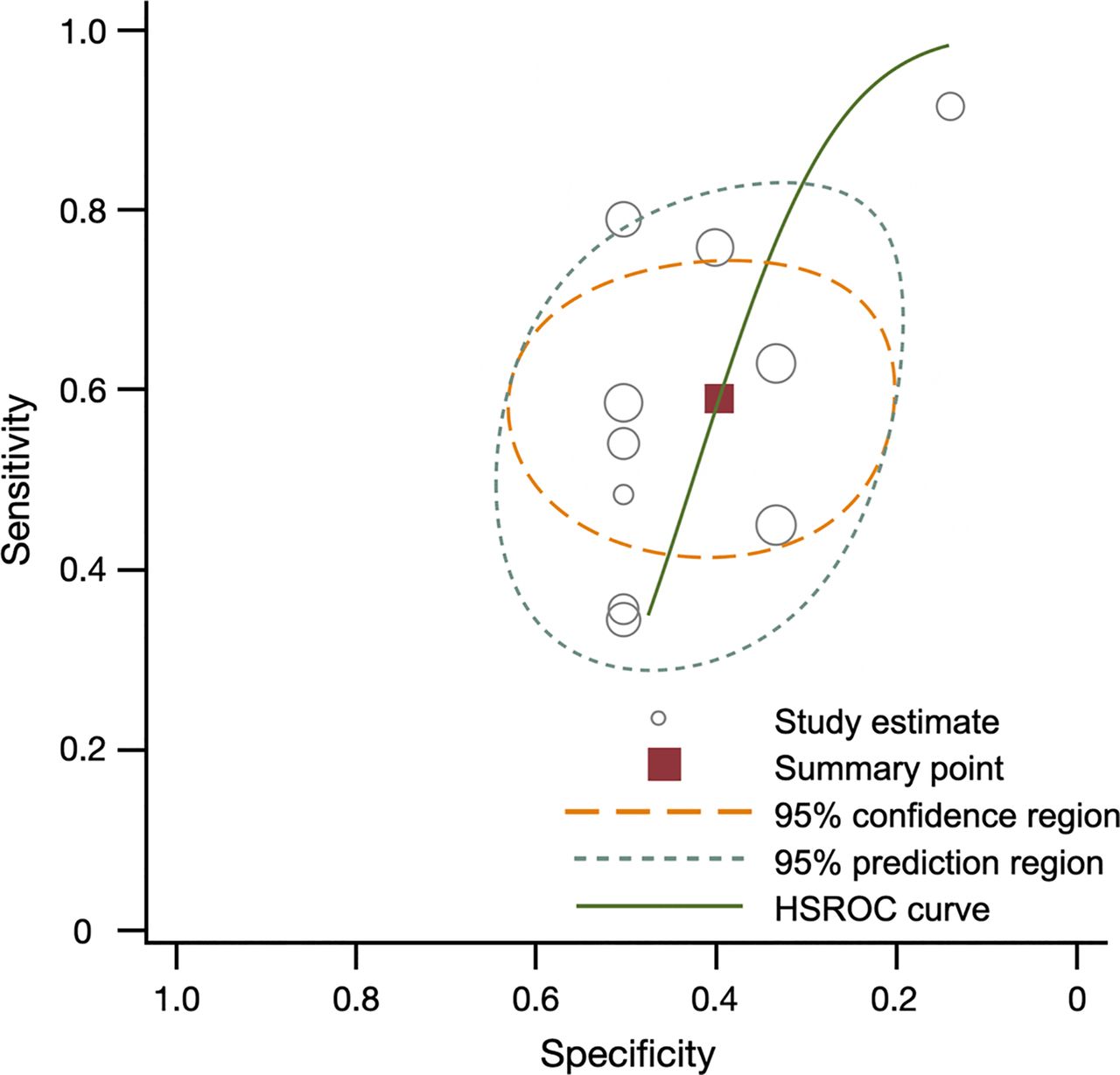
- FIGURE 3.
Summary of sensitivity, specificity, and hierarchic summary receiver-operating-characteristic (HSROC) plot of sensitivity and specificity for 99mTc-sestamibi vs. pathology overall. Effect sizes for sensitivity and specificity were 0.54 (0.29–0.79) and 0.43 (0.30–0.57), respectively. Size of circles represents size of individual studies.
- FIGURE 4.
Comparison of diagnostic sensitivities of 18F-FCH and 99mTc-sestamibi. Overall effect sizes (ES) were 0.96 (95% CI, 0.94–0.98) for 18F-FCH PET and 0.54 (95% CI, 0.29–0.79) for 99mTc-sestamibi. Size of squares represents size of individual studies. Reference numbers are in Supplemental Table 2.


Tables
First author Year Prospective or retrospective? NCT number Consent obtained Patients with 18F-FCH imaging Patients with parathyroidectomy Masked readers Readers Pathology correlation PET/CT or PET/MRI? Injected dose range (MBq) Injected dose average (MBq) Uptake time (min) Primary HPT only? Alharbi 2018 Retrospective No Yes 66 52 No 2 Yes Both NR 150 2 & 50 Yes Amadou 2019 Retrospective No No 41 23 No NR Yes PET/CT NR 231 60 Yes Bossert 2019 Prospective No Yes 34 17 Unclear 2 Yes PET/CT NR 3–3.5/kg 9 & 60 Yes Broos 2019 Prospective No Yes 271 139 Yes 3 Yes PET/CT NR 150 5 & 60 Yes Christakis 2019 Prospective No Yes 12 12 Yes 1 Yes PET/CT NR 300 60 & 90 Yes Fischli 2017 Retrospective No Yes 39 23 No 1 Yes PET/CT IQR 180–149 160 45 Yes Grimaldi 2018 Prospective No No 27 21 Unclear NR Yes PET/CT 77–230 100 30 Yes Hocevar 2017 Retrospective No No 151 151 No NR Yes PET/CT NR 100 5 & 60 Yes Huber 2018 Retrospective No Yes 26 26 Unclear NR Yes Both NR 151 45 No Khafif 2019 Prospective No Yes 19 19 No 2 Yes PET/MRI NR 93.75 16 Yes Kluijfhout 2017 Prospective No Yes 10 10 Yes 2 Yes PET/MRI 188 ± 26 188 0* Yes Kluijfhout 2016 Retrospective No Yes 33 33 Unclear NR Yes PET/CT NR 2/kg 30 No Lezaic 2014 Prospective No Yes 24 24 Unclear 2 Yes PET/CT NR 100 5 & 60 Yes López-Mora 2020 Prospective No Yes 33 33 Unclear 3 Yes PET/CT: digital vs. analog NR 0.1/kg Unclear Yes Michaud 2014 Prospective No Yes 12 12 No 1 Yes PET/CT NR 3/kg 0† No Piccardo 2019 Prospective No Yes 44 31 Unclear 2 Yes PET/CT NR 100 10 Yes Quak 2018 Prospective NCT02432599 Yes 25 24 Yes NR Yes PET/CT NR 1.5/kg 60 Yes Thanseer 2017 Prospective No Yes 54 54 Unclear NR Yes PET/CT 150–185 150–185 10–15 & 60 Yes Uslu-Beşli 2020 Retrospective No Yes 105 81 No 2 Yes PET/CT 325.1 ± 86.7 325.1 15 & 45 No Zajíčková 2018 Retrospective No Yes 13 13 Unclear 2 Yes PET/CT NR 180 30 Yes - TABLE 2
Overview of Studies Comparing Performance of 18F-FCH PET with Pathology in 20 Studies Reporting Total of 796 Patients
First author Year Patients TP FP TN FN Alharbi 2018 52 52 0 0 0 Amadou 2019 23 21 1 0 1 Bossert 2019 17 15 0 0 2 Broos 2019 139 131 0 2 6 Christakis 2019 12 7 5 0 0 Fischli 2017 23 21 1 NA 1 Grimaldi 2018 21 17 1 NA 3 Hocevar 2017 151 144 4 1 2 Huber 2018 26 25 0 0 1 Khafif 2019 19 19 0 0 0 Kluijfhout 2017 10 9 0 NA 1 Kluijfhout 2016 33 30 1 NA 2 Lezaic 2014 24 23 0 NA 1 López-Mora 2020 33 29 1 0 3 Michaud 2014 12 11 0 NA 1 Piccardo 2019 31 25 0 0 6 Quak 2018 24 19 3 NA 2 Thanseer 2017 54 52 2 NA 0 Uslu-Beşli 2020 79 76 NA NA 3 Zajíčková 2018 13 12 0 0 1 Total 796 738 19 3 33 TP = true positive; FP = false positive; TN = true negative; FN = false negative; NA = not applicable.
99mTc-sestamibi compared with pathology First author Year Patients with pathology TP FP TN FN Amadou 2019 23 9 1 0 13 Bossert 2019 17 3 0 0 14 Huber 2018 26 2 0 0 24 Khafif 2019 19 17 0 0 2 Kluijfhout 2017 33 8 0 0 21 Lezaic 2014 24 14 0 0 10 Michaud 2014 12 8 2 0 2 Thanseer 2017 54 42 1 1 10 Uslu-Beşli 2020 80 39 1 NA NA Zajíčková 2018 13 4 2 0 7 Total 301 146 7 1 103 TP = true positive; FP = false positive; TN = true negative; FN = false negative; NA = not applicable.
Supplemental Data
Files in this Data Supplement:


