


Article Figures & Data
Figures
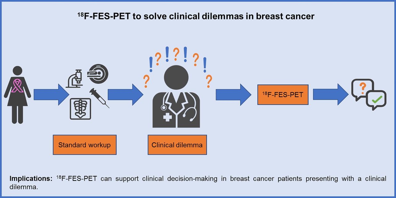
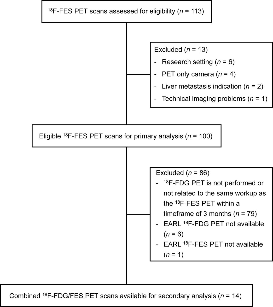
- FIGURE 1.
Consolidated Standards of Reporting Trials (CONSORT) diagram.
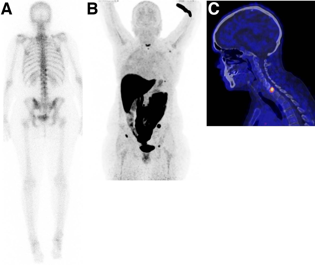
- FIGURE 2.
Equivocal lesions on standard workup. A 41-y-old woman known to have Bechterew disease was diagnosed with primary ER-positive BC 2 y previously. Conventional bone scanning was performed because of pain in neck region and showed heterogeneous uptake in spine and pelvis (A, static image posterior view). To differentiate between presence of bone metastases and lesions associated with Bechterew, 18F-FES PET scan was performed. Increased 18F-FES uptake was seen in multiple skeletal lesions: rib, left scapula, spine, and pelvis (B, maximum-intensity-projection view, and C, PET/CT sagittal view of cervical spine). On the basis of these findings, diagnosis was settled on metastatic BC, clinical dilemma was solved, and first-line endocrine treatment was started. In addition, patient received radiation to cervical spine.
- FIGURE 3.
Determination of ER status of disease. In 59-y-old woman diagnosed with ER-positive lobular BC 2 y previously and treated with tamoxifen, ER-positive bone metastases were identified 1 y after initial diagnosis. She was treated with first-line endocrine therapy in palliative setting. Thereafter, disease became progressive and palbociclib was added. However, after 2 wk of treatment, she presented with pancytopenia. 18F-FES PET was performed to determine whether bone metastases were still expressing ER and whether there was a rationale for another line of endocrine therapy. Increased 18F-FES uptake could be seen in lymph nodes above and below diaphragm and in multiple bone lesions (e.g., spine, costae, scapulae, sternum, and pelvis) (A, maximum-intensity-projection image; B, PET/CT sagittal view; C, PET/CT transversal view of left axillary region; D, PET/CT transversal view of pelvic region with positive inguinal lymph node). In addition, bone marrow involvement was visible. Diagnosis was settled on ER-positive metastatic disease, clinical dilemma was solved, and another line of endocrine therapy could be considered. However, because of bone marrow involvement, chemotherapy was indicated to achieve therapeutic effect more rapidly.
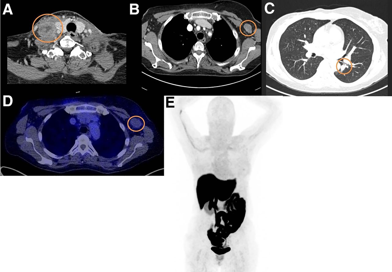
- FIGURE 4.
Inability to determine which primary tumor caused metastases. A 63-y-old woman known to have oral squamous cell carcinoma was recently diagnosed with ER-positive BC. At physical examination, a palpable mass was found in right neck region (level IV) and was also visible on CT (A). In addition, enlarged lymph node was visible in left axilla on CT (B), as well as abnormality in left lung (C). The dilemma was whether these metastases were associated with ER-positive BC or oral squamous cell carcinoma. 18F-FES PET was performed to evaluate whether these lesions were metastasis from BC (in case of 18F-FES–positive findings). However, 18F-FES PET did not show any significant tracer uptake in metastatic lesions (D and E). 18F-FES PET result did not solve dilemma, because there could be conversion from ER-positive to ER-negative status; therefore, biopsy of left axillary area was performed and confirmed presence of squamous cell carcinoma.
- FIGURE 5.
Value of 18F-FES PET in solving clinical dilemmas, per category.


Tables
Characteristic Data Mean age ± SD (y) 59 ± 11 Female (n) 99 (99%) BC stage at time of 18F-FES PET Metastatic disease* 51 (51%) Suspected metastatic disease 49 (49%) Time from primary tumor diagnosis to 18F-FES PET (y)† Median 6 Range 0–34 BC primary tumor ER expression (n = 94‡) Positive 92 (98%) Negative§ 2 (2%) Histology of primary tumor‖ (n = 87) Ductal 64 (74%) Lobular 21 (24%) Ductolobular 1 (1%) Micropapillary 1 (1%) ER expression in BC metastases¶ (n = 31)║ Positive 28 (90%) Negative# 3 (10%) Standard workup before 18F-FES PET At least 1 conventional technique** 90 (90%) CT scan 59 (59%) Bone scintigraphy 36 (36%) MRI 23 (23%) 18F-FDG PET 21 (21%) Biopsy 29 (29%) Breast lesion†† (n = 29) 12 (41%) Nonbreast lesion (n = 29) 17 (59%) ↵* Ultimately diagnosed with metastatic gastric carcinoma with breast metastases, instead of newly diagnosed metastatic BC (n = 1).
↵† If >1 primary BC, first diagnosis and histologic type of BC was included.
↵‡ In 5/6 unknown cases, metastatic lesion or secondary primary BC ER-positive.
↵§ One patient with ER-negative primary tumor presented with new palpable breast mass with metastases; it was unclear whether this new mass was secondary primary BC or recurrence, and biopsy was not possible. Another patient had mixed ER-negative and ER-positive primary tumor, which was treated as triple-negative BC.
↵‖ If >1 primary BC, first diagnosis and histologic type of BC was included.
↵¶ Metastasis biopsy was not always possible, was not performed, or was not representative; only cytology was available; or data were not available from medical records.
↵# Secondary (primary BC ER-positive).
↵** In 10 cases, standard workup could not or was not performed, for the following reasons: priority was to determine whole-body ER status for subsequent endocrine treatment (n = 4); previous tumor progression was detected only by 18F-FES PET, not by conventional imaging, so conventional imaging was deemed noninformative in present setting (n = 3); there was clinical and biochemical suspicion of tumor progression and presence of 2 different tumor types (n = 1); biopsy was not possible to determine ER status (n = 1); and after completion of chemotherapy, further diagnostic workup was required to clarify origin of cancer metastases (n = 1).
↵†† With or without axillary dissection.
Supplemental Data
Files in this Data Supplement:
In this issue


Jump to section
Related Articles
Cited By...
- Impact of 18F-FES PET/CT on Clinical Decisions in the Management of Recurrent or Metastatic Breast Cancer
- The Current and Future Roles of Precision Oncology in Advanced Breast Cancer
- Summary: Appropriate Use Criteria for Estrogen Receptor-Targeted PET Imaging with 16{alpha}-18F-Fluoro-17{beta}-Fluoroestradiol