


Article Figures & Data
Figures
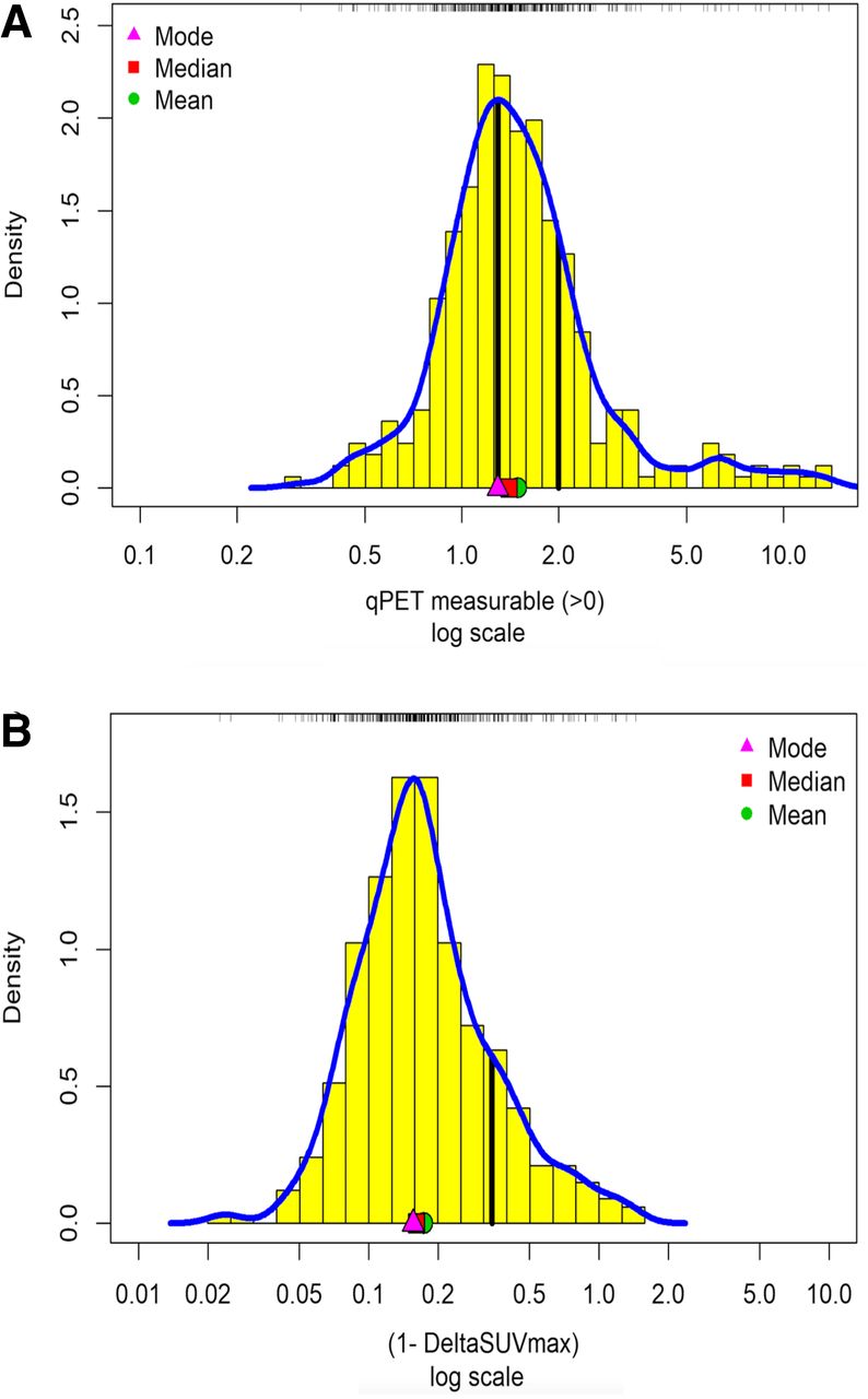
- FIGURE 1.
Density histograms for patients with measurable residual uptake at interim PET scanning (n = 332) evaluated by qPET (A) or ΔSUVmax (B) on log scale. First and second vertical lines in A indicate published thresholds between visual Deauville scores 3 and 4 (1.3) and 4 and 5 (2.0), respectively. ΔSUVmax in B is expressed as 1 − ΔSUVmax; vertical line indicates published threshold of 0.66, here 1–0.66 = 0.34.
- FIGURE 2.
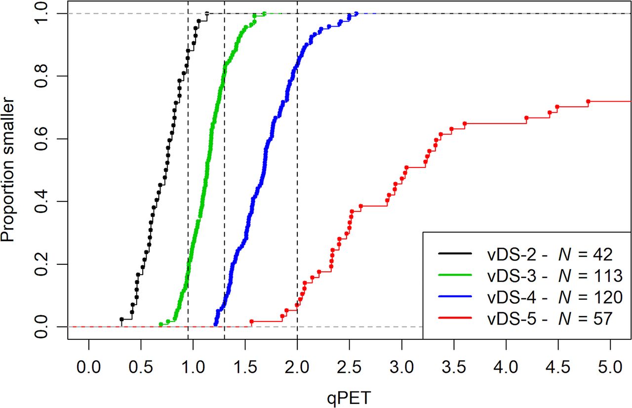
Empirical cumulative distribution functions of qPET measurements by visual Deauville categories. Vertical lines indicate published thresholds to map qPET values to individual categories. vDS = visual Deauville score.
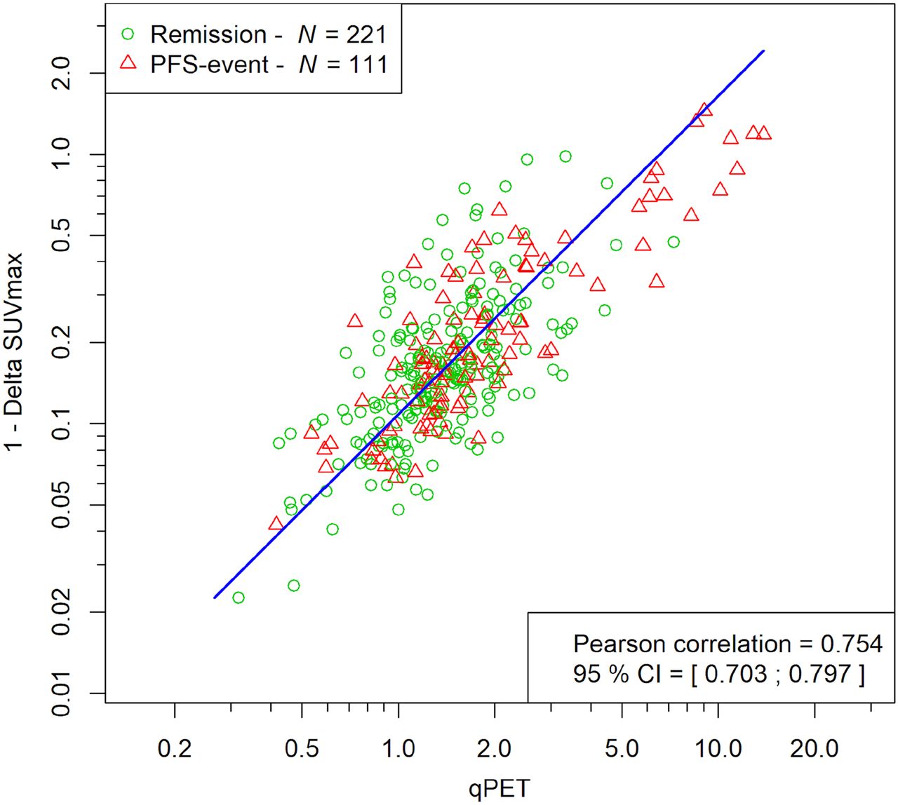
- FIGURE 3.
Scatterplot of qPET and ΔSUVmax. Triangles refer to patients experiencing treatment failure, whereas circles refer to patients who remained in remission. Blue line is the principal axis illustrating correlation. PFS = progression-free survival.
- FIGURE 4.
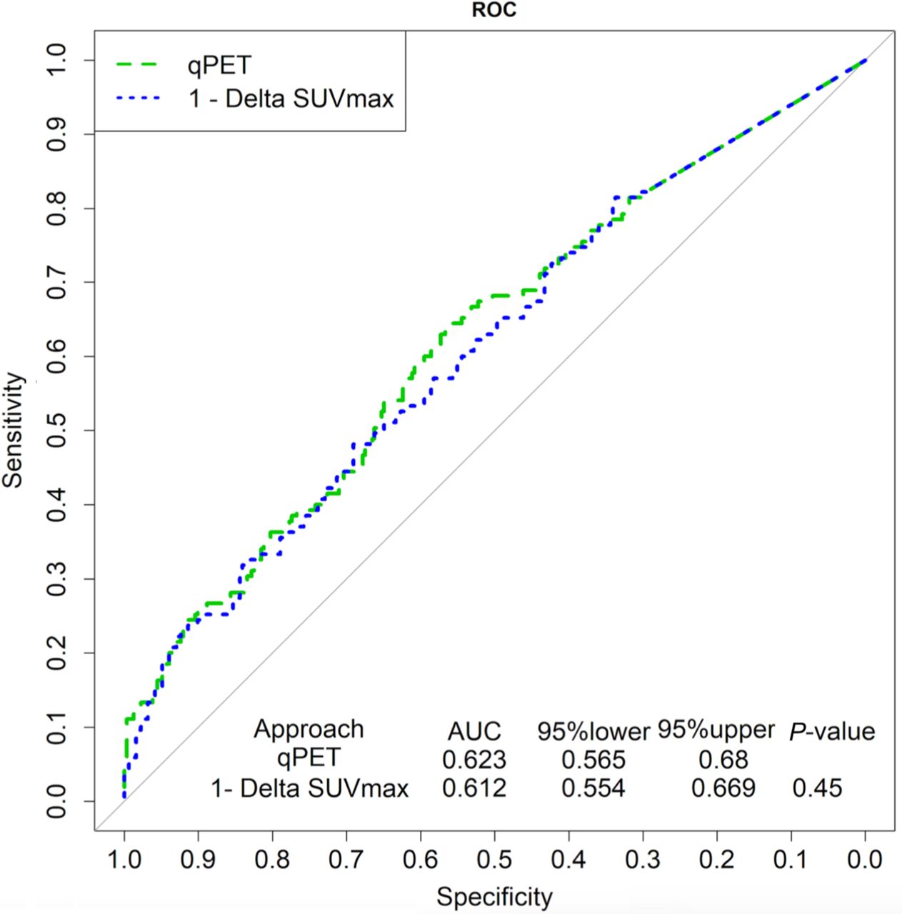
ROC curves of qPET and 1 − ΔSUVmax for progression-free survival.
- FIGURE 5.
Positive predictive value (A) and negative predictive value (B) of corresponding percentiles of qPET and ΔSUVmax measurements. Constant part of curves at low percentiles is due to inclusion of nonmeasurable values set at zero (n = 117).
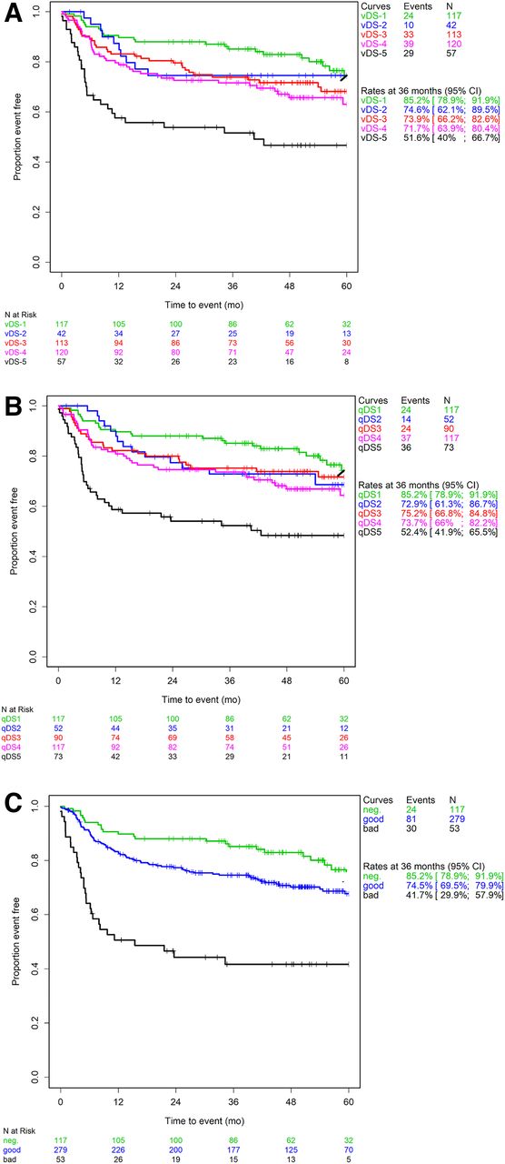
- FIGURE 6.
Progression-free survival in prognostic subgroups derived from visual Deauville scale (A), quantitative Deauville scale (B), or ΔSUVmax scale (C) (Kaplan–Meier analysis). vDS = visual Deauville score; qDS = quantitative Deauville score.
- FIGURE 7.
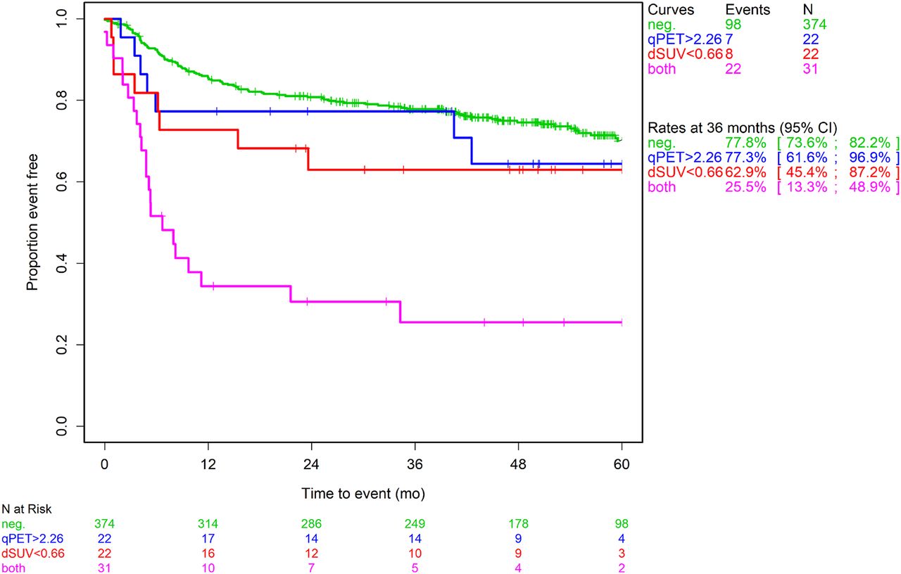
Progression-free survival in patients with good interim PET response according to both qPET and ΔSUVmax, only qPET, or only ΔSUVmax, or with poor interim PET response according to both methods (Kaplan–Meier analysis). dSUV = ΔSUVmax.


Tables
- TABLE 1
Baseline Characteristics of Patients Included in Present Analysis in Comparison to Excluded Patients and All DLBCL Patients Participating in PETAL Trial
Characteristic Patients included Patients excluded All patients No. of patients 449 160 609 Median age (y) 62 (range, 18–80) 59.5 (range, 18–79) 62 (range, 18–80) Age > 60 y 236 (52.6%) 78 (48.8%) 314 (51.6%) Male sex 249 (55.5%) 93 (58.1%) 342 (56.2%) ECOG performance status ≥ 2 48 (10.7%) 11 (6.9%) 59 (9.7%) Ann Arbor stage III or IV 258 (57.5%) 100 (62.5) 358 (58.8%) Extranodal sites > 1 148 (33.0%) 50 (31.2%) 198 (32.6%) Lactate dehydrogenase > ULN 257 (57.4%) 78 (48.8%) 335 (55.1%) International Prognostic Index Low risk 160 (35.7%) 64 (40.0%) 224 (36.8%) Low-intermediate risk 111 (24.8%) 47 (29.4%) 158 (26.0%) High-intermediate risk 102 (22.8%) 25 (15.6%) 127 (20.9%) High risk 75 (16.7%) 24 (15.0%) 99 (16.3%) ECOG = Eastern Cooperative Oncology Group; ULN = upper limit of normal.
Data are given as number of patients affected, followed by percentage of total number of patients with documented data, unless otherwise noted.
Quantitative Visual 1 2 3 4 5 Sum 1 117 0 0 0 0 117 2 0 35 7 0 0 42 3 0 17 74 22 0 113 4 0 0 9 91 20 120 5 0 0 0 4 53 57 Sum 117 52 90 117 73 449 - TABLE 3
Positive and Negative Predictive Values and Proportion of High-Risk Patients Identified by Interim PET: Comparison of Methods and Thresholds
Definition of high-risk patients PPV NPV Proportion of high-risk patients Visual Deauville score 4 or 5 38.4% 75.4% 39.4% Visual Deauville score 5 50.9% 73.0% 12.7% Quantitative Deauville score 4 or 5 38.4% 76.1% 42.3% Quantitative Deauville score 5 49.3% 73.7% 16.3% qPET ≥ 2.26 54.7% 73.2% 11.8% ΔSUVmax, ≤66% SUVmax reduction 56.6% 73.5% 11.8% PPV = positive predictive value; NPV = negative predictive value.
- TABLE 4
Selected Corresponding Thresholds in Categoric and Continuous Interim PET Response Scales
vDS qPET ΔSUVmax 2/3 0.95 91% 3/4 1.30 85% 4/5 2 73% 5 2.26 66% vDS = visual Deauville score.
Supplemental Data
Files in this Data Supplement:


