


Article Figures & Data
Figures
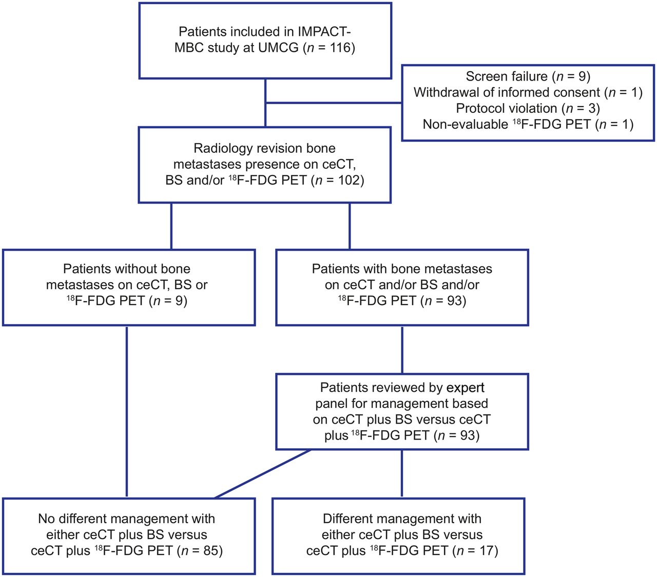
- FIGURE 1.
Flow chart visualizing selection of patients and how they are analyzed. UMCG = University Medical Center Groningen; CAD = calcium, vitamin D.
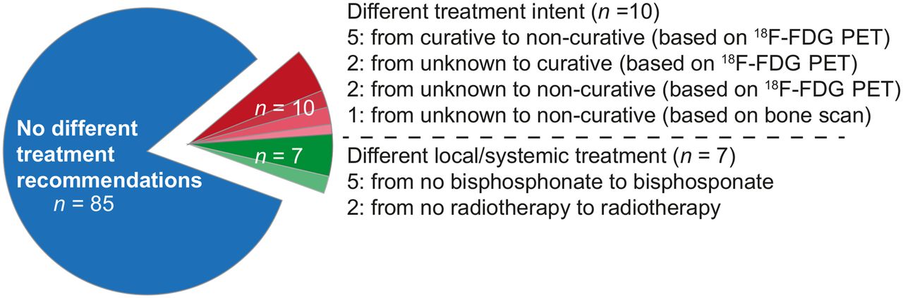
- FIGURE 2.
Pie chart visualizing number of patients with clinically relevant difference in management recommendations after 18F-FDG PET plus ceCT (n = 16) and BS plus ceCT (n = 1).
- FIGURE 3.
Example patient with clinically relevant difference in management recommendation after 18F-FDG PET plus ceCT compared with BS plus ceCT (patient 14 of Table 3). (A) ceCT visualized 1 bone metastasis in T5 (transversal section of ceCT through T5) and 3 equivocal lesions in iliac bone (1 on left, 2 on right). (B) BS visualized no bone lesions. (C) Maximum-intensity-projection 18F-FDG PET visualized 13 bone lesions (C) (C3, C5, T5, L3, L4, sacral bone [2], left acetabulum, right costa 7, left costa 5 [2], right humerus, and sternal bone). No equivocal lesions on ceCT were detected as metastases on 18F-FDG PET.


Tables
Parameter Data Breast cancer type Invasive carcinoma* 84 (82) Lobular 13 (13) Other 5 (5) Tumor characteristics, primary Elston grade 1 8 (8) Elston grade 2 59 (58) Elston grade 3 33 (32) Elston grade unknown 2 (2) HR-negative 17 (17) HR-positive 85 (83) HER2-negative 78 (76) HER2-positive 24 (24) Triple-negative 7 (7) Metastasis HR-negative 16 (16) HR-positive 81 (79) HER2-negative 77 (75) HER2-positive 20 (20) Triple-negative 7 (7) Unknown 5 (5) Time to tumor recurrence (mo) 77.5 (0.3–293.5) ↵* No special type; previously known as ductal.
HR = hormone receptor; HER2 = human epidermal growth factor receptor 2.
Qualitative data are numbers followed by percentages in parentheses; continuous data are median followed by range in parentheses. n = 102 patients.
- TABLE 2
Concordance of Management Recommendations by Expert Panel Based on Evaluation of Bone Lesions on BS Plus ceCT or 18F-FDG PET Plus ceCT
Recommendation 18F-FDG PET plus ceCT BS plus ceCT Concordant recommendation Treatment intent Curative 7 (6.9) 11 (10.8) 5 (4.9) Noncurative 83 (81.4) 77 (75.5) 76 (74.5) Unable to determine 3 (2.9) 5 (4.9) 1 (1.1) Not evaluated* 9 (8.8) 9 (8.8) 9 (8.8) Systemic therapy None 0 2 (2.0) 0 Antihormonal 57 (55.9) 53 (52.0) 51 (50) Chemotherapy 14 (13.7) 10 (9.8) 10 (9.8) Chemotherapy plus targeted therapy 17 (16.7) 16 (15.7) 16 (15.7) Unable to determine 5 (4.9) 12 (11.8) 3 (2.9) Not evaluated* 9 (8.8) 9 (8.8) 9 (8.8) CAD/bisphosphonate No 15 (14.7) 27 (26.5) 14 (13.7) Yes 77 (75.5) 62 (60.8) 62 (60.8) Unable to determine 1 (1.0) 4 (3.9) 0 Not evaluated* 9 (8.8) 9 (8.8) 9 (8.8) Radiotherapy None 62 (60.8) 56 (60.2) 39 (38.2) Curative 10 (9.8) 6 (6.5) 2 (2.0) Noncurative 20 (19.6) 25 (26.9) 8 (7.8) Unable to determine 1 (1.0) 6 (6.5) 1 (1.0) Not evaluated* 9 (8.8) 9 (8.8) 9 (8.8) Radiography/MRI No 63 (61.8) 65 (63.7) 53 (52.0) Yes 30 (29.4) 28 (27.5) 18 (17.6) Not evaluated* 9 (8.8) 9 (8.8) 9 (8.8) Other imaging† No 92 (90.2) 66 (64.7) 39 (38.2) Yes 1 (1.0) 27 (26.5) 1 (1.0) Not evaluated* 9 (8.8) 9 (8.8) 9 (8.8) ↵* For patients without bone lesions on any imaging modalities, it was assumed that management recommendations would be concordant between scenarios with 18F-FDG PET plus ceCT and BS plus ceCT and that there would be no requests for additional imaging to evaluate bone lesions, without evaluation of expert panel.
↵† Additional 18F-FDG PET in case of available BS and vice versa.
CAD = calcium, vitamin D.
Data are numbers followed by percentages in parentheses. n = 102 patients.
- TABLE 3
Predictors of Clinically Relevant Change in Management Recommendations After Evaluation of Bone Lesions by 18F-FDG PET Plus ceCT Instead of BS Plus ceCT
Univariable analysis Predictor Same treatment, n = 86 (84.3%) Difference in treatment, n = 16 (15.7%)* P AUC 95% CI Ductal histology of primary tumor (n) No 16 (88.9%) 2 (11.1%) 0.73 0.53 0.44–0.62 Yes 70 (83.3%) 14 (16.7%) Hormone receptor status of primary tumor (n) Negative 12 (70.6%) 5 (29.4%) 0.14 0.59 0.46–0.71 Positive 74 (87.1%) 11 (12.9%) HER2 status of primary tumor (n) Negative 67 (85.9%) 11 (14.1%) 0.52 0.55 0.42–0.67 Positive 19 (79.2%) 5 (20.8%) Grade of primary tumor (n) Grade 1 or 2 57 (85.1%) 10 (14.9%) 0.77 0.53 0.39–0.66 Grade 3 27 (81.8%) 6 (18.2%) Suspicion of bone metastases (n) No 63 (85.1%) 11 (14.9%) 0.76 0.52 0.40–0.65 Yes 23 (82.1%) 5 (17.9%) Relapse time (y) 0.70 0.53 0.37–0.69 Median 6 7 25th–75th percentiles 2%–9% 4%–10% Bone lesions on CT (n) 0.018 0.68 0.58–0.79 Median 4 1 25th–75th percentiles 0%–20% 0%–1% ↵* For this analysis of potential predictors of management recommendations based on 18F-FDG PET, 16 patients were included. Patient with changed recommendations based on BS was not included.
AUC = area under receiver-operating-characteristic curve.
Supplemental Data
Files in this Data Supplement:


Jump to section
Related Articles
Cited By...
- No citing articles found.