
Main menu

  • Home
  • Content
    • Current
    • Ahead of print
    • Past Issues
    • JNM Supplement
    • SNMMI Annual Meeting Abstracts
    • Continuing Education
    • JNM Podcasts
  • Subscriptions
    • Subscribers
    • Institutional and Non-member
    • Rates
    • Journal Claims
    • Corporate & Special Sales
  • Authors
    • Submit to JNM
    • Information for Authors
    • Assignment of Copyright
    • AQARA requirements
  • Info
    • Reviewers
    • Permissions
    • Advertisers
  • About
    • About Us
    • Editorial Board
    • Contact Information
  • More
    • Alerts
    • Feedback
    • Help
    • SNMMI Journals
  • SNMMI
    • JNM
    • JNMT
    • SNMMI Journals
    • SNMMI

User menu

  • Subscribe
  • My alerts
  • Log in
  • Log out
  • My Cart

Search

  • Advanced search
Journal of Nuclear Medicine
  • SNMMI
    • JNM
    • JNMT
    • SNMMI Journals
    • SNMMI
  • Subscribe
  • My alerts
  • Log in
  • Log out
  • My Cart
Journal of Nuclear Medicine

Advanced Search

  • Home
  • Content
    • Current
    • Ahead of print
    • Past Issues
    • JNM Supplement
    • SNMMI Annual Meeting Abstracts
    • Continuing Education
    • JNM Podcasts
  • Subscriptions
    • Subscribers
    • Institutional and Non-member
    • Rates
    • Journal Claims
    • Corporate & Special Sales
  • Authors
    • Submit to JNM
    • Information for Authors
    • Assignment of Copyright
    • AQARA requirements
  • Info
    • Reviewers
    • Permissions
    • Advertisers
  • About
    • About Us
    • Editorial Board
    • Contact Information
  • More
    • Alerts
    • Feedback
    • Help
    • SNMMI Journals
  • View or Listen to JNM Podcast
  • Visit JNM on Facebook
  • Join JNM on LinkedIn
  • Follow JNM on Twitter
  • Subscribe to our RSS feeds
Meeting ReportOncology: Clinical Therapy and Diagnosis

Prognostic value of pretreatment FDG PET/CT in uterine cervical cancer: significance of histopathological subtypes

Tomoaki Otani, Yuji Nakamoto, Takayoshi Ishimori, Tsuneo Saga and Kaori Togashi
Journal of Nuclear Medicine May 2020, 61 (supplement 1) 352;
Tomoaki Otani
1Radiology resident Kyoto Japan
  • Find this author on Google Scholar
  • Find this author on PubMed
  • Search for this author on this site
Yuji Nakamoto
2Kyoto University Kyoto Japan
  • Find this author on Google Scholar
  • Find this author on PubMed
  • Search for this author on this site
Takayoshi Ishimori
3Radiology, Diagnostic Imaging and Nuclear Medicine Kyoto University Kyoto Japan
  • Find this author on Google Scholar
  • Find this author on PubMed
  • Search for this author on this site
Tsuneo Saga
4Department of Daignostic Radiology Kyoto University Hospital Kyoto Japan
  • Find this author on Google Scholar
  • Find this author on PubMed
  • Search for this author on this site
Kaori Togashi
5Kyoto University Hospital Kyoto Japan
  • Find this author on Google Scholar
  • Find this author on PubMed
  • Search for this author on this site
  • Article
  • Figures & Data
  • Info & Metrics
Loading

Abstract

352

Objectives: There are two major histopathological subtypes in uterine cervical carcinoma: squamous cell carcinoma (SCC) and adenocarcinoma (AC). FDG-PET/CT (PET/CT) has been reported to have a prognostic value in various malignancies, including uterine cervical cancer, but its prognostic value for each histopathological type has not been fully investigated. The aim of this study was to assess the prognostic value of pretreatment PET/CT in cervical cancer according to histopathological types.

Methods: We retrospectively analyzed 83 SCC patients and 35 AC patients (age: 22-95 yr) who underwent pretreatment PET/CT. Quantitative indices, such as SUVmax, SUVmean, metabolic tumor volume (MTV), and total lesion Glycolysis (TLG) of primary tumor were calculated for each patient. These parameters and tumor size were compared using Mann-Whitney’s U test between AC and SCC. The receiver-operating-characteristic analysis was performed to determine the optimal cut-off values for SUVmax, SUVmean, MTV, and TLG. Kaplan-Meier analysis and log-rank test were used to estimate and compare the correlation between each PET parameter and progression-free survival (PFS)/Overall survival (OS). The prognostic value of the imaging parameters and clinical parameters including age, FIGO stage, histological subtype was assessed using univariate Cox proportional hazard model.

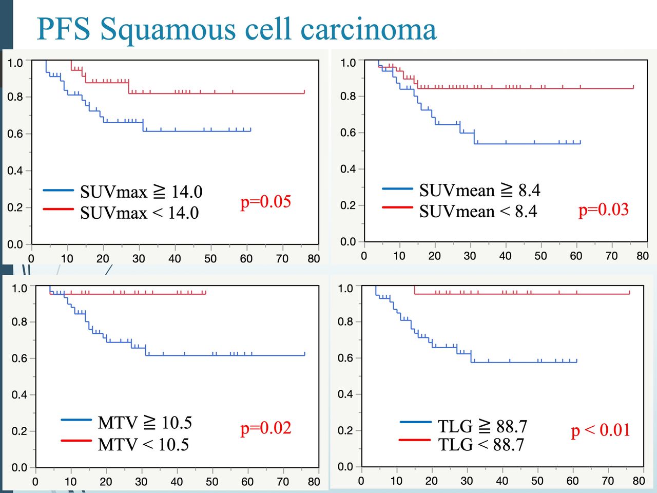
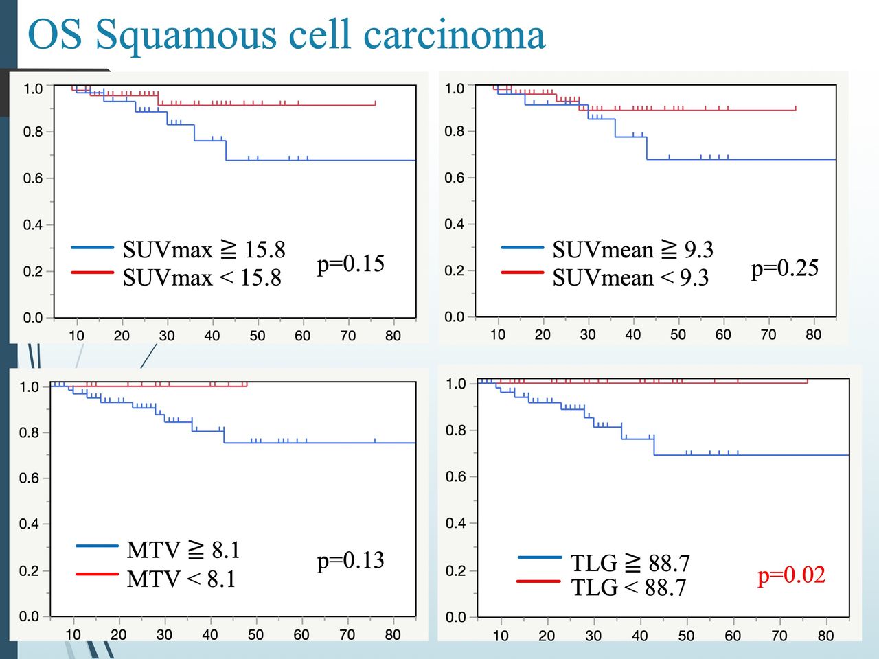
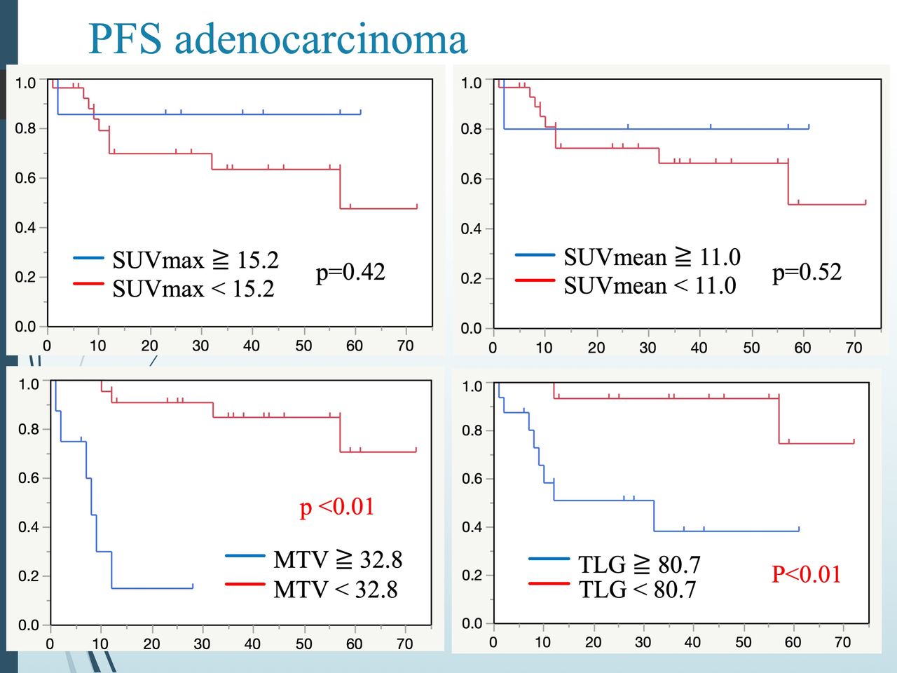
Results: The median follow-up duration was 27 (range: 6-85) months in SCC and 28 (range: 6-72) months in AC. In SCC, at the time of last follow-up, 64 (77 %) were alive without recurrence, 19 (23 %) were developed recurrence, and 10 (12 %) were died from the disease progression. In AC, 25 (71 %) patients were alive without recurrence, 10 (29 %) were developed recurrence, and 4 (11 %) were died from the disease progression. In SCC, 19 (23%) patients had PET positive pelvic lymph nodes, and 12 (14%) patients had para-aorta lymph nodes. In AC, 8 (23%) patients had PET positive pelvic lymph nodes, and 4 (11%) patients had para-aorta lymph nodes.The values of SUVmax, SUVmean, MTV and TLG of SCC patients were 14.9±6.4, 8.0±3.2, 32.2±37.1, and 283.9±345.5, respectively, and those of AC patients were 10.5±7.6, 6.3±4.7, 21.5±23.1, and 121.6±127.7, respectively. The SUVmax, SUVmean, and TLG of SCC were higher than those of AC (p<0.01, <0.01, and <0.01, respectively). There was no significant difference in MTV between the two groups (p=0.10). Tumor size in SCC was significantly larger than that in AC (42.5±16.5 mm vs 35.0±18.6 mm, p=0.02).In SCC, the Kaplan-Meier curves for PFS and OS indicated better outcomes for the lower groups in SUVmax, SUVmean, MTV and TLG (p=0.05, p=0.03, p=0.02, p<0.01 for PFS, and p=0.15, p=0.25, p=0.13, p=0.02 for OS, respectively). On the other hand, in AC, they indicated better outcomes for the lower groups in MTV and TLG (p<0.01, p<0.01 for PFS, and p<0.01, p<0.01 for OS), while SUVmax and SUVmean were not related to PSF and OS (p=0.42, p=0.52 for PSF, and p=0.73, p=0.97 for OS). As for univariate analysis, in SCC, higher SUVmax, SUVmean, MTV, and TLG correlated significantly with poorer PFS (p=0.04, 0.03, <0.01, and <0.01, respectively) and MTV, TLG, and tumor size correlated significantly with poorer OS (p=0.04, <0.01, and 0.02, respectively). In AC, MTV, TLG, tumor size, age, and FIGO stage correlated significantly with poorer PFS (p<0.01, <0.01, <0.01, <0.01, and <0.01, respectively) and MTV, TLG, and tumor size, and FIGO stage correlated significantly with poorer OS (p<0.01, <0.01, <0.01, and <0.01, respectively)

Conclusions: Our preliminary data suggest that PET/CT would be useful for predicting prognosis in cervical cancer, but clinical significance of quantitative values may be different according to histopathological types.

Figure
  • Download figure
  • Open in new tab
  • Download powerpoint
Figure
  • Download figure
  • Open in new tab
  • Download powerpoint
Figure
  • Download figure
  • Open in new tab
  • Download powerpoint
Figure
  • Download figure
  • Open in new tab
  • Download powerpoint
Previous
Back to top

In this issue

Journal of Nuclear Medicine
Vol. 61, Issue supplement 1
May 1, 2020
  • Table of Contents
  • Index by author
Article Alerts
Sign In to Email Alerts with your Email Address
Email Article

Thank you for your interest in spreading the word on Journal of Nuclear Medicine.

NOTE: We only request your email address so that the person you are recommending the page to knows that you wanted them to see it, and that it is not junk mail. We do not capture any email address.

Enter multiple addresses on separate lines or separate them with commas.
Prognostic value of pretreatment FDG PET/CT in uterine cervical cancer: significance of histopathological subtypes
(Your Name) has sent you a message from Journal of Nuclear Medicine
(Your Name) thought you would like to see the Journal of Nuclear Medicine web site.
Citation Tools
Prognostic value of pretreatment FDG PET/CT in uterine cervical cancer: significance of histopathological subtypes
Tomoaki Otani, Yuji Nakamoto, Takayoshi Ishimori, Tsuneo Saga, Kaori Togashi
Journal of Nuclear Medicine May 2020, 61 (supplement 1) 352;

Citation Manager Formats

  • BibTeX
  • Bookends
  • EasyBib
  • EndNote (tagged)
  • EndNote 8 (xml)
  • Medlars
  • Mendeley
  • Papers
  • RefWorks Tagged
  • Ref Manager
  • RIS
  • Zotero
Share
Prognostic value of pretreatment FDG PET/CT in uterine cervical cancer: significance of histopathological subtypes
Tomoaki Otani, Yuji Nakamoto, Takayoshi Ishimori, Tsuneo Saga, Kaori Togashi
Journal of Nuclear Medicine May 2020, 61 (supplement 1) 352;
Twitter logo Facebook logo LinkedIn logo Mendeley logo
  • Tweet Widget
  • Facebook Like
  • Google Plus One
Bookmark this article

Jump to section

  • Article
  • Figures & Data
  • Info & Metrics

Related Articles

  • No related articles found.
  • Google Scholar

Cited By...

  • No citing articles found.
  • Google Scholar

More in this TOC Section

Oncology: Clinical Therapy and Diagnosis

  • Added value of single photon emission computed tomography (SPECT) / Computed Tomography (CT) with radioiodine whole body scan in follow up of patients with differentiated thyroid cancer
  • Sequential occurrence of paraganglioma in abdomen, neck and head demonstrated by serial Ga68-DOTANOC PET-CT
  • The value of 18F-FDG PET / MR in skull-base bone invasion of nasopharyngeal carcinoma
Show more Oncology: Clinical Therapy and Diagnosis

Oncology Clinical Diagnosis: Gynecological

  • PREDICTION OF LYMPH NODE METASTASIS FROM 18F-FDG PET/CT RADIOMICS OF CERVICAL CANCER
  • Role of Fluorine-18 fluorodeoxyglucose PET/CT in post-surgery surveillance of uterine sarcoma
  • Radiomics analysis of uterine tumors in 18F-flourodeoxyglucose positron emission tomography for prediction of lymph node metastases in endometrial carcinoma
Show more Oncology Clinical Diagnosis: Gynecological

Similar Articles

SNMMI

© 2025 SNMMI

Powered by HighWire