
Main menu

  • Home
  • Content
    • Current
    • Ahead of print
    • Past Issues
    • JNM Supplement
    • SNMMI Annual Meeting Abstracts
    • Continuing Education
    • JNM Podcasts
  • Subscriptions
    • Subscribers
    • Institutional and Non-member
    • Rates
    • Journal Claims
    • Corporate & Special Sales
  • Authors
    • Submit to JNM
    • Information for Authors
    • Assignment of Copyright
    • AQARA requirements
  • Info
    • Reviewers
    • Permissions
    • Advertisers
  • About
    • About Us
    • Editorial Board
    • Contact Information
  • More
    • Alerts
    • Feedback
    • Help
    • SNMMI Journals
  • SNMMI
    • JNM
    • JNMT
    • SNMMI Journals
    • SNMMI

User menu

  • Subscribe
  • My alerts
  • Log in
  • Log out
  • My Cart

Search

  • Advanced search
Journal of Nuclear Medicine
  • SNMMI
    • JNM
    • JNMT
    • SNMMI Journals
    • SNMMI
  • Subscribe
  • My alerts
  • Log in
  • Log out
  • My Cart
Journal of Nuclear Medicine

Advanced Search

  • Home
  • Content
    • Current
    • Ahead of print
    • Past Issues
    • JNM Supplement
    • SNMMI Annual Meeting Abstracts
    • Continuing Education
    • JNM Podcasts
  • Subscriptions
    • Subscribers
    • Institutional and Non-member
    • Rates
    • Journal Claims
    • Corporate & Special Sales
  • Authors
    • Submit to JNM
    • Information for Authors
    • Assignment of Copyright
    • AQARA requirements
  • Info
    • Reviewers
    • Permissions
    • Advertisers
  • About
    • About Us
    • Editorial Board
    • Contact Information
  • More
    • Alerts
    • Feedback
    • Help
    • SNMMI Journals
  • View or Listen to JNM Podcast
  • Visit JNM on Facebook
  • Join JNM on LinkedIn
  • Follow JNM on Twitter
  • Subscribe to our RSS feeds
Meeting ReportOncology: Clinical Therapy and Diagnosis

The value of 18F-FDG PET / MR in skull-base bone invasion of nasopharyngeal carcinoma

Shifeng Wang, Yuanfan Xu, Qiang Li, Jiangtao Liang, YUE LIU and yanfang zhu
Journal of Nuclear Medicine May 2020, 61 (supplement 1) 1288;
Shifeng Wang
1Hangzhou Universal Imaging Diagnostic Center Hangzhou China
  • Find this author on Google Scholar
  • Find this author on PubMed
  • Search for this author on this site
Yuanfan Xu
1Hangzhou Universal Imaging Diagnostic Center Hangzhou China
  • Find this author on Google Scholar
  • Find this author on PubMed
  • Search for this author on this site
Qiang Li
1Hangzhou Universal Imaging Diagnostic Center Hangzhou China
  • Find this author on Google Scholar
  • Find this author on PubMed
  • Search for this author on this site
Jiangtao Liang
1Hangzhou Universal Imaging Diagnostic Center Hangzhou China
  • Find this author on Google Scholar
  • Find this author on PubMed
  • Search for this author on this site
YUE LIU
1Hangzhou Universal Imaging Diagnostic Center Hangzhou China
  • Find this author on Google Scholar
  • Find this author on PubMed
  • Search for this author on this site
yanfang zhu
1Hangzhou Universal Imaging Diagnostic Center Hangzhou China
  • Find this author on Google Scholar
  • Find this author on PubMed
  • Search for this author on this site
  • Article
  • Figures & Data
  • Info & Metrics
Loading

Abstract

1288

Objectives: To explore the value of 18F-FDG PET/MR in evaluating skull-base bone invasion of nasopharyngeal carcinoma.

Methods: One hundred and twenty-four patients with NPC who underwent PET/MR and enhanced MR scan before or after treatment were reviewed retrospectively. All images were analyzed by an experienced PET/MR doctor and a radiotherapy doctors. Difference of metabolism between the bone invasion region and tumor body was compared and the metabolic differences of different types of bone invasion was also compared. Difference between GTVPET(gross tumor volume), a tumor target region delineated with 30% of SUVmax as a threshold, and GTVMR, a tumor target region delineated with fat suppression enhanced T2 sequence was compared.

Results: 68 patients showed skull-base bone erosion, of the 68 patients, 24 showed osteolytic bone destruction and 44 showed bone marrow infiltration. The osteolytic bone destruction area is characterized by soft tissue mass signals. The bone marrow infiltration type is characterized by low signal intensity replaced high signal intensity in the bone marrow on the T1-weighted images or when hyperintensity was observed on the fat-suppressed T2WI, and a high signal was seen on DWI. All enhanced scans of the bone invasion area showed abnormal enhancement. The SUVmax [3.31(1.64,4.95)]of the skull base lesion area was significantly lower than that [9.77(6.47,11.54)]of the main body of the nasopharyngeal lesion (Z=-7.143,P<0.01). The SUVmax [3.22(2.44,3.60)]of bone lesions in bone marrow infiltrating cases was significantly lower than that[6.18(4.72,7.45)] of osteolytic bone destruction (Z=-6.776,P<0.01). Among the 44 cases of bone marrow infiltration, FDG metabolism in the visible lesion area was higher than that in the contralateral or surrounding normal bone in 23 cases, and no intense high metabolic region was observed in 21 cases. The area under the ROC curve (AUC) for the evaluation of skull base invasion by SUVmax was 0.866, the SUVmax corresponding to the most approximate index was 1.5, and the sensitivity and specificity were 79.4% and 46.0%, respectively. In osteolytic bone destruction cases, 4.17% (1/24) of the cases of skull-base bone lesions were located outside the GTVPET area, and 61.36% (27/44) of bone marrow infiltration cases were located outside the GTVPET area,there were statistical differences(χ2=20.97,P<0.01). GTVPET [35.46(25.27,43.56)] is smaller than GTVMR [41.27(30.45,29.58)] (Z=-5.316,P<0.01).

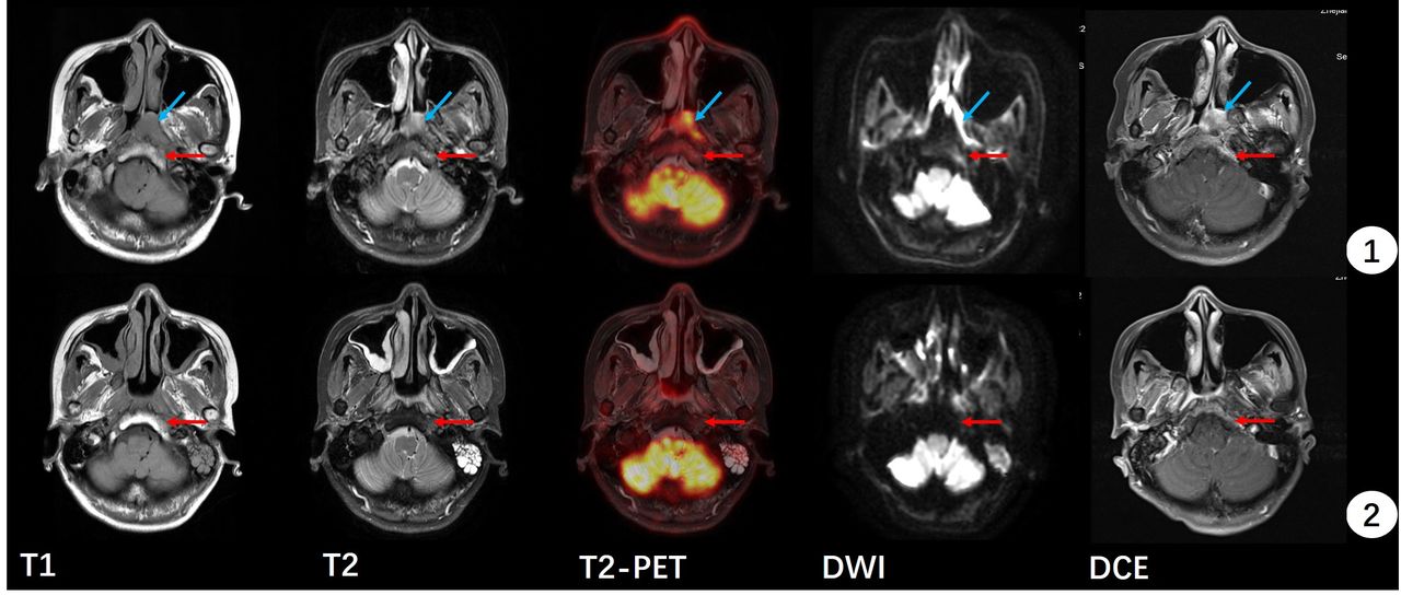
Conclusions: Although PET / MR is of great value for staging of nasopharyngeal carcinoma, FDG uptake in the area of &#8203;&#8203;skull base invasion is lower than that of nasopharyngeal lesions, and false negatives often occur in bone marrow infiltrating types. Evaluation of skull base bone invasion and radiotherapy target region with PET metabolic range may not be accurate, and enhanced MR is still a necessary detection method for pre-treatment evaluation and efficacy evaluation.Female, 40 years old. Diagnosis of nasopharyngeal cancer (blue arrow). 1) pre-treatment; 2) post- treatment. The clivus signal behind the nasopharyngeal mass was abnormal before treatment. It showed a low signal on T1WI, an asymmetric hyperintense on T2WI, a high signal on DWI ,no significant increase in FDG uptake and enhancement was visible and significantly enhanced. Post-treatment, the nasopharyngeal mass subsided, and the clivus lesions still showed a low signal on T1WI, lower signal on T2WI than pre-treatment, no significant increase in FDG uptake, no high signal on DWI and no significant enhancement was observed.

Figure
  • Download figure
  • Open in new tab
  • Download powerpoint
Previous
Back to top

In this issue

Journal of Nuclear Medicine
Vol. 61, Issue supplement 1
May 1, 2020
  • Table of Contents
  • Index by author
Article Alerts
Sign In to Email Alerts with your Email Address
Email Article

Thank you for your interest in spreading the word on Journal of Nuclear Medicine.

NOTE: We only request your email address so that the person you are recommending the page to knows that you wanted them to see it, and that it is not junk mail. We do not capture any email address.

Enter multiple addresses on separate lines or separate them with commas.
The value of 18F-FDG PET / MR in skull-base bone invasion of nasopharyngeal carcinoma
(Your Name) has sent you a message from Journal of Nuclear Medicine
(Your Name) thought you would like to see the Journal of Nuclear Medicine web site.
Citation Tools
The value of 18F-FDG PET / MR in skull-base bone invasion of nasopharyngeal carcinoma
Shifeng Wang, Yuanfan Xu, Qiang Li, Jiangtao Liang, YUE LIU, yanfang zhu
Journal of Nuclear Medicine May 2020, 61 (supplement 1) 1288;

Citation Manager Formats

  • BibTeX
  • Bookends
  • EasyBib
  • EndNote (tagged)
  • EndNote 8 (xml)
  • Medlars
  • Mendeley
  • Papers
  • RefWorks Tagged
  • Ref Manager
  • RIS
  • Zotero
Share
The value of 18F-FDG PET / MR in skull-base bone invasion of nasopharyngeal carcinoma
Shifeng Wang, Yuanfan Xu, Qiang Li, Jiangtao Liang, YUE LIU, yanfang zhu
Journal of Nuclear Medicine May 2020, 61 (supplement 1) 1288;
Twitter logo Facebook logo LinkedIn logo Mendeley logo
  • Tweet Widget
  • Facebook Like
  • Google Plus One
Bookmark this article

Jump to section

  • Article
  • Figures & Data
  • Info & Metrics

Related Articles

  • No related articles found.
  • Google Scholar

Cited By...

  • No citing articles found.
  • Google Scholar

More in this TOC Section

Oncology: Clinical Therapy and Diagnosis

  • Added value of single photon emission computed tomography (SPECT) / Computed Tomography (CT) with radioiodine whole body scan in follow up of patients with differentiated thyroid cancer
  • Sequential occurrence of paraganglioma in abdomen, neck and head demonstrated by serial Ga68-DOTANOC PET-CT
Show more Oncology: Clinical Therapy and Diagnosis

Head and Neck (Poster Session)

  • Thyroglobulin measurement in fine-needle aspiration optimize lateral neck dissection in patients with papillary thyroid cancer
  • Sequential occurrence of paraganglioma in abdomen, neck and head demonstrated by serial Ga68-DOTANOC PET-CT
  • Complementary role and spatial relationship of 18F-FAZA PET/CT, perfusion and diffusion brain MRI for hypoxia characterization in high-grade glioma
Show more Head and Neck (Poster Session)

Similar Articles

SNMMI

© 2025 SNMMI

Powered by HighWire