
Main menu

  • Home
  • Content
    • Current
    • Ahead of print
    • Past Issues
    • JNM Supplement
    • SNMMI Annual Meeting Abstracts
    • Continuing Education
    • JNM Podcasts
  • Subscriptions
    • Subscribers
    • Institutional and Non-member
    • Rates
    • Journal Claims
    • Corporate & Special Sales
  • Authors
    • Submit to JNM
    • Information for Authors
    • Assignment of Copyright
    • AQARA requirements
  • Info
    • Reviewers
    • Permissions
    • Advertisers
  • About
    • About Us
    • Editorial Board
    • Contact Information
  • More
    • Alerts
    • Feedback
    • Help
    • SNMMI Journals
  • SNMMI
    • JNM
    • JNMT
    • SNMMI Journals
    • SNMMI

User menu

  • Subscribe
  • My alerts
  • Log in
  • My Cart

Search

  • Advanced search
Journal of Nuclear Medicine
  • SNMMI
    • JNM
    • JNMT
    • SNMMI Journals
    • SNMMI
  • Subscribe
  • My alerts
  • Log in
  • My Cart
Journal of Nuclear Medicine

Advanced Search

  • Home
  • Content
    • Current
    • Ahead of print
    • Past Issues
    • JNM Supplement
    • SNMMI Annual Meeting Abstracts
    • Continuing Education
    • JNM Podcasts
  • Subscriptions
    • Subscribers
    • Institutional and Non-member
    • Rates
    • Journal Claims
    • Corporate & Special Sales
  • Authors
    • Submit to JNM
    • Information for Authors
    • Assignment of Copyright
    • AQARA requirements
  • Info
    • Reviewers
    • Permissions
    • Advertisers
  • About
    • About Us
    • Editorial Board
    • Contact Information
  • More
    • Alerts
    • Feedback
    • Help
    • SNMMI Journals
  • View or Listen to JNM Podcast
  • Visit JNM on Facebook
  • Join JNM on LinkedIn
  • Follow JNM on Twitter
  • Subscribe to our RSS feeds
Meeting ReportNeurosciences

Visualizing innate immune activation in a mouse model of Parkinson’s disease using a highly specific TREM1-PET tracer.

Katherine Lucot, Marc Stevens, Poorva Jain, T. Bonham, Ebony Webber, Jessica Klockow, E. Carmen Azevedo, Aisling Chaney, Edward Graves, Thomas Montine and Michelle James
Journal of Nuclear Medicine May 2020, 61 (supplement 1) 16;
Katherine Lucot
1Department of Pathology Stanford University Stanford CA United States
  • Find this author on Google Scholar
  • Find this author on PubMed
  • Search for this author on this site
Marc Stevens
2Department of Radiology, Molecular Imaging Program at Stanford Stanford University Stanford CA United States
  • Find this author on Google Scholar
  • Find this author on PubMed
  • Search for this author on this site
Poorva Jain
2Department of Radiology, Molecular Imaging Program at Stanford Stanford University Stanford CA United States
  • Find this author on Google Scholar
  • Find this author on PubMed
  • Search for this author on this site
T. Bonham
3Department of Comparative Medicine Stanford University Stanford CA United States
  • Find this author on Google Scholar
  • Find this author on PubMed
  • Search for this author on this site
Ebony Webber
3Department of Comparative Medicine Stanford University Stanford CA United States
  • Find this author on Google Scholar
  • Find this author on PubMed
  • Search for this author on this site
Jessica Klockow
4Department of Radiation Oncology, Molecular Imaging Program at Stanford Stanford University Stanford CA United States
  • Find this author on Google Scholar
  • Find this author on PubMed
  • Search for this author on this site
E. Carmen Azevedo
2Department of Radiology, Molecular Imaging Program at Stanford Stanford University Stanford CA United States
  • Find this author on Google Scholar
  • Find this author on PubMed
  • Search for this author on this site
Aisling Chaney
2Department of Radiology, Molecular Imaging Program at Stanford Stanford University Stanford CA United States
  • Find this author on Google Scholar
  • Find this author on PubMed
  • Search for this author on this site
Edward Graves
4Department of Radiation Oncology, Molecular Imaging Program at Stanford Stanford University Stanford CA United States
  • Find this author on Google Scholar
  • Find this author on PubMed
  • Search for this author on this site
Thomas Montine
1Department of Pathology Stanford University Stanford CA United States
  • Find this author on Google Scholar
  • Find this author on PubMed
  • Search for this author on this site
Michelle James
2Department of Radiology, Molecular Imaging Program at Stanford Stanford University Stanford CA United States
  • Find this author on Google Scholar
  • Find this author on PubMed
  • Search for this author on this site
  • Article
  • Figures & Data
  • Info & Metrics
Loading

Abstract

16

Objectives: Parkinson’s disease (PD) is the most prevalent movement disorder and one of the most common neurodegenerative conditions, second only to Alzheimer’s disease. The pathogenesis of PD is characterized by the loss of dopaminergic neurons in the substantia nigra and the accumulation of misfolded ⍺-synuclein protein within the cytoplasm. Importantly, PD is also strongly associated with aberrant adaptive and innate immune system responses, including microglial activation and infiltration of peripheral myeloid cells into the central nervous system (CNS). TREM1 is a promising new biomarker of proinflammatory innate immune cells including macrophages, microglia, monocytes, and neutrophils. Herein, we evaluate the utility of TREM1-PET for detecting innate immune activation in the CNS of mice after intra-striatal injection of the selective dopaminergic neurotoxin, 6-hydroxydopamine (6-OHDA) - a widely-used animal model of PD.

Methods: Twenty C57/BL6J mice were stereotaxically injected with either 6-OHDA (10 µg/µL in 1µL; n = 14) or saline (1 µL; n = 6) in their left striatum (coordinates: A/P = 0.5, L = 1.8, D/V = -3.5; Fig. 1). Additionally, three TREM1-knockout mice were injected with 6-OHDA (n = 3). On either day 7 or 14 post-surgery, mice were administered with [64Cu]TREM1-mAb (n = 18) or [64Cu]Isotype control-mAb (n = 5) and imaged by whole-body PET/CT after 19 hours. Ex vivoautoradiography (ARG) was performed to obtain high resolution images of tracer uptake within the striatum, and ratios of tracer signal in the ipsilateral versus contralateral striatum were quantified for both PET and ARG. Statistically, all groups were compared to the saline control cohort.

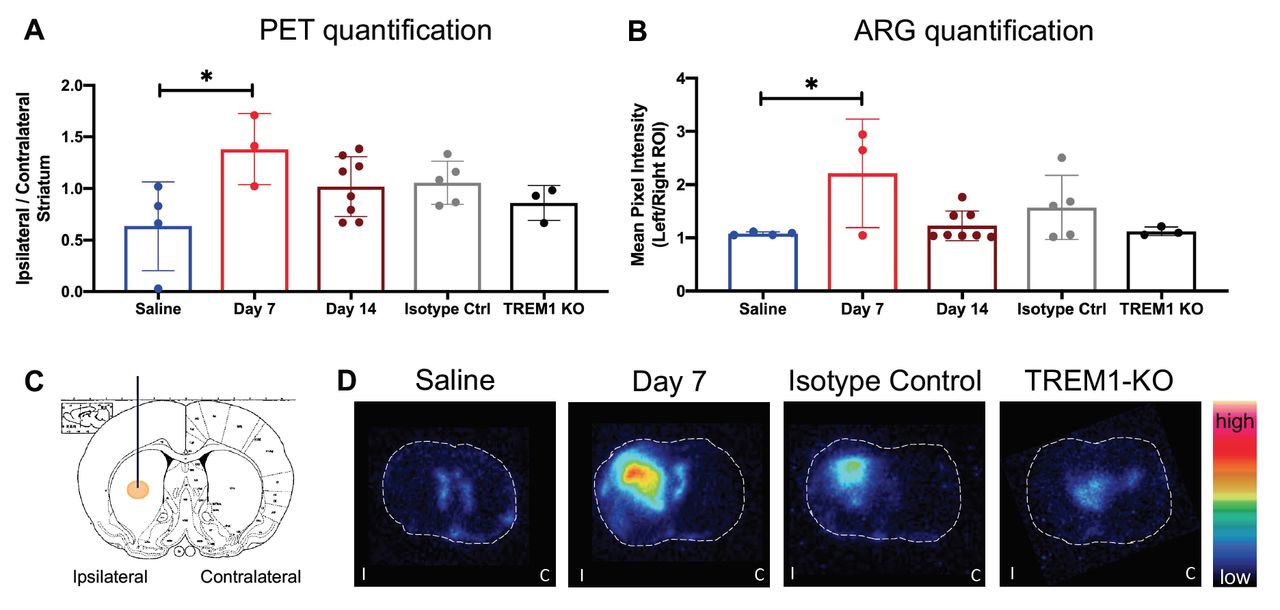
Results: Copper-64 radiolabeling of TREM1 mAb (chelator:mAb ratio = 0.96) and isotype control (chelator:mAb ratio = 1.9) resulted in 25% and 80% decay-corrected radiochemical yield, respectively (radiochemical purity > 99%). PET/CT images revealed TREM1-positive activated myeloid cells in the damaged brain hemisphere of 6-OHDA injected mice. PET image analysis verified significant tracer signal within the ipsilateral striatum of 6-OHDA compared to the saline-injected mice at 7 days post-surgery (P < 0.0146; Fig. 1a), which was corroborated by ex vivo ARG (P < 0.0202; Fig. 1b). Specificity of this signal was shown by the lack of significant binding in the ipsilateral striatum of TREM1-KO mice in addition to 6-OHDA mice imaged with [64Cu]Isotype control-mAb - all 7 days post-surgery. Conversely, there was no difference between saline mice and those injected with 6-OHDA at 14 days post-surgery, indicating that TREM1-PET may only be useful as an early biomarker of neuroinflammation in this model.

Conclusions: TREM1-PET represents a promising, highly specific, approach for visualizing the presence of early innate immune activation in PD rodent models. Notably, elevated levels of activated myeloid cells could be detected within the CNS of 6-OHDA injected mice in vivo using [64Cu]TREM1-mAb. Further investigation of this tracer is warranted in other PD rodent models and human postmortem tissue to assess its translational potential. Supporting Data: Figure 1. A) PET image analysis of mice injected with 6-OHDA or saline (N = 23; Saline n = 4, Day 7 n = 3, Day 14 n = 8, Isotype Ctrl n = 5, TREM1-KO n = 3), showing the ratio of tracer signal in the ipsilateral striatum over that in the contralateral striatum. B) Ex vivo autoradiography (ARG) of mice induced with 6-OHDA or saline, showing significant (P < 0.0202) tracer binding in the ipsilateral striatum over the contralateral striatum. C) Site of stereotaxic injection of 6-OHDA or saline in mouse brain - coronal view. D) Ex vivo ARG images of coronal mouse brain sections (40 μm thickness) showing tracer uptake in the striatum and injection site of 6-OHDA induced mice, compared to saline-injected mice, isotype controls, and TREM1-knockout mice. White dashed lines depict the outline of each coronal brain section.

Figure
  • Download figure
  • Open in new tab
  • Download powerpoint
Previous
Back to top

In this issue

Journal of Nuclear Medicine
Vol. 61, Issue supplement 1
May 1, 2020
  • Table of Contents
  • Index by author
Article Alerts
Sign In to Email Alerts with your Email Address
Email Article

Thank you for your interest in spreading the word on Journal of Nuclear Medicine.

NOTE: We only request your email address so that the person you are recommending the page to knows that you wanted them to see it, and that it is not junk mail. We do not capture any email address.

Enter multiple addresses on separate lines or separate them with commas.
Visualizing innate immune activation in a mouse model of Parkinson’s disease using a highly specific TREM1-PET tracer.
(Your Name) has sent you a message from Journal of Nuclear Medicine
(Your Name) thought you would like to see the Journal of Nuclear Medicine web site.
Citation Tools
Visualizing innate immune activation in a mouse model of Parkinson’s disease using a highly specific TREM1-PET tracer.
Katherine Lucot, Marc Stevens, Poorva Jain, T. Bonham, Ebony Webber, Jessica Klockow, E. Carmen Azevedo, Aisling Chaney, Edward Graves, Thomas Montine, Michelle James
Journal of Nuclear Medicine May 2020, 61 (supplement 1) 16;

Citation Manager Formats

  • BibTeX
  • Bookends
  • EasyBib
  • EndNote (tagged)
  • EndNote 8 (xml)
  • Medlars
  • Mendeley
  • Papers
  • RefWorks Tagged
  • Ref Manager
  • RIS
  • Zotero
Share
Visualizing innate immune activation in a mouse model of Parkinson’s disease using a highly specific TREM1-PET tracer.
Katherine Lucot, Marc Stevens, Poorva Jain, T. Bonham, Ebony Webber, Jessica Klockow, E. Carmen Azevedo, Aisling Chaney, Edward Graves, Thomas Montine, Michelle James
Journal of Nuclear Medicine May 2020, 61 (supplement 1) 16;
Twitter logo Facebook logo LinkedIn logo Mendeley logo
  • Tweet Widget
  • Facebook Like
  • Google Plus One
Bookmark this article

Jump to section

  • Article
  • Figures & Data
  • Info & Metrics

Related Articles

  • No related articles found.
  • Google Scholar

Cited By...

  • No citing articles found.
  • Google Scholar

More in this TOC Section

Neurosciences

  • PET CT Quantification to direct Alzheimer’s patients to anti-amyloid therapy
  • Multicenter trial study for usefulness of physical parameters toward the standardization of brain SPECT image: relation to visual analysis
  • Assessment of Brain Perfusion 123I-IMP SPECT imaging Using Low Energy High Resolution Collimator image reconstructed by Flash 3D
Show more Neurosciences

Brain Imaging Council YIA Symposium

  • Visualization of Flortaucipir Retention in Subjects Grouped According to their Autopsy-Confirmed Neurofibrillary Tangle Scores
  • Machine learning-derived multimodal neuroimaging of presurgical target area to predict individual’s seizure outcomes after epilepsy surgery
Show more Brain Imaging Council YIA Symposium

Similar Articles

SNMMI

© 2025 SNMMI

Powered by HighWire