
Main menu

  • Home
  • Content
    • Current
    • Ahead of print
    • Past Issues
    • JNM Supplement
    • SNMMI Annual Meeting Abstracts
    • Continuing Education
    • JNM Podcasts
  • Subscriptions
    • Subscribers
    • Institutional and Non-member
    • Rates
    • Journal Claims
    • Corporate & Special Sales
  • Authors
    • Submit to JNM
    • Information for Authors
    • Assignment of Copyright
    • AQARA requirements
  • Info
    • Reviewers
    • Permissions
    • Advertisers
  • About
    • About Us
    • Editorial Board
    • Contact Information
  • More
    • Alerts
    • Feedback
    • Help
    • SNMMI Journals
  • SNMMI
    • JNM
    • JNMT
    • SNMMI Journals
    • SNMMI

User menu

  • Subscribe
  • My alerts
  • Log in
  • Log out
  • My Cart

Search

  • Advanced search
Journal of Nuclear Medicine
  • SNMMI
    • JNM
    • JNMT
    • SNMMI Journals
    • SNMMI
  • Subscribe
  • My alerts
  • Log in
  • Log out
  • My Cart
Journal of Nuclear Medicine

Advanced Search

  • Home
  • Content
    • Current
    • Ahead of print
    • Past Issues
    • JNM Supplement
    • SNMMI Annual Meeting Abstracts
    • Continuing Education
    • JNM Podcasts
  • Subscriptions
    • Subscribers
    • Institutional and Non-member
    • Rates
    • Journal Claims
    • Corporate & Special Sales
  • Authors
    • Submit to JNM
    • Information for Authors
    • Assignment of Copyright
    • AQARA requirements
  • Info
    • Reviewers
    • Permissions
    • Advertisers
  • About
    • About Us
    • Editorial Board
    • Contact Information
  • More
    • Alerts
    • Feedback
    • Help
    • SNMMI Journals
  • View or Listen to JNM Podcast
  • Visit JNM on Facebook
  • Join JNM on LinkedIn
  • Follow JNM on Twitter
  • Subscribe to our RSS feeds
Meeting ReportNeurosciences

Visualization of Flortaucipir Retention in Subjects Grouped According to their Autopsy-Confirmed Neurofibrillary Tangle Scores

Vikas Kotari, Sudeepti Southekal, Ian Kennedy, Michael Navitsky, Michael Devous, Adam Fleisher and Michael Pontecorvo
Journal of Nuclear Medicine May 2020, 61 (supplement 1) 19;
Vikas Kotari
1Avid Radiopharmaceuticals Philadelphia PA United States
  • Find this author on Google Scholar
  • Find this author on PubMed
  • Search for this author on this site
Sudeepti Southekal
1Avid Radiopharmaceuticals Philadelphia PA United States
  • Find this author on Google Scholar
  • Find this author on PubMed
  • Search for this author on this site
Ian Kennedy
1Avid Radiopharmaceuticals Philadelphia PA United States
  • Find this author on Google Scholar
  • Find this author on PubMed
  • Search for this author on this site
Michael Navitsky
1Avid Radiopharmaceuticals Philadelphia PA United States
  • Find this author on Google Scholar
  • Find this author on PubMed
  • Search for this author on this site
Michael Devous
1Avid Radiopharmaceuticals Philadelphia PA United States
  • Find this author on Google Scholar
  • Find this author on PubMed
  • Search for this author on this site
Adam Fleisher
1Avid Radiopharmaceuticals Philadelphia PA United States
  • Find this author on Google Scholar
  • Find this author on PubMed
  • Search for this author on this site
Michael Pontecorvo
1Avid Radiopharmaceuticals Philadelphia PA United States
  • Find this author on Google Scholar
  • Find this author on PubMed
  • Search for this author on this site
  • Article
  • Figures & Data
  • Info & Metrics
Loading

Abstract

19

Introduction: Previously presented flortaucipir (FTP) image interpretation methodology demonstrated diagnostic power for predicting autopsy-confirmed neurofibrillary tangle (NFT) scores of B3 (Braak V/VI)1. Quantitation using an early tau volume of interest (ET)SUVr 2 improved sensitivity to intermediate (B2) Braak pathology3. In this work, we present groupwise surface maps for visualization of FTP retention based on NFT scores and compare with the proposed Braak staging pattern4,5.

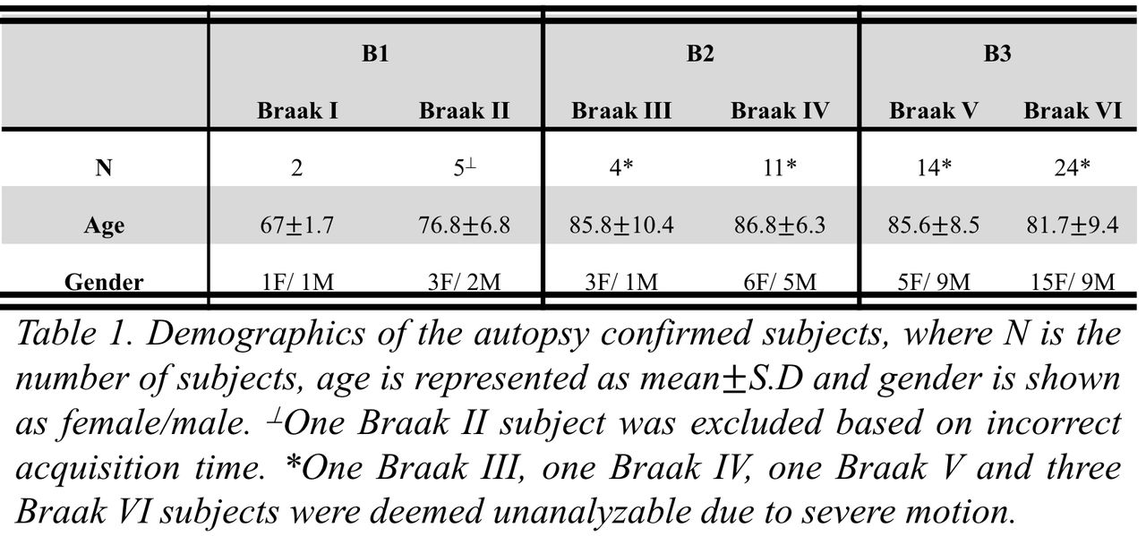
Methods: We included 60 subjects from a phase III autopsy study (Table 1) that underwent ante-mortem flortaucipir PET imaging. Neuropathological findings were recorded according to NIA-AA recommendations by two experts. Flortaucipir PET images were processed and spatially normalized to the MNI template according to procedures described previously6. SUVr values were calculated for each FTP image relative to a white matter reference region7. Subjects were grouped based on NFT score for groupwise mean and standard deviation SUVr image creation. These images were projected on the FreeSurfer FsAverage template8. Vertex-wise z-scores were estimated relative to a database of sixteen young controls from a different study6. Groupwise mean and z-score surface maps were created to represent groupwise flortaucipir retention.

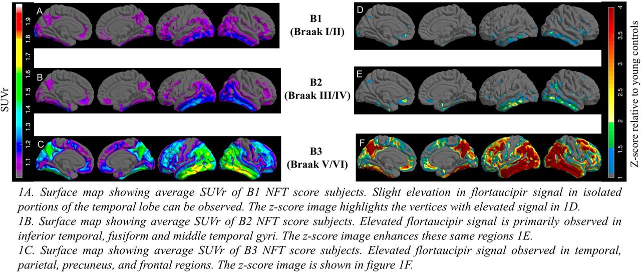
Results: The surface maps of mean SUVr and z-score values of subjects with a B1 (N=7) NFT score (Braak I/II) revealed slight elevation in flortaucipir signal in isolated vertices of the medial/lateral temporal lobe (Figure 1A and 1D). For B2 (N=15) NFT score subjects (Braak III/IV), we observed elevated flortaucipir signal in the inferior temporal, fusiform, middle temporal gyri and inferior parietal lobe (Figure 1B and 1E). In B3 (N=38) NFT score (Braak V/VI) group, we observed elevated flortaucipir signal in temporal, parietal, precuneus, and frontal regions (Figure 1C and 1F). These findings broadly align with the proposed Braak staging patterns.

Conclusions: In this first of its kind study, we present visualization of ante-mortem FTP retention. While the sample size is limited, the FTP retention patterns in the surface maps at a group level are in agreement with the neurofibrillary tangle staging scheme proposed by Braak. References: 1. Mintun, M. A. et al. Relationships between flortaucipir PET signal and tau neurofibrillary tangle pathology at autopsy. in Human Amyloid Imaging 304 (2019).2. Kotari, V. et al. Early tau detection and implications for disease progression. in Alzheimer’s & Dementia vols O5-01-06 (2019).3. Southekal, S. et al. Temporal Lobe Flortaucipir Quantitation may improve the detection of Intermediate Neurofibrillary Tangle Pathology in an Autopsy-Validated Cohort. in Alzheimer’s & Dementia (2019).4. Braak, H. & Braak, E. Staging of Alzheimer’s disease-related neurofibrillary changes. Neurobiol. Aging 16, 271-278; discussion 278-284 (1995).5. Braak, H., Alafuzoff, I., Arzberger, T., Kretzschmar, H. & Del Tredici, K. Staging of Alzheimer disease-associated neurofibrillary pathology using paraffin sections and immunocytochemistry. Acta Neuropathol. (Berl.) 112, 389-404 (2006).6. Pontecorvo, M. J. et al. Relationships between flortaucipir PET tau binding and amyloid burden, clinical diagnosis, age and cognition. Brain140, 748-763 (2017).7. Southekal, S. et al. Flortaucipir F 18 Quantitation Using Parametric Estimation of Reference Signal Intensity. J. Nucl. Med. Off. Publ. Soc. Nucl. Med. 59, 944-951 (2018).8. Dale, A. M., Fischl, B. & Sereno, M. I. Cortical surface-based analysis. I. Segmentation and surface reconstruction. NeuroImage 9, 179-194 (1999).

Figure
  • Download figure
  • Open in new tab
  • Download powerpoint
Figure
  • Download figure
  • Open in new tab
  • Download powerpoint
Previous
Back to top

In this issue

Journal of Nuclear Medicine
Vol. 61, Issue supplement 1
May 1, 2020
  • Table of Contents
  • Index by author
Article Alerts
Sign In to Email Alerts with your Email Address
Email Article

Thank you for your interest in spreading the word on Journal of Nuclear Medicine.

NOTE: We only request your email address so that the person you are recommending the page to knows that you wanted them to see it, and that it is not junk mail. We do not capture any email address.

Enter multiple addresses on separate lines or separate them with commas.
Visualization of Flortaucipir Retention in Subjects Grouped According to their Autopsy-Confirmed Neurofibrillary Tangle Scores
(Your Name) has sent you a message from Journal of Nuclear Medicine
(Your Name) thought you would like to see the Journal of Nuclear Medicine web site.
Citation Tools
Visualization of Flortaucipir Retention in Subjects Grouped According to their Autopsy-Confirmed Neurofibrillary Tangle Scores
Vikas Kotari, Sudeepti Southekal, Ian Kennedy, Michael Navitsky, Michael Devous, Adam Fleisher, Michael Pontecorvo
Journal of Nuclear Medicine May 2020, 61 (supplement 1) 19;

Citation Manager Formats

  • BibTeX
  • Bookends
  • EasyBib
  • EndNote (tagged)
  • EndNote 8 (xml)
  • Medlars
  • Mendeley
  • Papers
  • RefWorks Tagged
  • Ref Manager
  • RIS
  • Zotero
Share
Visualization of Flortaucipir Retention in Subjects Grouped According to their Autopsy-Confirmed Neurofibrillary Tangle Scores
Vikas Kotari, Sudeepti Southekal, Ian Kennedy, Michael Navitsky, Michael Devous, Adam Fleisher, Michael Pontecorvo
Journal of Nuclear Medicine May 2020, 61 (supplement 1) 19;
Twitter logo Facebook logo LinkedIn logo Mendeley logo
  • Tweet Widget
  • Facebook Like
  • Google Plus One
Bookmark this article

Jump to section

  • Article
  • Figures & Data
  • Info & Metrics

Related Articles

  • No related articles found.
  • Google Scholar

Cited By...

  • No citing articles found.
  • Google Scholar

More in this TOC Section

Neurosciences

  • PET CT Quantification to direct Alzheimer’s patients to anti-amyloid therapy
  • Multicenter trial study for usefulness of physical parameters toward the standardization of brain SPECT image: relation to visual analysis
  • Assessment of Brain Perfusion 123I-IMP SPECT imaging Using Low Energy High Resolution Collimator image reconstructed by Flash 3D
Show more Neurosciences

Brain Imaging Council YIA Symposium

  • Maximum Uptake and Hypermetabolic Volume of 18F-FDOPA PET Estimates Overall Survival in Low-Grade Gliomas
  • Regional associations between amyloid and glucose metabolism during the progression of Alzheimer’s disease in Down syndrome
Show more Brain Imaging Council YIA Symposium

Similar Articles

SNMMI

© 2025 SNMMI

Powered by HighWire