


Abstract
1263
Objectives: Limited data exist regarding the exact histopathological correlation between PSMA protein expression, tumor heterogeneity and functional uptake of 68Ga-PSMA-11 on PET. Aim of this study was, to identify immunohistochemical (IHC) PSMA patterns on radical prostatectomy specimens and correlate them to corresponding 68Ga-PSMA-PET examinations.
Methods: Retrospective, single center analysis of patients that underwent 68Ga-PSMA-11-PET and consecutive radical prostatectomy between 2016-2018 for prostate cancer (PCa). The dominant tumor lesion was identified and stained by IHC for PSMA. Maximal tumor diameter, Gleason Score (GS), PSMA expression on a semi-quantitative 4-tiered system and percentage of expressing tumor cells (PSMA%pos) were noted. Pathology specimens were correlated in an interdisciplinary read out with PSMA PET images and 68Ga-PSMA-11 uptake was quantified using SUVmax in the selected tumor area. Correlation between histopathological parameters and SUVmax was assessed using Spearman correlation.
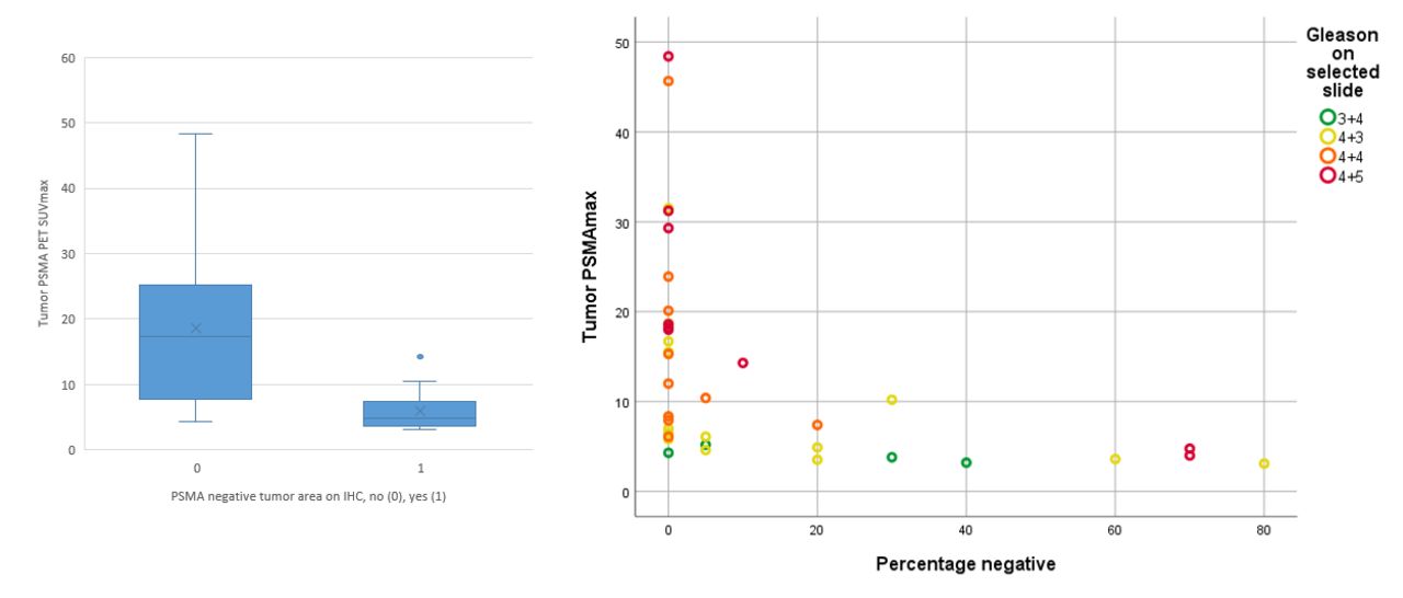
Results: 37 patients were available for this analysis. PSMA%pos and SUVmax had a significant correlation (r=0.715, p<0.001), while IHC intensity grading did not reach significance (r=0.193, p=0.252). The mean SUVmax in lesions with PSMA%pos ≤ 60% was 3.7 (SD 0.6, range 3.1-4.8), lesions with PSMA%pos > 60% had a mean SUVmax of 14.9 (SD 11.5, range 3.5-48.4). All lesions (n=5) with a SUVmax below 5 and PSMA%pos > 60% had a very heterogeneous and infiltrative growth pattern and two were smaller than 4 mm in diameter on histopathology. 25% of GS 7a lesions had PSMA%pos ≤60%, compared to 17% GS 7b and 11% GS≥8, respectively. Limitations include retrospective design and the low number of specimens.
Conclusions: PSMA-positive tumor area showed a strong correlation with SUVmax while PSMA intensity on IHC did not reach significance. Furthermore, our data confirms a higher rate of dominantly PSMA%pos tumors at higher GS.
In this issue


Jump to section
Related Articles
Cited By...
- No citing articles found.