
Main menu

  • Home
  • Content
    • Current
    • Ahead of print
    • Past Issues
    • JNM Supplement
    • SNMMI Annual Meeting Abstracts
    • Continuing Education
    • JNM Podcasts
  • Subscriptions
    • Subscribers
    • Institutional and Non-member
    • Rates
    • Journal Claims
    • Corporate & Special Sales
  • Authors
    • Submit to JNM
    • Information for Authors
    • Assignment of Copyright
    • AQARA requirements
  • Info
    • Reviewers
    • Permissions
    • Advertisers
  • About
    • About Us
    • Editorial Board
    • Contact Information
  • More
    • Alerts
    • Feedback
    • Help
    • SNMMI Journals
  • SNMMI
    • JNM
    • JNMT
    • SNMMI Journals
    • SNMMI

User menu

  • Subscribe
  • My alerts
  • Log in
  • My Cart

Search

  • Advanced search
Journal of Nuclear Medicine
  • SNMMI
    • JNM
    • JNMT
    • SNMMI Journals
    • SNMMI
  • Subscribe
  • My alerts
  • Log in
  • My Cart
Journal of Nuclear Medicine

Advanced Search

  • Home
  • Content
    • Current
    • Ahead of print
    • Past Issues
    • JNM Supplement
    • SNMMI Annual Meeting Abstracts
    • Continuing Education
    • JNM Podcasts
  • Subscriptions
    • Subscribers
    • Institutional and Non-member
    • Rates
    • Journal Claims
    • Corporate & Special Sales
  • Authors
    • Submit to JNM
    • Information for Authors
    • Assignment of Copyright
    • AQARA requirements
  • Info
    • Reviewers
    • Permissions
    • Advertisers
  • About
    • About Us
    • Editorial Board
    • Contact Information
  • More
    • Alerts
    • Feedback
    • Help
    • SNMMI Journals
  • View or Listen to JNM Podcast
  • Visit JNM on Facebook
  • Join JNM on LinkedIn
  • Follow JNM on Twitter
  • Subscribe to our RSS feeds
Meeting ReportPhysics, Instrumentation & Data Sciences

NEMA Performance Evaluation of Long Axial Field-of-View Brain PET Insert with 7T MRI Compatibility

Haewook Park, Seungeun Lee, Jun Yeon Won and Jae Sung Lee
Journal of Nuclear Medicine May 2020, 61 (supplement 1) 12;
Haewook Park
1Seoul National University Chongno-Gu Korea, Republic of
  • Find this author on Google Scholar
  • Find this author on PubMed
  • Search for this author on this site
Seungeun Lee
1Seoul National University Chongno-Gu Korea, Republic of
  • Find this author on Google Scholar
  • Find this author on PubMed
  • Search for this author on this site
Jun Yeon Won
1Seoul National University Chongno-Gu Korea, Republic of
  • Find this author on Google Scholar
  • Find this author on PubMed
  • Search for this author on this site
Jae Sung Lee
1Seoul National University Chongno-Gu Korea, Republic of
  • Find this author on Google Scholar
  • Find this author on PubMed
  • Search for this author on this site
  • Article
  • Figures & Data
  • Info & Metrics
Loading

Abstract

12

Objectives: We have developed a human brain PET insert that is compatible with 7T MRI scanners. The brain PET insert has a 16.7-cm-long axial field-of-view (FOV) that provides simultaneous 7T brain PET/MR images without a bed movement. Here, we present the scanner design and system performance of the brain PET insert in accordance with NEMA NU 2-2018 and NU 4-2008 standards.

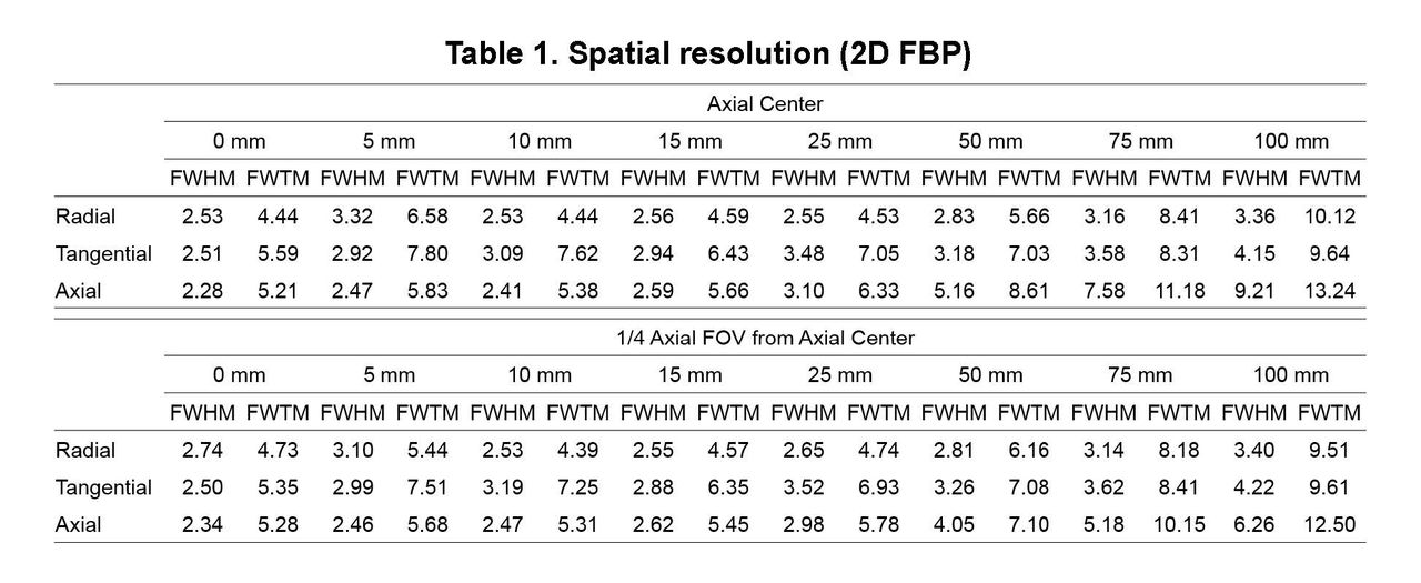
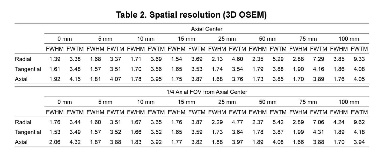
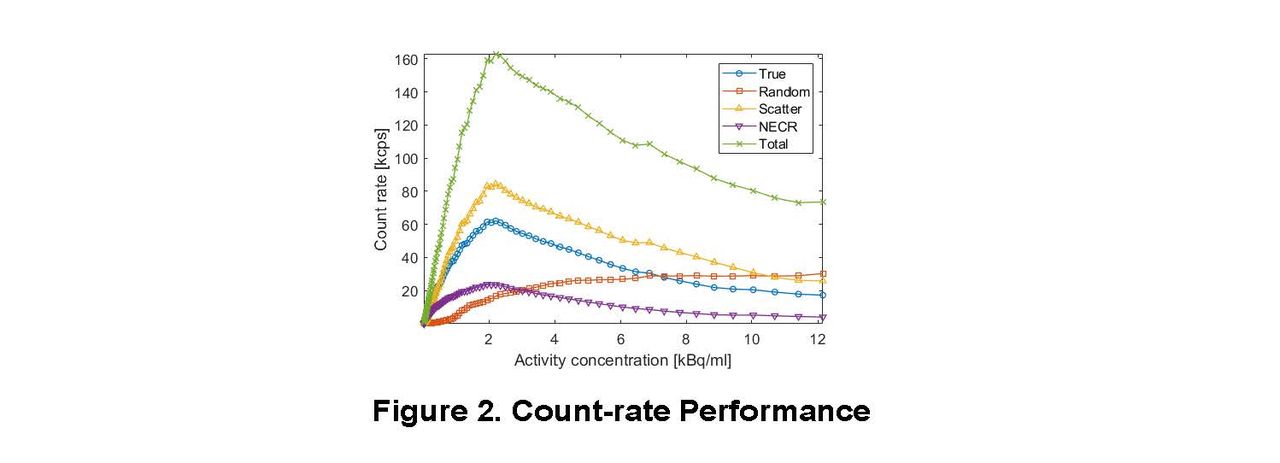
Methods: Our brain PET insert is comprised of 18 sectors with a ring diameter of 33 cm. Each sector is encapsulated with a copper-based RF shielding box with non-magnetic components to operate within the 7T MRI scanners. A block detector consists of a dual-layer staggered DOI crystal array coupled with a 2×2 array of 16-channel Hamamatsu MPPCs. The upper layer is an 11×11 array of 2.09×2.09×8 mm3 LSO crystals and the lower layer is a 12×12 array of 2.09×2.09×12 mm3 LSO crystals. All the PET data were collected using an in-house developed FPGA DAQ system. The energy channels consist of gated charge-to-time converters (QTCs) combined with single-ended memory interface (SeMI) of FPGA, and the timing channels are based on 16-ps-resolution time-to-digital converters (TDCs) implemented within the same FPGA. The system performance was evaluated using an energy window of 350-650 keV and a fixed coincidence time-window of 4 ns, unless otherwise noted. The spatial resolution was measured based on the NEMA NU 4 standard using a 22Na point source embedded in a 1 cm3 acrylic cube. The point source was placed at the center of transverse FOV and sequentially stepped in a radial direction up to 100 mm with a 5-mm interval. The measurement was repeated at the axial center and 1/4-axial-offset position. The acquired data were reconstructed using both a 2D FBP and 3D OSEM. The sensitivity specified in the NEMA NU 4 standard was measured using the same 22Na point source. The point source was placed at the center of transverse FOV and axially stepped with a 2.17-mm interval to cover the entire axial FOV. The sensitivity specified in the NEMA NU 2 standard was measured using a [18F]-FDG line source with five aluminum tubes. The acquisition was repeated with the source axially shifted by 0 cm, 5 cm, and 10 cm from the isocenter of the PET scanner. The background count-rate was measured without locating the source and subtracted from each dataset. The sensitivity was measured at three different energy windows of 250-750 keV, 350-650 keV, and 450-550 keV. The count-rate performance specified in the NEMA NU 2 standard was measured using a 70-cm-long [18F]-FDG line source. The line source was inserted into a cylindrical polyethylene phantom and placed at the center of transverse FOV. The peak noise equivalent count rate (NECR) and scatter fraction were then estimated.

Results: The radial/tangential/axial resolution at the isocenter of the PET scanner was 2.53/2.51/2.28 mm FWHM (2D FBP) and 1.39/1.61/1.92 mm FWHM (3D OSEM). The spatial resolutions at each source position are summarized in Tables 1 (2D FBP) and 2 (3D OSEM). The sensitivity was 18.87/20.64/23.72 kcps/MBq at 0 cm, 5 cm, and 10 cm off-center (NEMA NU 2) and 6.19% (NEMA NU 4) at the energy window of 350-650 keV. The axial sensitivity profiles at three different energy windows are illustrated in Figures 1a (NEMA NU 2) and 1b (NEMA NU 4). The count-rate performance as a function of activity concentration was shown in Figure 2. The peak NECR was 11.9 kcps at 1.94 kBq/mL with the scatter fraction of 57%. An FPGA firmware upgrade is in progress to improve the count-rate performance.

Conclusions: In this study, we measured the system performance of our newly-developed 7T MR-compatible brain PET insert based on the NEMA standards. Our next milestone is to perform in-vivo human brain studies and assess the image quality using a cylindrical phantom that contains hot and cold spheres with six different diameters. Research Support: This work was supported by grants from National Research Foundation of Korea (NRF) funded by Korean Ministry of Science, ICT, and Future Planning (grant no. NRF-2014M3C7034000).

Figure
  • Download figure
  • Open in new tab
  • Download powerpoint
Figure
  • Download figure
  • Open in new tab
  • Download powerpoint
Figure
  • Download figure
  • Open in new tab
  • Download powerpoint
Figure
  • Download figure
  • Open in new tab
  • Download powerpoint
Previous
Back to top

In this issue

Journal of Nuclear Medicine
Vol. 61, Issue supplement 1
May 1, 2020
  • Table of Contents
  • Index by author
Article Alerts
Sign In to Email Alerts with your Email Address
Email Article

Thank you for your interest in spreading the word on Journal of Nuclear Medicine.

NOTE: We only request your email address so that the person you are recommending the page to knows that you wanted them to see it, and that it is not junk mail. We do not capture any email address.

Enter multiple addresses on separate lines or separate them with commas.
NEMA Performance Evaluation of Long Axial Field-of-View Brain PET Insert with 7T MRI Compatibility
(Your Name) has sent you a message from Journal of Nuclear Medicine
(Your Name) thought you would like to see the Journal of Nuclear Medicine web site.
Citation Tools
NEMA Performance Evaluation of Long Axial Field-of-View Brain PET Insert with 7T MRI Compatibility
Haewook Park, Seungeun Lee, Jun Yeon Won, Jae Sung Lee
Journal of Nuclear Medicine May 2020, 61 (supplement 1) 12;

Citation Manager Formats

  • BibTeX
  • Bookends
  • EasyBib
  • EndNote (tagged)
  • EndNote 8 (xml)
  • Medlars
  • Mendeley
  • Papers
  • RefWorks Tagged
  • Ref Manager
  • RIS
  • Zotero
Share
NEMA Performance Evaluation of Long Axial Field-of-View Brain PET Insert with 7T MRI Compatibility
Haewook Park, Seungeun Lee, Jun Yeon Won, Jae Sung Lee
Journal of Nuclear Medicine May 2020, 61 (supplement 1) 12;
Twitter logo Facebook logo LinkedIn logo Mendeley logo
  • Tweet Widget
  • Facebook Like
  • Google Plus One
Bookmark this article

Jump to section

  • Article
  • Figures & Data
  • Info & Metrics

Related Articles

  • No related articles found.
  • Google Scholar

Cited By...

  • No citing articles found.
  • Google Scholar

More in this TOC Section

Physics, Instrumentation & Data Sciences

  • Exploration of Multi-objective Optimization with Genetic Algorithms for PET Image Reconstruction
  • AI-based methods for nuclear-medicine imaging: Need for objective task-specific evaluation
  • Ultra-Fast Reconstruction of Short List-Mode PET Data Frames for Real-Time Visualization and Processing
Show more Physics, Instrumentation & Data Sciences

Physics, Instrumentation & Data Sciences Countil YIA Symposium

  • Anatomy-guided PET image reconstruction with deep neural network
  • Aortic valve imaging using 18F-Sodium Fluoride: Implications of triple motion correction on test-retest reproducibility
  • Performance Characterization of an SiPM-based Time-of-Flight Canon PET/CT Scanner
Show more Physics, Instrumentation & Data Sciences Countil YIA Symposium

Similar Articles

SNMMI

© 2025 SNMMI

Powered by HighWire