
Main menu

  • Home
  • Content
    • Current
    • Ahead of print
    • Past Issues
    • JNM Supplement
    • SNMMI Annual Meeting Abstracts
    • Continuing Education
    • JNM Podcasts
  • Subscriptions
    • Subscribers
    • Institutional and Non-member
    • Rates
    • Journal Claims
    • Corporate & Special Sales
  • Authors
    • Submit to JNM
    • Information for Authors
    • Assignment of Copyright
    • AQARA requirements
  • Info
    • Reviewers
    • Permissions
    • Advertisers
  • About
    • About Us
    • Editorial Board
    • Contact Information
  • More
    • Alerts
    • Feedback
    • Help
    • SNMMI Journals
  • SNMMI
    • JNM
    • JNMT
    • SNMMI Journals
    • SNMMI

User menu

  • Subscribe
  • My alerts
  • Log in
  • Log out
  • My Cart

Search

  • Advanced search
Journal of Nuclear Medicine
  • SNMMI
    • JNM
    • JNMT
    • SNMMI Journals
    • SNMMI
  • Subscribe
  • My alerts
  • Log in
  • Log out
  • My Cart
Journal of Nuclear Medicine

Advanced Search

  • Home
  • Content
    • Current
    • Ahead of print
    • Past Issues
    • JNM Supplement
    • SNMMI Annual Meeting Abstracts
    • Continuing Education
    • JNM Podcasts
  • Subscriptions
    • Subscribers
    • Institutional and Non-member
    • Rates
    • Journal Claims
    • Corporate & Special Sales
  • Authors
    • Submit to JNM
    • Information for Authors
    • Assignment of Copyright
    • AQARA requirements
  • Info
    • Reviewers
    • Permissions
    • Advertisers
  • About
    • About Us
    • Editorial Board
    • Contact Information
  • More
    • Alerts
    • Feedback
    • Help
    • SNMMI Journals
  • View or Listen to JNM Podcast
  • Visit JNM on Facebook
  • Join JNM on LinkedIn
  • Follow JNM on Twitter
  • Subscribe to our RSS feeds
Meeting ReportPhysics, Instrumentation & Data Sciences

Attenuation Correction for Amyloid PET Imaging Using Deep Learning Based on 3D UTE/Multi-Echo Dixon MR images

Kuang Gong, Paul Han, Keith Johnson, Georges El Fakhri, Chao Ma and Quanzheng Li
Journal of Nuclear Medicine May 2020, 61 (supplement 1) 111;
Kuang Gong
1Massachusetts General Hospital Boston MA United States
  • Find this author on Google Scholar
  • Find this author on PubMed
  • Search for this author on this site
Paul Han
1Massachusetts General Hospital Boston MA United States
  • Find this author on Google Scholar
  • Find this author on PubMed
  • Search for this author on this site
Keith Johnson
2MGH Brookline MA United States
  • Find this author on Google Scholar
  • Find this author on PubMed
  • Search for this author on this site
Georges El Fakhri
3Massachusetts General Hospital, Harvard Medical Sc Boston MA United States
  • Find this author on Google Scholar
  • Find this author on PubMed
  • Search for this author on this site
Chao Ma
1Massachusetts General Hospital Boston MA United States
  • Find this author on Google Scholar
  • Find this author on PubMed
  • Search for this author on this site
Quanzheng Li
1Massachusetts General Hospital Boston MA United States
  • Find this author on Google Scholar
  • Find this author on PubMed
  • Search for this author on this site
  • Article
  • Figures & Data
  • Info & Metrics
Loading

Abstract

111

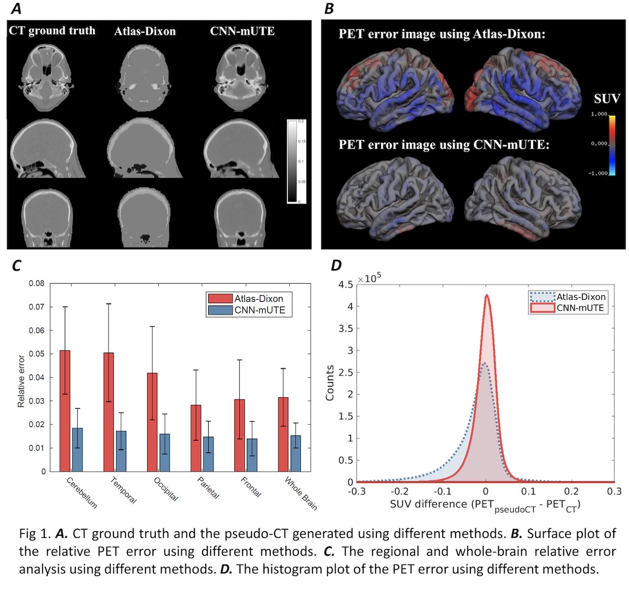
Objectives: The cortical uptake of amyloid PET imaging is a powerful biomarker for the diagnosis and treatment monitoring of Alzheimer’s disease (AD). As the cortical regions are close to the bone, quantitative accuracy of amyloid PET imaging can be hugely influenced by the inaccuracy of attenuation correction (AC). This will be an issue if amyloid imaging is acquired from PET/MR systems, where attenuation maps cannot be obtained from MR images through simple transform. Previously we have developed a 3D mUTE sequence for PET/MR AC, which combines 3D UTE with multi-echo Dixon acquisitions, demonstrated to better resolve the bone and air regions. In this work, the mUTE sequence and the convolutional neural network (CNN) method were integrated together, denoted as CNN-mUTE, to derive accurate and continuous attenuation maps for amyloid PET imaging. Methods: Thirty-five subjects were scanned with approval from the local IRB. MR acquisitions were performed on a 3T MR scanner using the in-house developed 3D mUTE sequence, with different TE time (70, 2110, 2310, 3550, 3750, 4990, 5190 µs), voxel size of 1.875 x 1.875 x 1.875 mm3 and FOV of 240 mm3. A separate 11-C-PiB PET/CT scanning (15 mCi injection followed by a dynamic scan for 70 min) was performed on a whole-body PET/CT scanner for the same subjects. Only data from 40-50 min were used in this work for the quantification of different AC methods. CT images were acquired during the PET/CT scanning and were used as the training labels. For the CNN method, 3D U-net was employed as the network structure and L1 difference between the ground-truth CT and the generated CT was used as the training function. When preparing the network training pairs, we first registered mUTE images to the CT images through rigid transformation using the ANTs software. Random rotation and permutation was performed on the training pairs to avoid over-fitting. The network input has seven channels as each mUTE acquisition will produce seven 3D MR images. Quantitative analysis was based on five-fold cross-validation. The network structure was implemented in TensorFlow 1.12. The training batch size was set to 1 and 500 epochs were run. The atlas method based on Dixon MR images (Atlas-Dixon) was adopted as the baseline method. Regional as well as global relative PET error were used to compare the performance of different methods. Results: The CNN-mUTE method can generate pseudo-CT images with more structure details (as shown in Fig 1(a)). We can observe the inhomogeneity inside the bone region due to the signals present in mUTE MR images and the strong approximation power of CNN. Quantitative analysis in Table 1 shows that CNN-mUTE can achieve a Dice coefficient of 0.92 for the bone region above the eye. Surface maps of the PET error images shown in Fig 1(b) demonstrate that the CNN-mUTE method can produce smaller errors for the cortical regions. Regional analysis in cerebellum and brain lobes in Fig 1(c) and the histogram analysis shown in Fig 1(d) also demonstrates the improvement by CNN-mUTE over the Atlas-Dixon method. Conclusion: We have combined the mUTE sequence and the CNN method to generate accurate attenuation map for amyloid PET imaging. Analysis based on real data sets shows that the CNN-mUTE method has the potential for accurate PET/MR amyloid imaging. Acknowledgements: This work was supported by NIH grants R01 AG052653 and P41 EB022544.

Figure
  • Download figure
  • Open in new tab
  • Download powerpoint
View this table:
  • View inline
  • View popup

Comparisons of the generated pseudo-CT images based on 35 datasets. The Dice index of bone regions w

Previous
Back to top

In this issue

Journal of Nuclear Medicine
Vol. 61, Issue supplement 1
May 1, 2020
  • Table of Contents
  • Index by author
Article Alerts
Sign In to Email Alerts with your Email Address
Email Article

Thank you for your interest in spreading the word on Journal of Nuclear Medicine.

NOTE: We only request your email address so that the person you are recommending the page to knows that you wanted them to see it, and that it is not junk mail. We do not capture any email address.

Enter multiple addresses on separate lines or separate them with commas.
Attenuation Correction for Amyloid PET Imaging Using Deep Learning Based on 3D UTE/Multi-Echo Dixon MR images
(Your Name) has sent you a message from Journal of Nuclear Medicine
(Your Name) thought you would like to see the Journal of Nuclear Medicine web site.
Citation Tools
Attenuation Correction for Amyloid PET Imaging Using Deep Learning Based on 3D UTE/Multi-Echo Dixon MR images
Kuang Gong, Paul Han, Keith Johnson, Georges El Fakhri, Chao Ma, Quanzheng Li
Journal of Nuclear Medicine May 2020, 61 (supplement 1) 111;

Citation Manager Formats

  • BibTeX
  • Bookends
  • EasyBib
  • EndNote (tagged)
  • EndNote 8 (xml)
  • Medlars
  • Mendeley
  • Papers
  • RefWorks Tagged
  • Ref Manager
  • RIS
  • Zotero
Share
Attenuation Correction for Amyloid PET Imaging Using Deep Learning Based on 3D UTE/Multi-Echo Dixon MR images
Kuang Gong, Paul Han, Keith Johnson, Georges El Fakhri, Chao Ma, Quanzheng Li
Journal of Nuclear Medicine May 2020, 61 (supplement 1) 111;
Twitter logo Facebook logo LinkedIn logo Mendeley logo
  • Tweet Widget
  • Facebook Like
  • Google Plus One
Bookmark this article

Jump to section

  • Article
  • Figures & Data
  • Info & Metrics

Related Articles

  • No related articles found.
  • Google Scholar

Cited By...

  • No citing articles found.
  • Google Scholar

More in this TOC Section

Physics, Instrumentation & Data Sciences

  • Deep Learning Based 3D Dose Estimation from Prompt Gamma Distribution for Proton Therapy Monitoring
  • Data-driven respiratory signal estimation from finely sampled projection data in conventional cardiac perfusion SPECT imaging.
  • Clinical feasibility of quantitative holmium-166 SPECT in presence of technetium-99m colloid
Show more Physics, Instrumentation & Data Sciences

Attenuation, Scatter and Resolution Compensation (PET & SPECT)

  • Reconstruction using Depth of Interaction Information of Curved and Flat Detector Designs for Quantitative Multi-Pinhole Brain SPECT
  • Clinical evaluation of Poisson maximum likelihood-based scatter subtraction for 68Ga-PSMA imaging
  • PET Attenuation Correction Using MRI-aided Two-Stream Pyramid Attention Network
Show more Attenuation, Scatter and Resolution Compensation (PET & SPECT)

Similar Articles

SNMMI

© 2025 SNMMI

Powered by HighWire