


Abstract
1055
Objectives: ALK-positive NSCLC is an important subtype of lung cancer, accounting for about 4~7%. Other than pathological biopsy, a noninvasively molecular imaging method for ALK-positive NSCLC is urgently needed. Therefore, we tried to develop a new PET probe targeting ALK-TK for NSCLC patients.
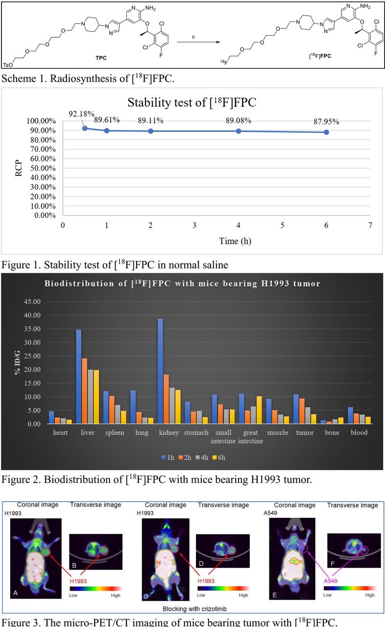
Methods: After attaching the TsO-PEG4- group to the N1 position of the terminal piperidine ring of crizotinib, we obtained the precursor TPC for F-18 labeling with TsO- as the leaving group. Next, we synthesized [18F]FPC from precursor TPC with a one-step reaction in extra-dry MeCN at 90℃ to give the desired [18F]FPC (Scheme 1). The in vitro stability in normal saline was evaluated at 0.5, 1, 2, 4, and 6 h with HPLC. The biodistribution study of [18F]FPC was carried out using male BALB/c nude mice (7 weeks old; weight 17~24 g each). Mice (n = 3) was injected with 0.37 MBq of [18F]FPC and were anesthetized and sacrificed at four time points (1 h, 2 h, 4 h, and 6 h). The micro-PET/CT imaging of mice bearing H1973 tumor or A549 tumor was carried out at 4 h postinjection. Blocking imaging of mice bearing H1993 tumor was performed with co-injection of [18F]FPC and crizotinib (1000 times excess).
Results: [18F]FPC was obtained in a radiochemical yield of 3.5 ± 1.2% [n = 5, end of synthesis (EOS), from [18F]KF after preparative HPLC. The radiochemical purity (RCP) and the specific activity (SA) exceeded 92% and 111±74 GBq/μmol, respectively. Stability test showed that [18F]FPC was stable in normal saline at 37℃, it had not decomposed much for 6 h (Figure 1). The high uptake of [18F]FPC in liver and intestinal tissues, especially the uptake value in the large intestine increasing from 4.9% ID/g (2 h post injection) to 10.1% ID/g (6 h post injection), indicated that elimination of [18F]FPC is mainly via the hepatobiliary pathway. The radio activity of [18F]FPC in tumors kept up for at least 4 h (10.8% ID/g at 1 h post injection, 6.0 %ID/g at 4 h post-injection). The tumor-to-lung ratio increased over time and reach the highest value of 2.61 at 4 h post-injection (Figure 2). The micro-PET/CT imaging of [18F]FPC showed that H1993 tumor was visualized more clearly than A549 tumor (2.51 vs 1.15, %ID/g), and the uptake of H1993 tumor could be inhibited by crizotinib (1.54 %ID/g, Figure 3).
Conclusions: We designed and synthesized a F-18 labeling crizotinib derivative, [18F]FPC, which was targeted ALK-positive NSCLC tumor. [18F]FPC had a potential to act as a PET imaging probe to discriminate ALK-positive tumor in NSCLC and evaluate of ALK-TK expression in it.
In this issue


Jump to section
Related Articles
Cited By...
- No citing articles found.