
Main menu

  • Home
  • Content
    • Current
    • Ahead of print
    • Past Issues
    • JNM Supplement
    • SNMMI Annual Meeting Abstracts
    • Continuing Education
    • JNM Podcasts
  • Subscriptions
    • Subscribers
    • Institutional and Non-member
    • Rates
    • Journal Claims
    • Corporate & Special Sales
  • Authors
    • Submit to JNM
    • Information for Authors
    • Assignment of Copyright
    • AQARA requirements
  • Info
    • Reviewers
    • Permissions
    • Advertisers
  • About
    • About Us
    • Editorial Board
    • Contact Information
  • More
    • Alerts
    • Feedback
    • Help
    • SNMMI Journals
  • SNMMI
    • JNM
    • JNMT
    • SNMMI Journals
    • SNMMI

User menu

  • Subscribe
  • My alerts
  • Log in
  • My Cart

Search

  • Advanced search
Journal of Nuclear Medicine
  • SNMMI
    • JNM
    • JNMT
    • SNMMI Journals
    • SNMMI
  • Subscribe
  • My alerts
  • Log in
  • My Cart
Journal of Nuclear Medicine

Advanced Search

  • Home
  • Content
    • Current
    • Ahead of print
    • Past Issues
    • JNM Supplement
    • SNMMI Annual Meeting Abstracts
    • Continuing Education
    • JNM Podcasts
  • Subscriptions
    • Subscribers
    • Institutional and Non-member
    • Rates
    • Journal Claims
    • Corporate & Special Sales
  • Authors
    • Submit to JNM
    • Information for Authors
    • Assignment of Copyright
    • AQARA requirements
  • Info
    • Reviewers
    • Permissions
    • Advertisers
  • About
    • About Us
    • Editorial Board
    • Contact Information
  • More
    • Alerts
    • Feedback
    • Help
    • SNMMI Journals
  • View or Listen to JNM Podcast
  • Visit JNM on Facebook
  • Join JNM on LinkedIn
  • Follow JNM on Twitter
  • Subscribe to our RSS feeds
Research ArticleTheranostics

Quantitative Test–Retest Measurement of 68Ga-PSMA-HBED-CC in Tumor and Normal Tissue

Janet H. Pollard, Caleb Raman, Yousef Zakharia, Chad R. Tracy, Kenneth G. Nepple, Tim Ginader, Patrick Breheny and John J. Sunderland
Journal of Nuclear Medicine August 2020, 61 (8) 1145-1152; DOI: https://doi.org/10.2967/jnumed.119.236083
Janet H. Pollard
1Department of Radiology, University of Iowa Carver College of Medicine, Iowa City, Iowa
2Iowa City Veterans Healthcare Center, Iowa City, Iowa
  • Find this author on Google Scholar
  • Find this author on PubMed
  • Search for this author on this site
Caleb Raman
3College of Arts and Sciences, University of Iowa, Iowa City, Iowa
  • Find this author on Google Scholar
  • Find this author on PubMed
  • Search for this author on this site
Yousef Zakharia
4Department of Internal Medicine, University of Iowa Carver College of Medicine, Iowa City, Iowa
  • Find this author on Google Scholar
  • Find this author on PubMed
  • Search for this author on this site
Chad R. Tracy
5Department of Urology, University of Iowa Carver College of Medicine, Iowa City, Iowa
  • Find this author on Google Scholar
  • Find this author on PubMed
  • Search for this author on this site
Kenneth G. Nepple
5Department of Urology, University of Iowa Carver College of Medicine, Iowa City, Iowa
  • Find this author on Google Scholar
  • Find this author on PubMed
  • Search for this author on this site
Tim Ginader
6Biostatistics Core, University of Iowa Carver College of Medicine, Iowa City, Iowa; and
  • Find this author on Google Scholar
  • Find this author on PubMed
  • Search for this author on this site
Patrick Breheny
7Department of Biostatistics, University of Iowa College of Public Health, Iowa City, Iowa
  • Find this author on Google Scholar
  • Find this author on PubMed
  • Search for this author on this site
John J. Sunderland
1Department of Radiology, University of Iowa Carver College of Medicine, Iowa City, Iowa
  • Find this author on Google Scholar
  • Find this author on PubMed
  • Search for this author on this site
  • Article
  • Figures & Data
  • Supplemental
  • Info & Metrics
  • PDF
Loading

Article Figures & Data

Figures

  • Tables
  • Additional Files
  • FIGURE 1.
    • Download figure
    • Open in new tab
    • Download powerpoint
    FIGURE 1.

    68Ga-PSMA-HBED-CC CC PET/CT for subject 2 for scan 1 (baseline) and scan 2, performed 2 d later, showing segmentation of normal tissues (salivary glands and spleen) and sites of abnormal uptake in 2 left pelvic lymph nodes (1 and 2) and left prostate bed (3).
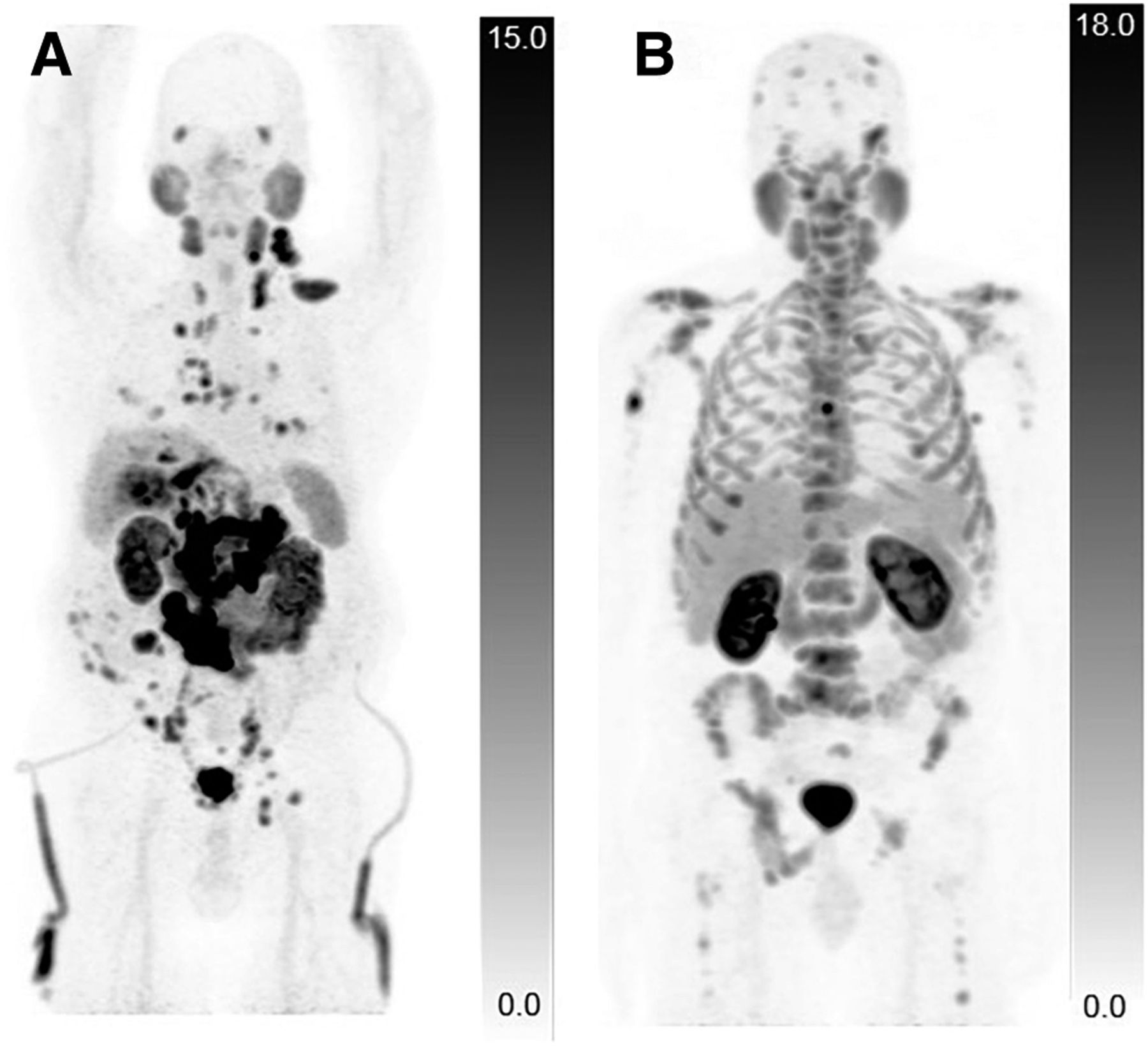
  • FIGURE 2.
    • Download figure
    • Open in new tab
    • Download powerpoint
    FIGURE 2.

    68Ga-PSMA-HBED-CC CC PET/CT for 2 subjects demonstrating different distribution of metastatic lesions. (A) Predominantly soft-tissue metastatic prostate cancer is seen with visceral metastases in lungs and liver and nodal metastases in neck, chest, abdomen, and pelvis. Percutaneous nephrostomy tubing is also visible. (B) Extensive metastatic prostate cancer is seen involving only skeleton.
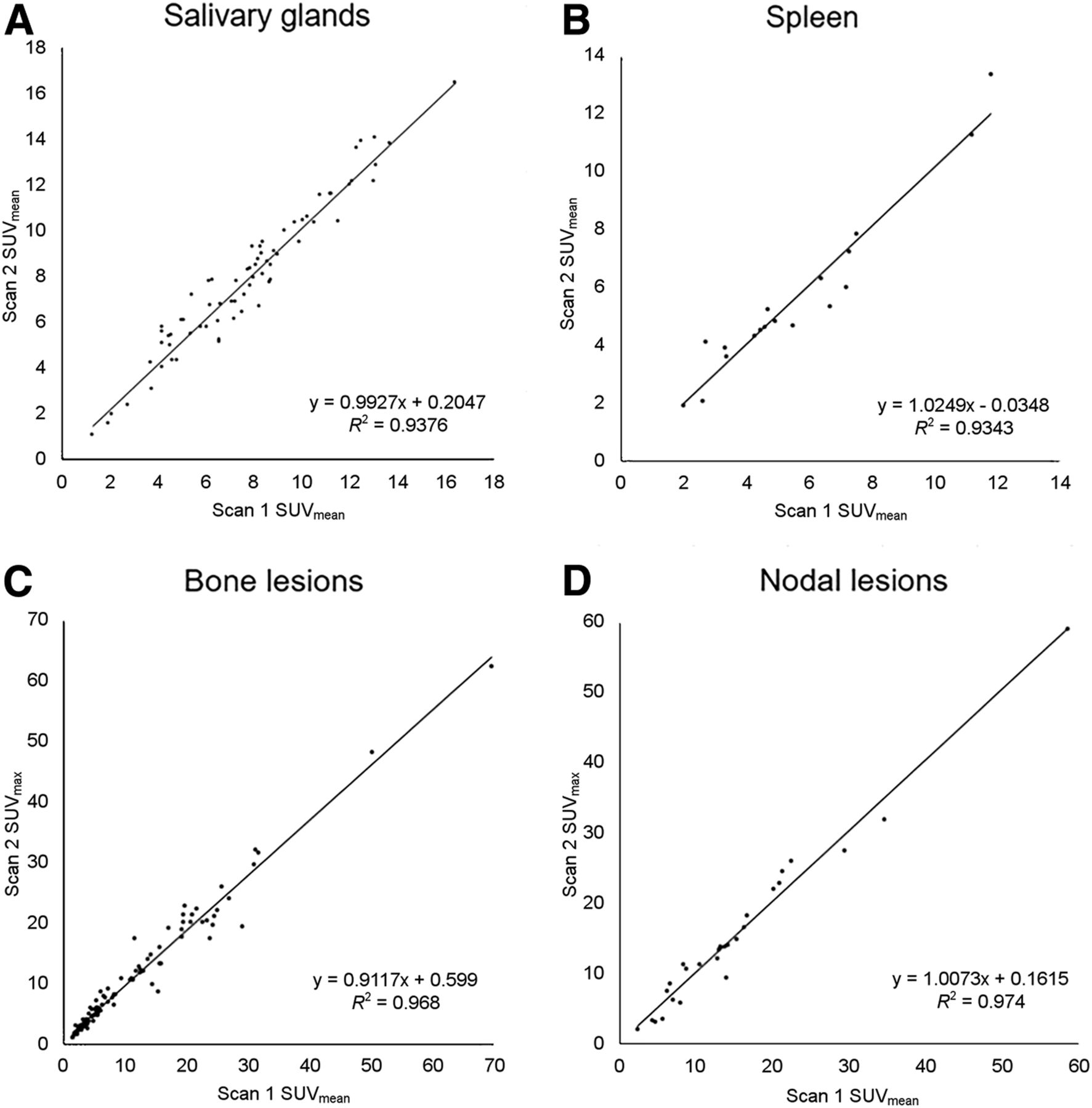
  • FIGURE 3.
    • Download figure
    • Open in new tab
    • Download powerpoint
    FIGURE 3.

    Correlation between test–retest measures of 68Ga-PSMA-HBED-CC uptake. Plots show SUVmean of normal salivary glands and spleen (A and B) and SUVmax for bone and nodal lesions (C and D).
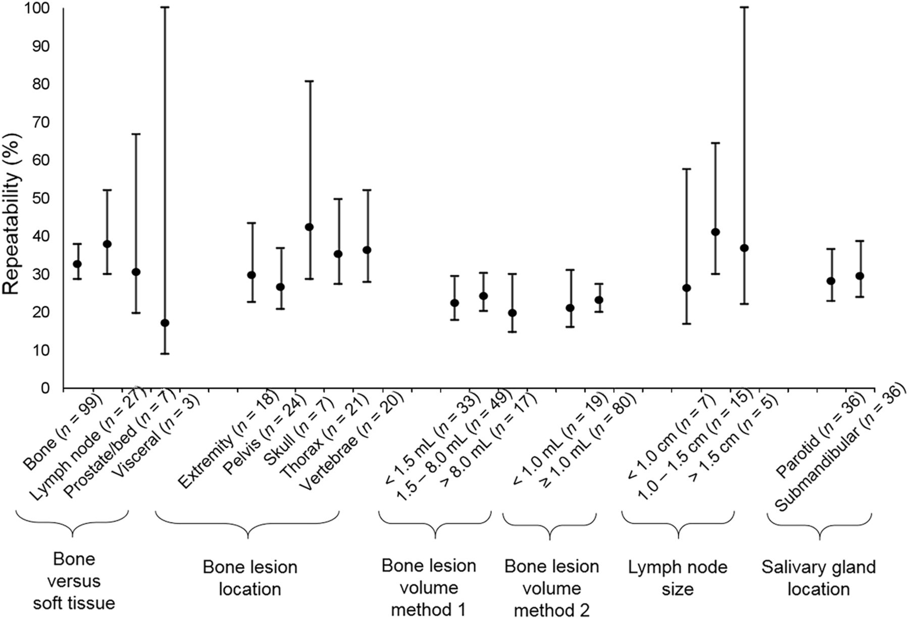
  • FIGURE 4.
    • Download figure
    • Open in new tab
    • Download powerpoint
    FIGURE 4.

    Comparison of SUV RC point estimates; 95% CIs (y-axis) show substantial overlap, indicating no significant difference in repeatability for different categories of measurements (x-axis).
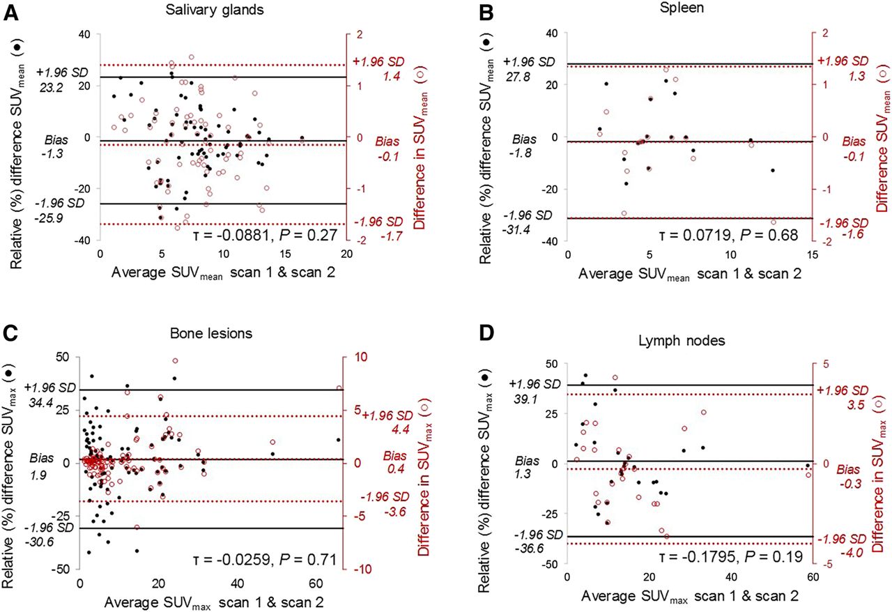
  • FIGURE 5.
    • Download figure
    • Open in new tab
    • Download powerpoint
    FIGURE 5.

    Bland–Altman scatterplots show difference between test–retest SUV measurements and average lesion or organ intensity. Plots suggest no association between mean lesion or organ intensity (x-axis) and test–retest relative percentage differences (primary y-axis) based on Kendall τ analysis. Both relative percentage difference (●, primary vertical axis) and absolute difference (○, right vertical axis) are plotted together for salivary glands (A), spleen (B), bone lesions (C), and lymph nodes (D). Horizontal lines represent upper limits of agreement (+1.96 SD), lower limits of agreement (−1.96 SD), and bias (or mean difference). Kendall τ coefficient and P values were calculated for relative percentage difference data and are included in field of graphs.

Tables

  • Figures
  • Additional Files
    • View popup
    TABLE 1

    Subject Characteristics

    SubjectPSA within ≤90 d (ng/cm3)Gleason score at diagnosisTotal lesions measuredProstate or prostate bedBonesNodesViscera
    10.157 (4 + 5)—————
    24.356 (3 + 3)31—2—
    3104.59 (4 + 5)15—15——
    40.149 (4 + 5)1019——
    50.669 (5 + 4)4121—
    60.229 (5 + 4)1—1——
    756.3Presumptive diagnosis12—12——
    895.57 (4 + 3)23—2183
    9276.39 (4 + 5)15—15——
    100.04Presumptive diagnosis—————
    110.649 (4 + 5)5—5——
    122.8Lymph node biopsy12183—
    1340.110 (5 + 5)131102—
    1419.77 (3 + 4)2—2——
    152.5Bone biopsy11———
    1654.19 (5 + 4)15—15——
    172.59 (5 + 4)211——
    182.59 (5 + 4)3—21—
    • PSA = prostate-specific membrane antigen.

    • View popup
    TABLE 2

    Repeatability Data (SUVmax)

    Organ or lesion typewCV (%)Symmetric RC*Asymmetric RC
    LRC (%)URC (%)
    Bone lesions, total (n = 99)11.7± 32.5 [28.5, 37.8]−28.0+38.8
    Location
     Extremities (n = 20)10.7±29.6 [22.5, 43.3]−25.8+34.7
     Pelvis (n = 25)9.5±26.3 [20.6, 36.6]−23.2+30.3
     Skull (n = 9)15.2±42.1 [28.4, 80.6]−34.8+53.3
     Thorax (n = 23)12.7±35.1 [27.1, 49.7]−29.9+42.6
     Vertebrae (n = 22)13.1±36.2 [27.9, 51.8]−30.7+44.2
    Lesion volume, method 1
     <1.5 cm3 (n = 33)8.0±22.1 [17.8, 29.3]−19.9+24.8
     1.5–8.0 cm3 (n = 49)8.7±24.0 [20.1, 30.0]−21.4+27.3
     >8.0 cm3 (n = 17)7.1±19.6 [14.6, 29.8]−17.8+21.7
    Lesion volume, method 2
     <1.0 cm3 (n = 19)7.5±20.9 [15.8, 30.9]−18.9+23.3
     ≥1.0 cm3 (n = 80)8.3±22.9 [19.9, 27.2]−20.5+25.9
     Nodal lesions (n = 27)13.7±37.9 [29.8, 51.9]−31.9+46.8
    Size, long axis
     <1.0 cm (n = 7)9.4±26.1 [16.8, 57.4]−23.1+30.0
     1.0–1.5 cm (n = 15)14.7±40.8 [29.8, 64.3]−33.9+51.3
     >1.5 cm (n = 5)13.3±36.7 [22.0, 100.0]−31.0+44.9
    Prostate or prostate bed (n = 7)10.9±30.3 [19.5, 66.7]−26.3+35.7
    Visceral lesions (n = 3)6.1±16.9 [8.8,100.0]−15.6+18.5
    • ↵* Data are percentages followed by 95% CIs in brackets.

    • LRC = lower RC; URC = upper RC’

    • View popup
    TABLE 3

    Repeatability Data (SUVmean)

    Organ or lesion typewCV (%)Symmetric RC*
    Salivary glands, total (n = 72)8.9±24.6 [21.1, 29.4]
    Parotid glands (n = 36)9.6±26.5 [21.5, 34.5]
    Submandibular glands (n = 36)8.2±22.8 [18.5, 29.8]
    Spleen (n = 18)10.7±29.6 [22.2, 44.4]
    Bone lesions, total (n = 99)11.2±30.9 [27.2, 36.0]
    Location
     Extremities (n = 20)13.6±37.8 [28.7, 55.2]
     Pelvis (n = 25)9.6±26.7 [20.8, 37.1]
     Skull (n = 9)13.4±37.0 [25.0, 70.9]
     Thorax (n = 23)11.0±30.4 [23.5, 43.1]
     Vertebrae (n = 22)9.7±26.7 [20.7, 38.4]
    Lesion volume, method 1
     <1.5 cm3 (n = 33)6.3±17.4 [14.0, 23.0]
     1.5–8.0 cm3 (n = 49)7.4±20.5 [17.1, 25.6]
     >8.0 cm3 (n = 17)6.2±17.1 [12.7, 26.0]
    Lesion volume, method 2
     <1.0 cm3 (n = 19)7.1±19.6 [17.0, 23.2]
     ≥1.0 cm3 (n = 80)6.2±17.1 [12.9, 25.3]
    Nodal lesions (n = 27)13.2±36.6 [28.9, 50.2]
    Size, long axis
     <1.0 cm (n = 7)8.3±23.0 [14.8, 50.7]
     1.0–1.5 cm (n = 15)13.7±37.8 [27.7, 59.7]
     >1.5 cm (n = 5)17.1±47.3 [28.4, 100.0]
    Prostate or prostate bed (n = 7)8.3±22.9 [14.8, 50.4]
    Visceral lesions (n = 3)2.7±7.6 [4.0, 47.8]
    • ↵* Data are percentages followed by 95% CIs in brackets.

    • LRC = lower RC; URC = upper RC.

Additional Files

  • Figures
  • Tables
  • Supplemental Data

    Files in this Data Supplement:

    • Supplemental Data
PreviousNext
Back to top

In this issue

Journal of Nuclear Medicine: 61 (8)
Journal of Nuclear Medicine
Vol. 61, Issue 8
August 1, 2020
  • Table of Contents
  • Table of Contents (PDF)
  • About the Cover
  • Index by author
Print
Download PDF
Article Alerts
Sign In to Email Alerts with your Email Address
Email Article

Thank you for your interest in spreading the word on Journal of Nuclear Medicine.

NOTE: We only request your email address so that the person you are recommending the page to knows that you wanted them to see it, and that it is not junk mail. We do not capture any email address.

Enter multiple addresses on separate lines or separate them with commas.
Quantitative Test–Retest Measurement of 68Ga-PSMA-HBED-CC in Tumor and Normal Tissue
(Your Name) has sent you a message from Journal of Nuclear Medicine
(Your Name) thought you would like to see the Journal of Nuclear Medicine web site.
Citation Tools
Quantitative Test–Retest Measurement of 68Ga-PSMA-HBED-CC in Tumor and Normal Tissue
Janet H. Pollard, Caleb Raman, Yousef Zakharia, Chad R. Tracy, Kenneth G. Nepple, Tim Ginader, Patrick Breheny, John J. Sunderland
Journal of Nuclear Medicine Aug 2020, 61 (8) 1145-1152; DOI: 10.2967/jnumed.119.236083

Citation Manager Formats

  • BibTeX
  • Bookends
  • EasyBib
  • EndNote (tagged)
  • EndNote 8 (xml)
  • Medlars
  • Mendeley
  • Papers
  • RefWorks Tagged
  • Ref Manager
  • RIS
  • Zotero
Share
Quantitative Test–Retest Measurement of 68Ga-PSMA-HBED-CC in Tumor and Normal Tissue
Janet H. Pollard, Caleb Raman, Yousef Zakharia, Chad R. Tracy, Kenneth G. Nepple, Tim Ginader, Patrick Breheny, John J. Sunderland
Journal of Nuclear Medicine Aug 2020, 61 (8) 1145-1152; DOI: 10.2967/jnumed.119.236083
Twitter logo Facebook logo LinkedIn logo Mendeley logo
  • Tweet Widget
  • Facebook Like
  • Google Plus One
Bookmark this article

Jump to section

  • Article
    • Abstract
    • MATERIALS AND METHODS
    • RESULTS
    • DISCUSSION
    • CONCLUSION
    • DISCLOSURE
    • Footnotes
    • REFERENCES
  • Figures & Data
  • Supplemental
  • Info & Metrics
  • PDF

Related Articles

  • This Month in JNM
  • PubMed
  • Google Scholar

Cited By...

  • No citing articles found.
  • Google Scholar

More in this TOC Section

Theranostics

  • Determination of the Intralesional Distribution of Theranostic 124I-Omburtamab Convection-Enhanced Delivery in Treatment of Diffuse Intrinsic Pontine Glioma
  • Evidence-Based Clinical Protocols to Monitor Efficacy of [177Lu]Lu-PSMA Radiopharmaceutical Therapy in Metastatic Castration-Resistant Prostate Cancer Using Real-World Data
  • 177Lu-Labeled Anticlaudin 6 Monoclonal Antibody for Targeted Therapy in Esophageal Cancer
Show more Theranostics

Clinical

  • Addition of 131I-MIBG to PRRT (90Y-DOTATOC) for Personalized Treatment of Selected Patients with Neuroendocrine Tumors
  • SUVs Are Adequate Measures of Lesional 18F-DCFPyL Uptake in Patients with Low Prostate Cancer Disease Burden
  • Hypermetabolism on Pediatric PET Scans of Brain Glucose Metabolism: What Does It Signify?
Show more Clinical

Similar Articles

Keywords

  • repeatability
  • SUV
  • PSMA HBED-CC
  • PSMA-11
  • PET/CT
SNMMI

© 2025 SNMMI

Powered by HighWire