


Article Figures & Data
Figures
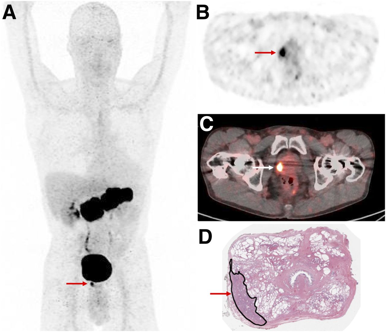
- FIGURE 1.
A 64-y-old man with newly diagnosed high-risk prostate cancer (PSA, 6.42 ng/mL). Intense focal 68Ga-RM2 uptake is seen in prostate gland (arrows) on maximum-intensity-projection (A), axial PET (B), and PET/CT (C) images, correlating with location of cancer marked in black ink on postprostatectomy histopathology slide (D).
- FIGURE 2.
A 72-y-old man with BCR prostate cancer (PSA, 0.72 ng/mL). (A, B, and D) Intense 68Ga-RM2 uptake is seen in right prostate bed (arrows) on early maximum-intensity-projection (A), axial PET (B), and PET/MR (D) images. (C) Corresponding axial T1-weighted MR image is also shown.
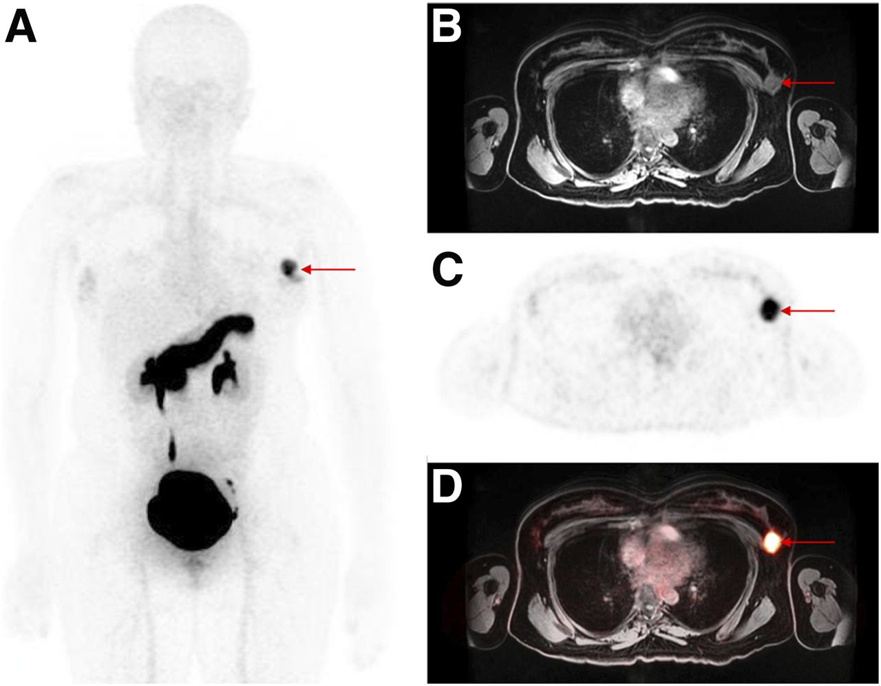
- FIGURE 3.
A 36-y-old woman with newly diagnosed ER-positive breast cancer. Intense 68Ga-RM2 uptake is seen in left breast (arrows) on maximum-intensity-projection (A), axial T1-weighted MR (B), axial PET (C), and PET/MR (D) images.
- FIGURE 4.
A 54-y-old man with newly diagnosed intermediate-risk prostate cancer (PSA, 5.09 ng/mL). Focal 68Ga-RM2 uptake is seen in right prostate gland (arrow) on axial fused PET/CT and PET images (top row), whereas focal 68Ga-PSMA11 uptake is seen in left prostate gland (arrow) on axial PET/MR and PET images (bottom row). Both were prostate cancer on postprostatectomy histopathology.


Additional Files
Supplemental Data
Files in this Data Supplement:


