


Article Figures & Data
Figures
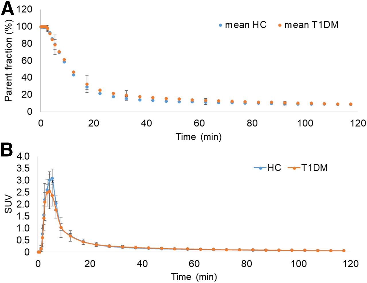
- FIGURE 1.
(A) Mean ± SD of parent fraction curves from HC and T1DM individuals. (B) Mean ± SD of metabolite-corrected arterial input functions from HC and T1DM individuals *Significant difference (P = 0.02) was seen between groups only at 5 min.
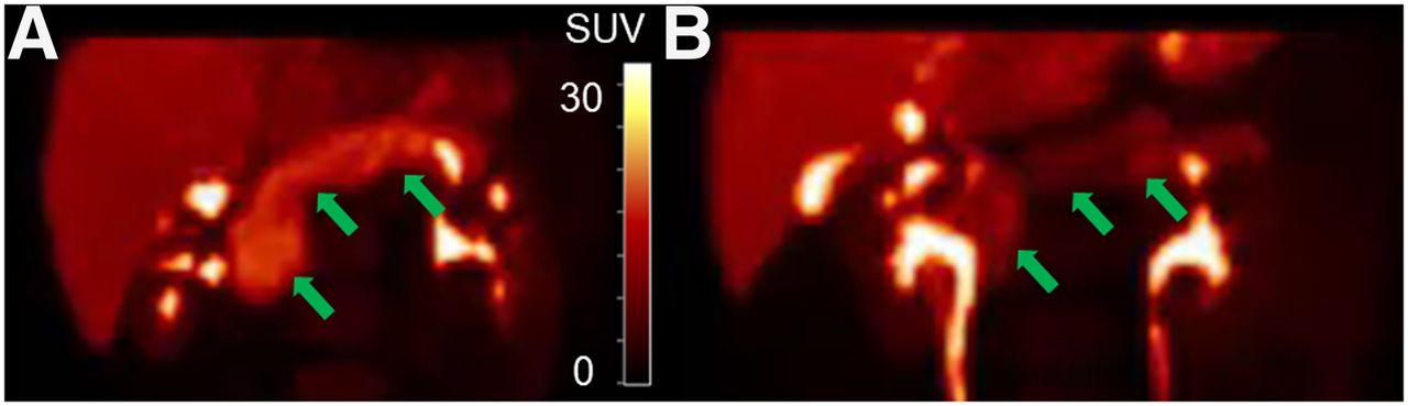
- FIGURE 2.
Representative maximum-intensity projections of PET SUV images (20–30 min) of 11C-(+)-PHNO pancreas (arrows) with uptake similar to SUV group means for HC (A) and type 1 diabetic mellitus (B) individuals. SUV scale = 0–30.
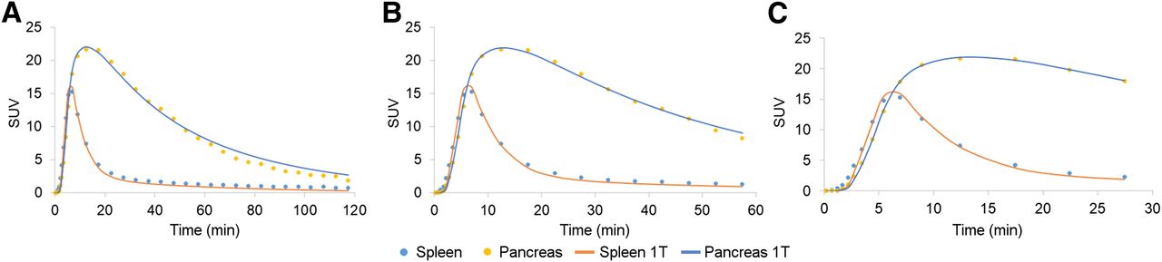
- FIGURE 3.
Representative time–activity curves for pancreas and spleen and 1TC model fits for tmax of 120 min (A), 60 min (B), and 30 min (C).
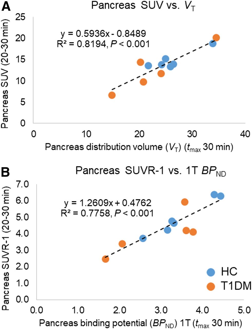
- FIGURE 4.
(A) Correlation of 1TC model (tmax, 30 min) pancreas VT values with pancreas SUV summed from 20 to 30 min. (B) Correlation of 1TC model (tmax, 30 min) pancreas BPND values (spleen as reference region) with pancreas SUVR-1 summed from 20 to 30 min.
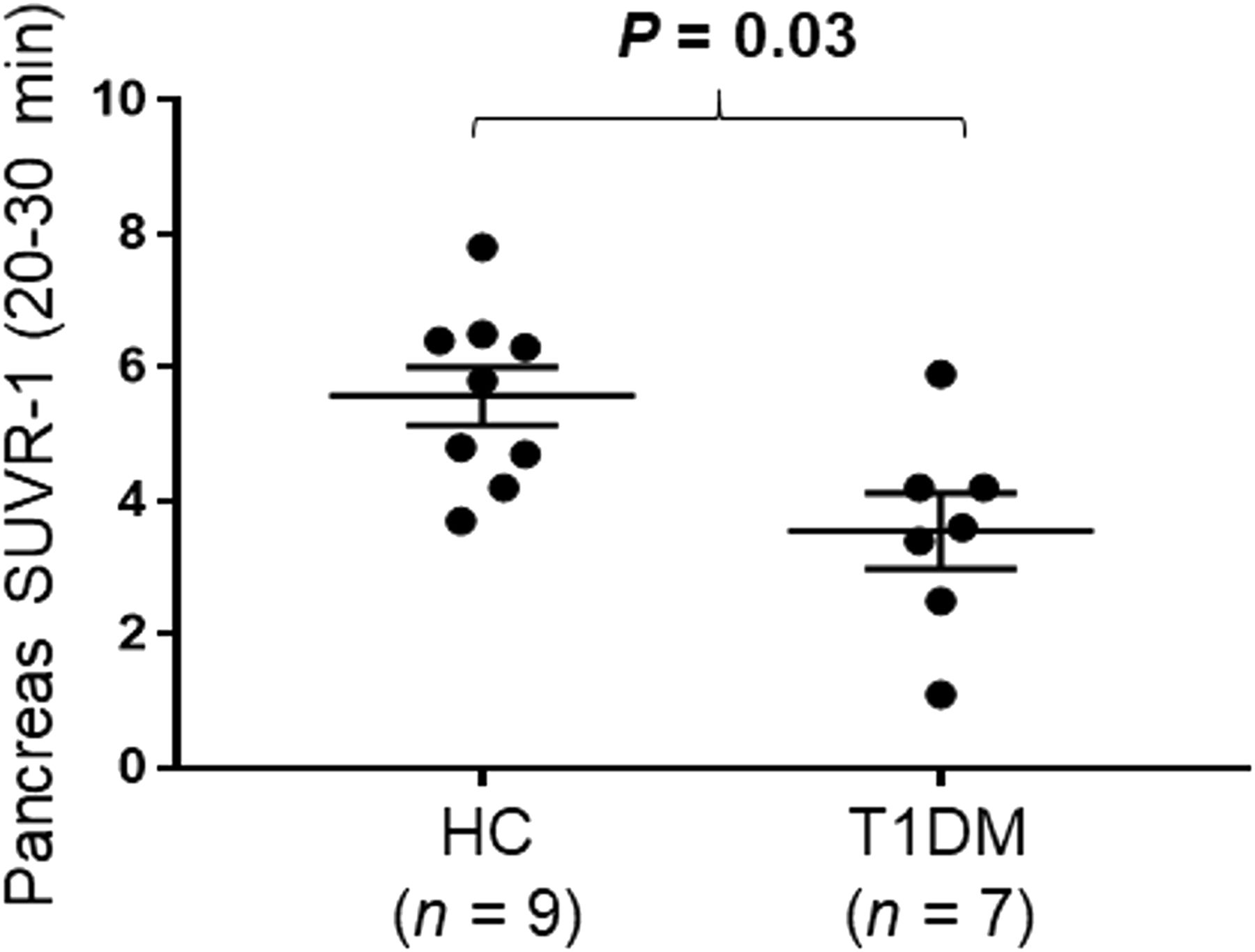
- FIGURE 5.
Dot plot demonstrating group differences between HCs and T1DMs for pancreas SUVR-1 (20–30 min). Data are mean ± SEM.
- FIGURE 6.
(A and B) Pearson correlations of age at diagnosis with pancreas BPND SRTM (tmax, 30 min) (A) and age at diagnosis with pancreas SUVR-1 (20–30 min) (B). (C and D) Spearman rank correlation of C-peptide release from arginine stimulus test with SUVR-1 (20–30 min) (C) and proinsulin release from arginine stimulus test with SUVR-1 (20–30 min) (D). All methods use spleen as reference region.


Tables
Diagnosis Age at diagnosis (y) T1DM duration (y) HbA1c Fasting levels before PET scan C-peptide (ng/mL)* Proinsulin (pM)* C-peptide (ng/dL) Blood glucose (mg/dL) Baseline (−5 to 0 min) Acute phase (2–10 min minus baseline) Steady-state (100–120 min) Maximal glycemic potentiation) (120–122 min) Baseline (−5 to 0 min) Steady-state (100–120 min) Maximal glycemic potentiation) (120–122 min) T1DM 14 16 5.3 0 135 0 0 0 0 1.0 1.0 2.0 T1DM 9 17 7.7 0 169 0 0.02 0 0 5.0 3.5 3.1 T1DM 35 19 7.2 0.05 141 0 0 0 0 8.0 3.8 6.3 T1DM 7 17 5.9 0 145 0 0 0 0 4.2 5.4 5.5 T1DM 35 14 7.8 0 217 0 0 0 0 1.0 1.0 1.0 T1DM 2 30 11.1 0 147 T1DM 8 14 7.6 0.38 95 HC NA NA 0.86 1.5 7.4 5.6 10.5 60.4 77.5 HC NA NA 78 1.21 2.5 11.5 9.7 8.5 51.8 68.0 HC NA NA 87 HC NA NA 70 HC NA NA 72 HC NA NA 85 HbA1c = hemoglobin A1c, in percentage (%); NA = not applicable; empty cells = data not available.
↵* C-peptide release and proinsulin data are from plasma samples during glucose clamp studies for HC (n = 2) and T1DM individuals (n = 5). For C-peptide, values < 0.02 were undetectable and listed as 0 ng/mL. For proinsulin, values < 2.0 were undetectable and listed as 1 pM.
Supplemental Data
Files in this Data Supplement:


Jump to section
Related Articles
Cited By...
- No citing articles found.