


Article Figures & Data
Figures
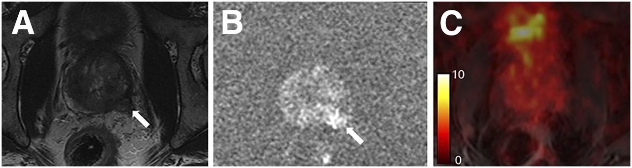
- FIGURE 1.
A 66-y-old man with biopsy-proven PC (prostate-specific antigen, 5.0 ng/mL; Gleason score, 4 + 4). T2-weighted image (A), diffusion-weighted image (B), and fused PET/MR image (C) are shown. Primary tumor (arrows) in right peripheral zone showed low and high signal intensity on T2-weighted image and diffusion-weighted image, respectively. Although all readers pointed out 68Ga-PSMA11 uptake corresponding to this tumor, readers’ judgments based on EANM criteria were discordant (positive vs. equivocal). In such a case, a difference in each reader’s recognition of “focal intense” and “moderate,” referred to in EANM criteria, might lead to interreader disagreement.
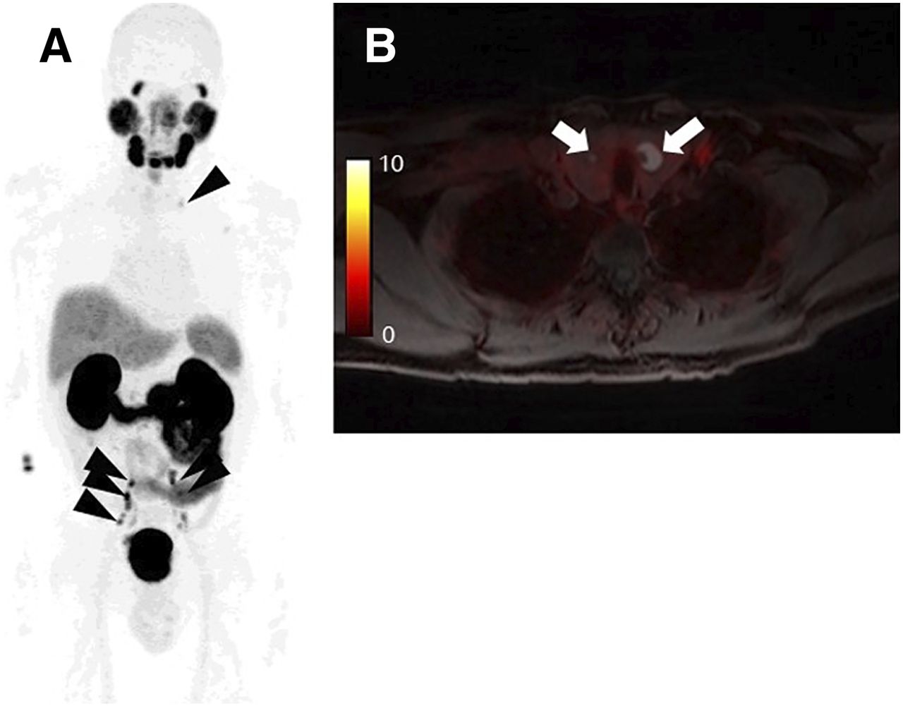
- FIGURE 2.
A 63-y-old man with biopsy-proven PC (prostate-specific antigen, 50.4 ng/mL; Gleason score, 4 + 5). (A) Maximum-intensity projection image showed multiple lymph node metastases in pelvic and left supraclavicular regions (arrowheads). (B) One reader pointed out thyroid nodules without focal 68Ga-PSMA11 uptake (arrows) and judged them as equivocal (PSMA-RADS-3D), whereas other readers did not refer to these lesions and judged them as negative. Further assessment has not been performed for thyroid.
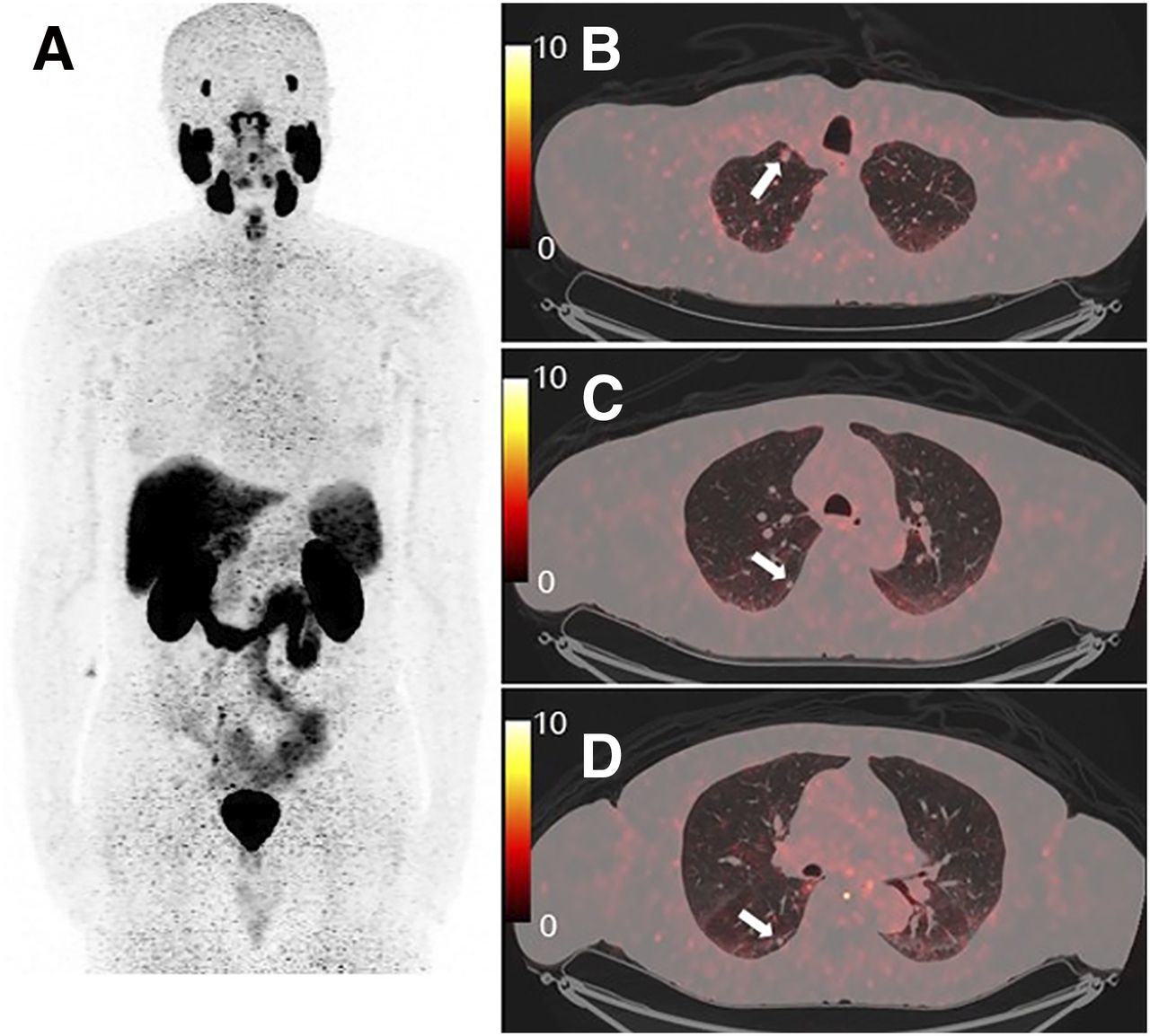
- FIGURE 3.
A 75-y-old man with BCR PC after radical prostatectomy (prostate-specific antigen, 0.2 ng/mL). (A) Maximum-intensity projection image did not reveal any abnormal 68Ga-PSMA11 uptake. (B–D) However, fused PET/CT images showed multiple lung nodules (arrows). One reader judged these nodules as PSMA-RADS-3D (lesion suggestive of malignancy but lacking uptake), whereas other readers judged them as negative. These nodules were judged as negative by all readers based on EANM and PROMISE criteria because of lack of 68Ga-PSMA11 uptake. Prostate-specific antigen stayed <1.0 ng/mL under bicalutamide treatment.
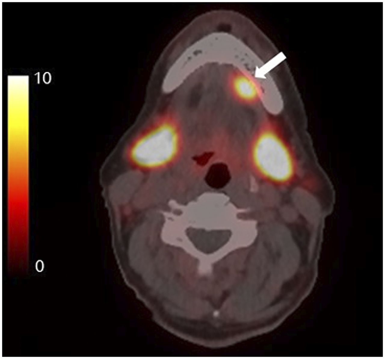
- FIGURE 4.
A 73-y-old man with BCR PC after high-dose external radiotherapy (prostate-specific antigen, 3.8 ng/mL). One reader pointed out asymmetric 68Ga-PSMA11 uptake in left sublingual gland (arrow). Judgments were nonpathologic (other malignancy may be considered), equivocal (“consider positive” because of higher uptake than that of parotid glands), and PSMA-RADS-3C (intense uptake in site highly atypical of PC) based on EANM criteria, PROMISE, and PSMA-RADS, respectively. Further assessment of this uptake was not performed.
- FIGURE 5.
A 60-y-old man with biopsy-proven PC (prostate-specific antigen, 9.4 ng/mL; Gleason score, 4 + 5). (A and B) Primary tumor with invasion to left seminal vesicle (arrows) was clearly detected on T2-weighted (A) and diffusion-weighted (B) MR images. (C) However, this lesion did not have elevated 68Ga-PSMA-11 uptake on fused PET/MR image. Although histopathologic evidence was unavailable, PSMA-ligand–negative PC was suspected. This primary tumor could be judged as negative (no uptake), positive (PI-RADS class 5), and equivocal (PSMA-RADS-3D) based on EANM criteria, PROMISE, and PSMA-RADS, respectively.


Tables
Parameter EANM (9) PROMISE (10) PSMA-RADS (11) Summary All areas of increased PSMA uptake in sites not expected to show physiologic uptake are to be reported as “anomalous,” followed by subclassification to 3 categories Both CT/MRI appearance and PSMA uptake are considered, and diagnosis is judged as “positive,” “equivocal,” or “negative” for each site All abnormal findings are classified by 5-point scale based on possibility of cancerous lesion Definition of significant uptake Focal uptake higher than adjacent background Basically, uptake equal to or above liver Not clearly defined Lesion site Local sites, local lymph nodes, distant lymph nodes, skeletal, other Local sites before and after treatment, lymph nodes, bone/visceral organ Bone, soft tissue (including lymph nodes) Classification in each site Anomalous, pathologic, uncertain, nonpathologic, normal Positive, equivocal, negative 5: PC almost certainly present; 4: PC highly likely; 3: equivocal (3A–D); 2: likely benign; 1: benign (1A/B) Final judgment Abnormal (pathologic), normal Positive, equivocal, or negative, plus miTNM classification Highest PSMA-RADS score among detected lesions miTNM = molecular imaging TNM.
Characteristic PET/MRI for initial staging PET/CT due to BCR n 47 57 Age (y) 64.2 ± 6.1 (44–74) 70.5 ± 6.7 (58–89) Prostate-specific antigen (ng/mL) 10.4 ± 8.4 (3.3–50.4) 35.7 ± 172.2 (0.2–1.170) Injected dose (MBq) 155.4 ± 30.7 (91.4–236.4) 145.8 ± 14.8 (111–199.8) Uptake time (min) 49.9 ± 5.3 (41–69) 61.4 ± 13.6 (44–90) Treatment before PET/CT Not applicable Prostatectomy, 41; radiotherapy, 32; brachytherapy, 6; hormonal therapy, 36; chemotherapy, 3; 223Ra, 1 Continuous data are expressed as mean ± SD, followed by range in parentheses.
Parameter Reference* EANM PROMISE PSMA-RADS Each lesion site Positive Pathologic Positive PSMA-RADS-4/5 Equivocal Uncertain Equivocal PSMA-RADS-3 Negative Nonpathologic/normal Negative PSMA-RADS-1/2 Final judgment per patient Positive Abnormal Positive PSMA-RADS-4/5 Equivocal Not applicable Equivocal PSMA-RADS-3 Negative Normal Negative PSMA-RADS-1/2 ↵* These judgments were used for statistical analyses in this study.
Group Site EANM PROMISE PSMA-RADS PET/MRI for initial staging Local sites 0.70 0.75 0.73 Lymph node metastases 0.93 0.93 0.93 Distant metastases 0.96 0.97 0.89 Final judgment 0.89 0.79 0.72 PET/CT due to BCR Local sites 0.69 0.73 0.77 Lymph node metastases 0.80 0.79 0.78 Distant metastases 0.84 0.80 0.57* Final judgment 0.79 0.67 0.64 ↵* Moderate agreement.
Group Site EANM PROMISE PSMA-RADS PET/MRI for initial staging Local sites 0.95/0.63 0.93/0.74 0.98/0.70 Lymph node metastases 0.93/0.98 0.79/0.98 0.93/0.98 Distant metastases 0.93/1.00 0.96/0.98 0.98/0.98 Final judgment 0.95/0.94 0.91/0.77 0.88/0.75 PET/CT due to BCR Local sites 0.93/0.78 0.91/0.78 0.93/0.78 Lymph node metastases 0.90/0.79 0.86/0.73 0.86/0.69 Distant metastases 0.92/0.83 0.83/0.81 0.75/0.81 Final judgment 0.91/0.73 0.93/0.52* 0.91/0.49* ↵* Moderate agreement.
Each Gwet AC is expressed as reader 1/reader 2.
Group Site Gwet AC PET/MRI for initial staging Local 0.92 Lymph node 0.97 Distant 0.96 Final judgment 0.93 PET/CT due to BCR Local 0.97 Lymph node 0.98 Distant 0.72 Final judgment 0.91


Jump to section
Related Articles
Cited By...
- Reproducibility of PSMA PET/CT Imaging for Primary Staging of Treatment-Naive Prostate Cancer Patients Depends on the Applied Radiotracer: A Retrospective Study
- 18F-DCFPyL PET Acquisition, Interpretation, and Reporting: Suggestions After Food and Drug Administration Approval
- aPROMISE: A Novel Automated PROMISE Platform to Standardize Evaluation of Tumor Burden in 18F-DCFPyL Images of Veterans with Prostate Cancer
- High Interobserver Agreement for the Standardized Reporting System SSTR-RADS 1.0 on Somatostatin Receptor PET/CT
- Automated analysis of PSMA-PET/CT studies using convolutional neural networks