


Article Figures & Data
Figures
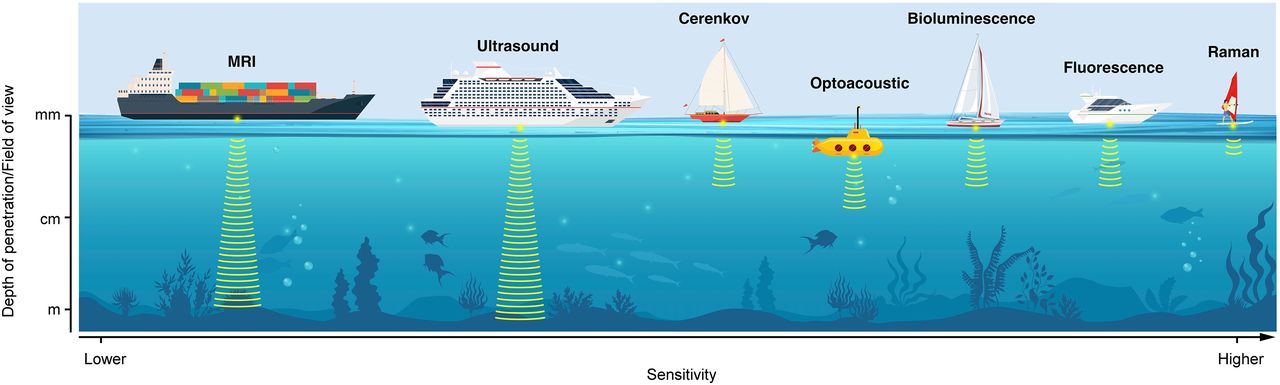
- FIGURE 1.
Schematic representation of various optical imaging modalities discussed in this review. Different modalities are represented as different boats, each with its individual pros and cons and its specific utility. (Printed with permission of Memorial Sloan Kettering Cancer Center.)
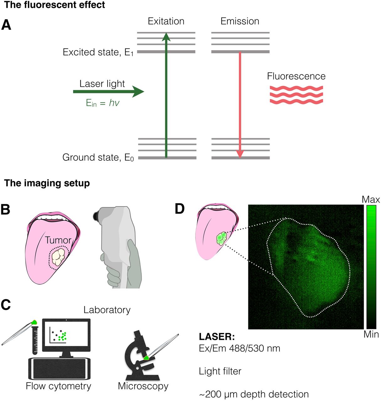
- FIGURE 2.
Fluorescence disease imaging. (A) Incident laser light can excite electron to E1; relaxation of acquired excited state emits light known as fluorescence. Ein is the incident photon energy, v is its frequency, and h is the Plank constant. (B) One potential application for fluorescence molecules targeting tumors is margin delineation of surface lesions, such as in oropharyngeal cancer. (C) Fluorescent dyes have broad application in laboratory settings, such as in flow cytometry sorting or fluorescent microscopy. (D) Example of use of PARPi-FL, a fluorescent molecule targeting poly(adenosine diphosphate ribose) polymerase 1, for tumor detection in clinic. Ex/Em = excitation/emission. (Adapted with permission of (22).)
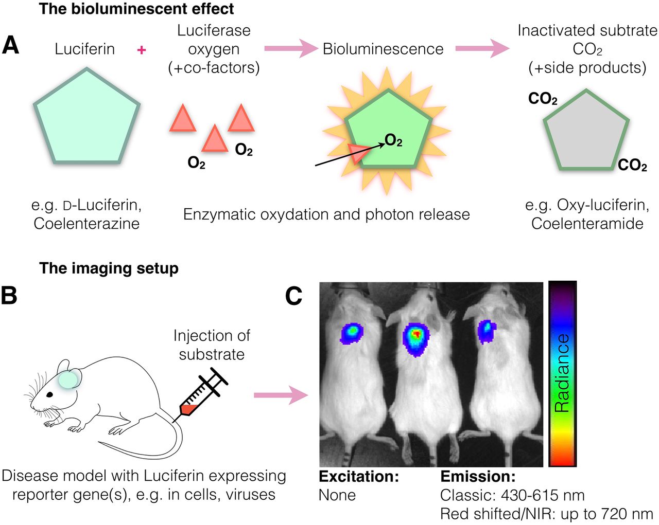
- FIGURE 3.
Bioluminescence method for disease imaging. (A) Bioluminescence emission occurs on oxidation of substrate (luciferin) by enzyme (luciferase), in some cases requiring cofactors such as adenosine triphosphate and magnesium. (B) In BLI animal models, transplanted cells or genetically modified tissues express luciferin, whereas luciferase is delivered systemically to induce bioluminescence. (C) In small animals, bioluminescence detection usually occurs using charge-coupled-device cameras, which are suitable for low-light detection. NIR = near infrared.
- FIGURE 4.
Optoacoustic methods for cancer detection. (A) Excitation from light absorption causes absorber to undergo radiative relaxation, which generates local heating. Thermal relaxation generates pressure waves and, in turn, thermoelastic expansion, known as photoacoustic effect. (B) Example of optoacoustic imaging setup (multispectral optoacoustic tomography, or MSOT), which surrounds tissue of interest with ring laser and ultrasound transducer in 270° array. MSOT has tunable laser (680–900 nm) and allows for multispectral unmixing. (C) Representative MSOT images after multispectral unmixing before (top) and after (bottom) intravenous injection of NIR dye (green) and overlaid with optoacoustic background (900 nm). Ein is the incident photon energy, ER is the relaxation energy, ET1 and ET2 represent thermal relaxation energy, and Ed is the difference of relaxation energy. v and v′ refer to the photon frequency, and h represents the Plank constant. a.u. = arbitrary units; i.v. = intravenous; US = ultrasound. (Adapted with permission of (41,90).)
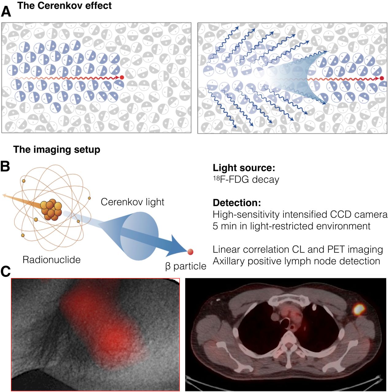
- FIGURE 5.
Cerenkov luminescence light for disease detection. (A) Left: Charged particle (red dot) traveling more quickly than light in medium polarizes medium. Right: As medium returns to ground state, blue-weighted light is emitted in forward direction. (B) Cerenkov light is emitted (blue cone and arrow) by medium in which charged particle travels. Radionuclides that emit β-particles with energies greater than Cerenkov threshold (261 keV in water) result in CL. (C) Left: White-light photograph from left axilla, overlaid with significant CL signal. Right: This signal colocalized with PET/CT finding. CCD = charge-coupled device. (Adapted with permission of (68,83).)

Tables
Parameter Fluorescence Bioluminescence Optoacoustic CL Principle Absorption of light excites status of dye and its relaxation emits light Oxidation of substrate by enzyme emits light Light is absorbed and causes molecular vibrations that emit sound waves Charged particles travel quickly through medium and emit light Advantage Easy to perform; large variety of dyes No incident radiation needed; no background Safe Safe and informative Disadvantage Needs excitation source Is not yet clinical Needs complex image reconstruction Needs radioactive preinjection Translatable? Yes Not currently Yes Yes Ease to implement Medium Medium Medium Difficult Ease to use Easy Medium Medium Medium Ease to detect Easy Easy Medium Medium Detectable range Visible spectrum and NIR 470–750 nm 400–800 nm (RSOM), 680–980 nm (MSOT) Ultraviolet-to-visible spectrum Preclinical applications In vitro and in vivo molecular imaging In vitro and in vivo molecular imaging In vivo tumor and vasculature imaging In vivo tumor imaging Potential clinical applications Screening, diagnostic, intraoperative Stem cell or chimeric antigen receptor T-cell tracking Muscular dystrophy, vasculature, cancer imaging Positive lymph nodes and cancer detection Cost Low Low Medium High


