


Abstract
555
Objectives: Endometrial cancers have been divided into estrogen-dependent type I and the less common clinically aggressive estrogen-independent type II. The aim of this study was to evaluate the capability of integrated FDG-PET/MRI to characterize the distinct phenotypes of endometrial cancer.
Methods: 30 patients with endometrial cancer (22 with type I including 17 G1 and 5 G2 endometrioid adenocarcinomas, and 8 with type II including 3 G3 endometrioid adenocarcinomas, 2 carcinosarcomas and 3 serous carcinomas) underwent pretreatment FDG-PET/MRI scan with simultaneous high-resolution, small field-of-view diffusion weighted imaging. SUV, apparent diffusion coefficient (ADC), and SUV-to-ADC ratio were compared between type I and II cancers and between low-risk (type I and stage IA) and high-risk (type II or stage IB ≤) cancers. The diagnostic accuracy for discriminating the cancer phenotypes was compared using receiver-operating-characteristic (ROC) analysis.
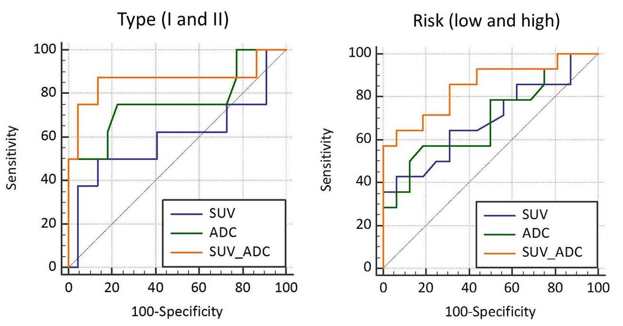
Results: Although SUV was not significantly different between type I and II endometrial cancers, type II cancer showed a significantly lower ADC (667±276 x 10-6) and a greater SUV-to-ADC ratio (24.5±8.1 x 10⁹) than did type I cancer (924±145 x 10-6, p=0.04, and 14.1±4.9 x 10⁹, p<0.001, respectively). High-risk endometrial cancer showed a significantly higher SUV (15.6±5.8), a lower ADC (758±242 x 10-6), and a greater SUV-to-ADC ratio (21.7±7.5 x 10⁹) than did low-risk cancer (11.7±3.5, p=0.04, 941±153 x 10-6, p=0.02, and 12.7±4.1 x 10⁹, p=0.001, respectively). In ROC analysis, the most accurate diagnostic index for predicting type I and II cancers and for predicting low-risk and high-risk cancers was SUV-to-ADC ratio. The optimal SUV-to-ADC cutoff value of 18.5 x 10⁹ for predicting type II cancer revealed 88% sensitivity, 86% specificity, and 87% accuracy, and the optimal cutoff of 13.6 x 10⁹ for predicting high-risk cancer revealed 86% sensitivity, 69% specificity, and 77% accuracy, that were significantly better than the 77% and 67% accuracy for SUV, respectively.
Conclusions: Endometrial cancer tends to increase tumor cellularity with accelerated glucose metabolism in higher histological grade or stage. The SUV-to-ADC ratio obtained by integrated FDG-PET/MRI reflects tumor aggressiveness and will be useful for noninvasive diagnosis and for deciding on the appropriate therapeutic strategy for patients with endometrial cancer.

In this issue


Jump to section
Related Articles
Cited By...
- No citing articles found.